3D models of Cainotheriids Ossicular chain
Explodable 3D Dog Skull for Veterinary Education
3D models of Kalakocetus, the earliest Cetacea
3D GM dataset of bird skeletal variation
Skeletal embryonic development in the catshark
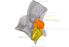
Bony connexions of the petrosal bone of extant hippos
bony labyrinth (14) , inner ear (11) , Eocene (11) , geometric morphometrics (10) , CT-scan (10) , Oligocene (9) , Micro-CT (9)
Lionel Hautier (25) , Maëva Judith Orliac (24) , Laurent Marivaux (18) , Renaud Lebrun (15) , Rodolphe Tabuce (15) , Pierre-Olivier Antoine (13) , Bastien Mennecart (13)
|
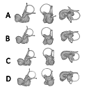
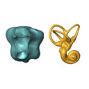
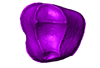
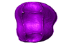
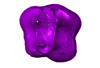
3D models related to the publication: Size Variation under Domestication: Conservatism in the inner ear shape of wolves, dogs and dingoesAnita V. Schweizer, Renaud Lebrun
Published online: 17/10/2017 |

|
M3#2293D virtual endocast of the left inner ear Type: "3D_surfaces"doi: 10.18563/m3.sf.229 state:published |
Download 3D surface file |
Canis lupus familiaris NMBE-LAT-1136 View specimen

|
M3#2423D virtual endocast of the left inner ear Type: "3D_surfaces"doi: 10.18563/m3.sf.242 state:published |
Download 3D surface file |
Canis lupus familiaris NMBE-LAT-1119 View specimen

|
M3#2433D virtual endocast of the left inner ear Type: "3D_surfaces"doi: 10.18563/m3.sf.243 state:published |
Download 3D surface file |
Canis lupus familiaris NMBE-BUR-1057 View specimen

|
M3#2443D virtual endocast of the left inner ear Type: "3D_surfaces"doi: 10.18563/m3.sf.244 state:published |
Download 3D surface file |
Canis lupus familiaris NMBE-LUS-1102 View specimen

|
M3#2453D virtual endocast of the left inner ear Type: "3D_surfaces"doi: 10.18563/m3.sf.245 state:published |
Download 3D surface file |
Canis lupus familiaris NMBE-LUS-1095 View specimen

|
M3#2463D virtual endocast of the left inner ear Type: "3D_surfaces"doi: 10.18563/m3.sf.246 state:published |
Download 3D surface file |
Canis lupus familiaris NMBE-DUR-1124 View specimen

|
M3#2473D virtual endocast of the left inner ear Type: "3D_surfaces"doi: 10.18563/m3.sf.247 state:published |
Download 3D surface file |
Canis lupus chanco ZMUZH 17603 View specimen

|
M3#2483D virtual endocast of the left inner ear Type: "3D_surfaces"doi: 10.18563/m3.sf.248 state:published |
Download 3D surface file |
Canis lupus chanco ZMUZH 20201 View specimen

|
M3#2493D virtual endocast of the left inner ear Type: "3D_surfaces"doi: 10.18563/m3.sf.249 state:published |
Download 3D surface file |
Canis lupus chanco ZMUZH 17602 View specimen

|
M3#2503D virtual endocast of the left inner ear Type: "3D_surfaces"doi: 10.18563/m3.sf.250 state:published |
Download 3D surface file |
Canis lupus ZMUZH 13854 View specimen

|
M3#2403D virtual endocast of the left inner ear Type: "3D_surfaces"doi: 10.18563/m3.sf.240 state:published |
Download 3D surface file |
Canis lupus chanco ZMUZH 20202 View specimen

|
M3#2393D virtual endocast of the left inner ear Type: "3D_surfaces"doi: 10.18563/m3.sf.239 state:published |
Download 3D surface file |
Canis lupus chanco ZMUZH 17612 View specimen

|
M3#2303D virtual endocast of the left inner ear Type: "3D_surfaces"doi: 10.18563/m3.sf.230 state:published |
Download 3D surface file |
Canis lupus chanco ZMUZH 18082 View specimen

|
M3#2313D virtual endocast of the left inner ear Type: "3D_surfaces"doi: 10.18563/m3.sf.231 state:published |
Download 3D surface file |
Canis lupus ZMUZH 17118 View specimen

|
M3#2323D virtual endocast of the left inner ear Type: "3D_surfaces"doi: 10.18563/m3.sf.232 state:published |
Download 3D surface file |
Canis lupus ZMUZH 15858 View specimen

|
M3#2333D virtual endocast of the left inner ear Type: "3D_surfaces"doi: 10.18563/m3.sf.233 state:published |
Download 3D surface file |
Canis lupus familiaris ZMUZH 17712 View specimen

|
M3#2343D virtual endocast of the left inner ear Type: "3D_surfaces"doi: 10.18563/m3.sf.234 state:published |
Download 3D surface file |
Canis lupus familiaris ZMUZH 17713 View specimen

|
M3#2353D virtual endocast of the left inner ear Type: "3D_surfaces"doi: 10.18563/m3.sf.235 state:published |
Download 3D surface file |
Canis lupus familiaris ZMUZH 10166 View specimen

|
M3#2363D virtual endocast of the left inner ear Type: "3D_surfaces"doi: 10.18563/m3.sf.236 state:published |
Download 3D surface file |
Canis lupus familiaris ZMUZH 10175 View specimen

|
M3#2373D virtual endocast of the left inner ear Type: "3D_surfaces"doi: 10.18563/m3.sf.237 state:published |
Download 3D surface file |
Canis lupus familiaris ZMUZH 14842 View specimen

|
M3#2383D virtual endocast of the left inner ear Type: "3D_surfaces"doi: 10.18563/m3.sf.238 state:published |
Download 3D surface file |
Canis lupus familiaris ZMUZH 10342 View specimen

|
M3#2513D virtual endocast of the left inner ear Type: "3D_surfaces"doi: 10.18563/m3.sf.251 state:published |
Download 3D surface file |
Canis lupus familiaris ZMUZH 10343 View specimen

|
M3#2523D virtual endocast of the left inner ear Type: "3D_surfaces"doi: 10.18563/m3.sf.252 state:published |
Download 3D surface file |
Canis lupus familiaris ZMUZH 13766 View specimen

|
M3#2533D virtual endocast of the left inner ear Type: "3D_surfaces"doi: 10.18563/m3.sf.253 state:published |
Download 3D surface file |
Canis lupus familiaris ZMUZH 17717 View specimen

|
M3#2653D virtual endocast of the left inner ear Type: "3D_surfaces"doi: 10.18563/m3.sf.265 state:published |
Download 3D surface file |
Canis lupus familiaris ZMUZH 17711 View specimen

|
M3#2663D virtual endocast of the left inner ear Type: "3D_surfaces"doi: 10.18563/m3.sf.266 state:published |
Download 3D surface file |
Canis lupus familiaris ZMUZH 17714 View specimen

|
M3#2673D virtual endocast of the left inner ear Type: "3D_surfaces"doi: 10.18563/m3.sf.267 state:published |
Download 3D surface file |
Canis lupus familiaris ZMUZH 17715 View specimen

|
M3#2683D virtual endocast of the left inner ear Type: "3D_surfaces"doi: 10.18563/m3.sf.268 state:published |
Download 3D surface file |
Canis lupus familiaris PIMUZ A/V 2835 View specimen

|
M3#2693D virtual endocast of the left inner ear Type: "3D_surfaces"doi: 10.18563/m3.sf.269 state:published |
Download 3D surface file |
Canis lupus familiaris PIMUZ A/V 2834 View specimen

|
M3#2703D virtual endocast of the left inner ear Type: "3D_surfaces"doi: 10.18563/m3.sf.270 state:published |
Download 3D surface file |
Canis lupus familiaris PIMUZ A/V 2837 View specimen

|
M3#2713D virtual endocast of the left inner ear Type: "3D_surfaces"doi: 10.18563/m3.sf.271 state:published |
Download 3D surface file |
Canis lupus familiaris PIMUZ A/V 2831 View specimen

|
M3#2723D virtual endocast of the left inner ear Type: "3D_surfaces"doi: 10.18563/m3.sf.272 state:published |
Download 3D surface file |
Canis lupus familiaris PIMUZ A/V 2845 View specimen

|
M3#2733D virtual endocast of the left inner ear Type: "3D_surfaces"doi: 10.18563/m3.sf.273 state:published |
Download 3D surface file |
Canis lupus familiaris PIMUZ A/V 3001 View specimen

|
M3#2643D virtual endocast of the left inner ear Type: "3D_surfaces"doi: 10.18563/m3.sf.264 state:published |
Download 3D surface file |
Canis lupus familiaris PIMUZ A/V 2832 View specimen

|
M3#2633D virtual endocast of the left inner ear Type: "3D_surfaces"doi: 10.18563/m3.sf.263 state:published |
Download 3D surface file |
Canis lupus familiaris PIMUZ A/V 3000 View specimen

|
M3#2543D virtual endocast of the left inner ear Type: "3D_surfaces"doi: 10.18563/m3.sf.254 state:published |
Download 3D surface file |
Canis lupus familiaris PIMUZ A/V 2847 View specimen

|
M3#2553D virtual endocast of the left inner ear Type: "3D_surfaces"doi: 10.18563/m3.sf.255 state:published |
Download 3D surface file |
Canis lupus familiaris PIMUZ A/V 2846 View specimen

|
M3#2563D virtual endocast of the left inner ear Type: "3D_surfaces"doi: 10.18563/m3.sf.256 state:published |
Download 3D surface file |
Canis lupus familiaris PIMUZ A/V 2836 View specimen

|
M3#2573D virtual endocast of the left inner ear Type: "3D_surfaces"doi: 10.18563/m3.sf.257 state:published |
Download 3D surface file |
Canis lupus familiaris NMB 12080 View specimen

|
M3#2583D virtual endocast of the left inner ear Type: "3D_surfaces"doi: 10.18563/m3.sf.258 state:published |
Download 3D surface file |
Canis lupus familiaris NMB 12081 View specimen

|
M3#2593D virtual endocast of the left inner ear Type: "3D_surfaces"doi: 10.18563/m3.sf.259 state:published |
Download 3D surface file |
Canis lupus familiaris NMB 12079 View specimen

|
M3#2603D virtual endocast of the left inner ear Type: "3D_surfaces"doi: 10.18563/m3.sf.260 state:published |
Download 3D surface file |
Canis lupus familiaris NMB 12078 View specimen

|
M3#2613D virtual endocast of the left inner ear Type: "3D_surfaces"doi: 10.18563/m3.sf.261 state:published |
Download 3D surface file |
Canis lupus familiaris NMBE 1051209 View specimen

|
M3#2623D virtual endocast of the left inner ear Type: "3D_surfaces"doi: 10.18563/m3.sf.262 state:published |
Download 3D surface file |
Canis lupus familiaris NMBE 1051226 View specimen

|
M3#2283D virtual endocast of the left inner ear Type: "3D_surfaces"doi: 10.18563/m3.sf.228 state:published |
Download 3D surface file |
Canis lupus familiaris NMBE 1051381 View specimen

|
M3#2213D virtual endocast of the left inner ear Type: "3D_surfaces"doi: 10.18563/m3.sf.221 state:published |
Download 3D surface file |
Canis lupus familiaris NMBE 1051418 View specimen

|
M3#1843D virtual endocast of the left inner ear Type: "3D_surfaces"doi: 10.18563/m3.sf.184 state:published |
Download 3D surface file |
Canis lupus familiaris ZMUZH A.II. View specimen

|
M3#1973D virtual endocast of the left inner ear Type: "3D_surfaces"doi: 10.18563/m3.sf.197 state:published |
Download 3D surface file |
Canis lupus familiaris ZMUZH A.VII. View specimen

|
M3#1983D virtual endocast of the left inner ear Type: "3D_surfaces"doi: 10.18563/m3.sf.198 state:published |
Download 3D surface file |
Canis lupus familiaris ZMUZH We.6. View specimen

|
M3#1993D virtual endocast of the left inner ear Type: "3D_surfaces"doi: 10.18563/m3.sf.199 state:published |
Download 3D surface file |
Canis lupus familiaris ZMUZH Ez.2. View specimen

|
M3#2003D virtual endocast of the left inner ear Type: "3D_surfaces"doi: 10.18563/m3.sf.200 state:published |
Download 3D surface file |
Canis lupus familiaris ZMUZH Ez.E. View specimen

|
M3#2013D virtual endocast of the left inner ear Type: "3D_surfaces"doi: 10.18563/m3.sf.201 state:published |
Download 3D surface file |
Canis lupus familiaris ZMUZH A.6. View specimen

|
M3#2023D virtual endocast of the left inner ear Type: "3D_surfaces"doi: 10.18563/m3.sf.202 state:published |
Download 3D surface file |
Canis lupus familiaris ZMUZH Wyn.9. View specimen

|
M3#2033D virtual endocast of the left inner ear Type: "3D_surfaces"doi: 10.18563/m3.sf.203 state:published |
Download 3D surface file |
Canis lupus familiaris ZMUZH F.48. View specimen

|
M3#2043D virtual endocast of the left inner ear Type: "3D_surfaces"doi: 10.18563/m3.sf.204 state:published |
Download 3D surface file |
Canis lupus familiaris ZMUZH Terp.1. View specimen

|
M3#2053D virtual endocast of the left inner ear Type: "3D_surfaces"doi: 10.18563/m3.sf.205 state:published |
Download 3D surface file |
Canis lupus familiaris ZMUZH A.VIII. View specimen

|
M3#1963D virtual endocast of the left inner ear Type: "3D_surfaces"doi: 10.18563/m3.sf.196 state:published |
Download 3D surface file |
Canis lupus familiaris ZMUZH A.VI. View specimen

|
M3#1953D virtual endocast of the left inner ear Type: "3D_surfaces"doi: 10.18563/m3.sf.195 state:published |
Download 3D surface file |
Canis lupus familiaris ZMUZH A.IV. View specimen

|
M3#1853D virtual endocast of the left inner ear Type: "3D_surfaces"doi: 10.18563/m3.sf.185 state:published |
Download 3D surface file |
Canis lupus familiaris NMBE A.403. View specimen

|
M3#1873D virtual endocast of the left inner ear Type: "3D_surfaces"doi: 10.18563/m3.sf.187 state:published |
Download 3D surface file |
Canis lupus familiaris NMBE A.5.a. View specimen

|
M3#1883D virtual endocast of the left inner ear Type: "3D_surfaces"doi: 10.18563/m3.sf.188 state:published |
Download 3D surface file |
Canis lupus NMB 8381 View specimen

|
M3#1893D virtual endocast of the left inner ear Type: "3D_surfaces"doi: 10.18563/m3.sf.189 state:published |
Download 3D surface file |
Canis lupus lycaon NMB C.1362 View specimen

|
M3#1903D virtual endocast of the left inner ear Type: "3D_surfaces"doi: 10.18563/m3.sf.190 state:published |
Download 3D surface file |
Canis lupus NMB Z309 View specimen

|
M3#1913D virtual endocast of the left inner ear Type: "3D_surfaces"doi: 10.18563/m3.sf.191 state:published |
Download 3D surface file |
Canis lupus NMB 2761 View specimen

|
M3#1923D virtual endocast of the left inner ear Type: "3D_surfaces"doi: 10.18563/m3.sf.192 state:published |
Download 3D surface file |
Canis lupus occidentalis NMB No Nb View specimen

|
M3#1933D virtual endocast of the left inner ear Type: "3D_surfaces"doi: 10.18563/m3.sf.193 state:published |
Download 3D surface file |
Canis lupus NMB 5258 View specimen

|
M3#1943D virtual endocast of the left inner ear Type: "3D_surfaces"doi: 10.18563/m3.sf.194 state:published |
Download 3D surface file |
Canis lupus NMB SCM320 View specimen

|
M3#2063D virtual endocast of the left inner ear Type: "3D_surfaces"doi: 10.18563/m3.sf.206 state:published |
Download 3D surface file |
Canis lupus arabs NMB 11019 View specimen

|
M3#2073D virtual endocast of the left inner ear Type: "3D_surfaces"doi: 10.18563/m3.sf.207 state:published |
Download 3D surface file |
Canis lupus UMZC K.3141 View specimen

|
M3#2083D virtual endocast of the left inner ear Type: "3D_surfaces"doi: 10.18563/m3.sf.208 state:published |
Download 3D surface file |
Canis lupus UMZC K.3150.1 View specimen

|
M3#2193D virtual endocast of the left inner ear Type: "3D_surfaces"doi: 10.18563/m3.sf.219 state:published |
Download 3D surface file |
Canis lupus UMZC K.3152 View specimen

|
M3#2203D virtual endocast of the left inner ear Type: "3D_surfaces"doi: 10.18563/m3.sf.220 state:published |
Download 3D surface file |
Canis lupus UMZC K.3149 View specimen

|
M3#2223D virtual endocast of the left inner ear Type: "3D_surfaces"doi: 10.18563/m3.sf.222 state:published |
Download 3D surface file |
Canis lupus familiaris UMZC K.3016 View specimen

|
M3#2233D virtual endocast of the left inner ear Type: "3D_surfaces"doi: 10.18563/m3.sf.223 state:published |
Download 3D surface file |
Canis lupus occidentalis ZMUZH 17210 View specimen

|
M3#2243D virtual endocast of the left inner ear Type: "3D_surfaces"doi: 10.18563/m3.sf.224 state:published |
Download 3D surface file |
Canis lupus familiaris SZ 7961 View specimen

|
M3#2253D virtual endocast of the left inner ear Type: "3D_surfaces"doi: 10.18563/m3.sf.225 state:published |
Download 3D surface file |
Canis lupus familiaris SZ 7959 View specimen

|
M3#2263D virtual endocast of the left inner ear Type: "3D_surfaces"doi: 10.18563/m3.sf.226 state:published |
Download 3D surface file |
Canis lupus familiaris SZ 7958 View specimen

|
M3#2173D virtual endocast of the left inner ear Type: "3D_surfaces"doi: 10.18563/m3.sf.217 state:published |
Download 3D surface file |
Canis lupus familiaris SZ 7930 View specimen

|
M3#2163D virtual endocast of the left inner ear Type: "3D_surfaces"doi: 10.18563/m3.sf.216 state:published |
Download 3D surface file |
Canis lupus familiaris SZ 7926 View specimen

|
M3#2183D virtual endocast of the left inner ear Type: "3D_surfaces"doi: 10.18563/m3.sf.218 state:published |
Download 3D surface file |
Canis lupus familiaris SZ 7929 View specimen

|
M3#2093D virtual endocast of the left inner ear Type: "3D_surfaces"doi: 10.18563/m3.sf.209 state:published |
Download 3D surface file |
Canis lupus dingo M6297 View specimen

|
M3#1863D virtual endocast of the left inner ear Type: "3D_surfaces"doi: 10.18563/m3.sf.186 state:published |
Download 3D surface file |
Canis lupus dingo M24153 View specimen

|
M3#2103D virtual endocast of the left inner ear Type: "3D_surfaces"doi: 10.18563/m3.sf.210 state:published |
Download 3D surface file |
Canis lupus dingo M33608 View specimen

|
M3#2113D virtual endocast of the left inner ear Type: "3D_surfaces"doi: 10.18563/m3.sf.211 state:published |
Download 3D surface file |
Canis lupus dingo M38587 View specimen

|
M3#2123D virtual endocast of the left inner ear Type: "3D_surfaces"doi: 10.18563/m3.sf.212 state:published |
Download 3D surface file |
Canis lupus dingo Blumenbach UMZC K.3221 View specimen

|
M3#2133D virtual endocast of the left inner ear Type: "3D_surfaces"doi: 10.18563/m3.sf.213 state:published |
Download 3D surface file |
Canis lupus dingo Blumenbach UMZC K.3223 View specimen

|
M3#2143D virtual endocast of the left inner ear Type: "3D_surfaces"doi: 10.18563/m3.sf.214 state:published |
Download 3D surface file |
Canis lupus dingo UniSyd FVS 45 View specimen

|
M3#2153D virtual endocast of the left inner ear Type: "3D_surfaces"doi: 10.18563/m3.sf.215 state:published |
Download 3D surface file |
Canis lupus dingo UNSW Z354 View specimen

|
M3#2273D virtual endocast of the left inner ear Type: "3D_surfaces"doi: 10.18563/m3.sf.227 state:published |
Download 3D surface file |
Canis lupus familiaris TMM M-150 View specimen

|
M3#2413D virtual endocast of the left inner ear Type: "3D_surfaces"doi: 10.18563/m3.sf.241 state:published |
Download 3D surface file |
Canis lupus M39960 View specimen

|
M3#2743D virtual endocast of the left inner ear Type: "3D_surfaces"doi: 10.18563/m3.sf.274 state:published |
Download 3D surface file |
Canis lupus NMB 8635 View specimen

|
M3#2753D virtual endocast of the left inner ear Type: "3D_surfaces"doi: 10.18563/m3.sf.275 state:published |
Download 3D surface file |
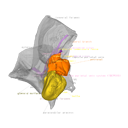
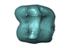
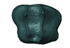
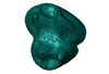
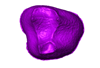
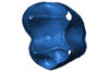
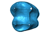
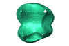
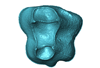
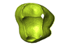
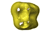
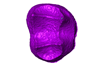
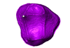
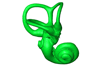
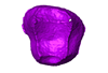
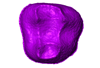
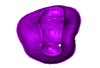
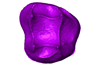
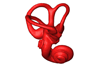
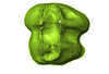
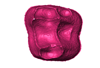
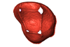
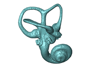
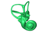
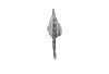
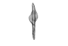
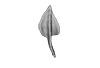
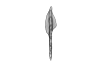
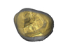
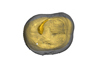
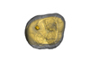
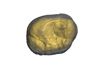
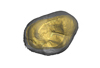
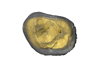
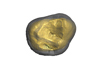
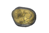
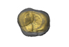
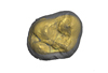
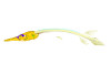
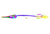
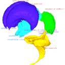
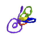
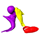
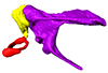
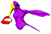
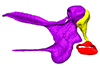
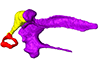
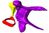
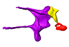
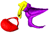
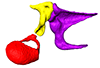
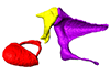
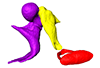
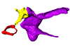
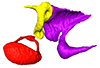
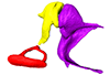
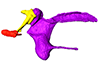
This project presents the osteological connexions of the petrosal bone of the extant Hippopotamidae Hippopotamus amphibius and Choeropsis liberiensis by a virtual osteological dissection of the ear region. The petrosal, the bulla, the sinuses and the major morphological features surrounding the petrosal bone are labelled, both in situ and in an exploded model presenting disassembly views. The directional underwater hearing mode of Hippopotamidae is discussed based on the new observations.
Choeropsis liberiensis UPPal-M09-5-005a View specimen
|
M3#1Labelled compact model of the right ear region of Choeropsis liberiensis (UPPal-M09-5-005a) Type: "3D_surfaces"doi: 10.18563/m3.sf1 state:published |
Download 3D surface file |

|
M3#2Labelled exploded model of the right ear region of Choeropsis liberiensis (UPPal-M09-5-005a) Type: "3D_surfaces"doi: 10.18563/m3.sf2 state:published |
Download 3D surface file |
Hippopotamus amphibius UM N179 View specimen

|
M3#3Labelled compact model of the right ear region of Hippopotamus amphibius (UM N 179) Type: "3D_surfaces"doi: 10.18563/m3.sf3 state:published |
Download 3D surface file |

|
M3#4Labelled exploded model of the right ear region of Hippopotamus amphibius (UM N 179) Type: "3D_surfaces"doi: 10.18563/m3.sf4 state:published |
Download 3D surface file |
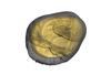
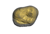
The present 3D Dataset contains the 3D models of the enamel-dentine junctions of upper third molars and of the bony labyrinths of the extant cercopithecoid specimens analyzed in the following publication: Beaudet, A., Dumoncel, J., Thackeray, J.F., Bruxelles, L., Duployer, B., Tenailleau, C., Bam, L., Hoffman, J., de Beer, F., Braga, J.: Upper third molar internal structural organization and semicircular canal morphology in Plio-Pleistocene South African cercopithecoids. Journal of Human Evolution 95, 104-120. https://doi.org/10.1016/j.jhevol.2016.04.004
Cercocebus atys 81.007-M-0041 View specimen
|
M3#4453D model of the enamel-dentine junction of the right upper third molar. Type: "3D_surfaces"doi: 10.18563/m3.sf.445 state:published |
Download 3D surface file |
Cercocebus torquatus 73.018-M-0359 View specimen
|
M3#4463D model of the enamel-dentine junction of the right upper third molar. Type: "3D_surfaces"doi: 10.18563/m3.sf.446 state:published |
Download 3D surface file |

|
M3#4963D model of the left bony labyrinth. Type: "3D_surfaces"doi: 10.18563/m3.sf.496 state:published |
Download 3D surface file |
Mandrillus leucophaeus 73.029-M-0106 View specimen
|
M3#4473D model of the enamel-dentine junction of the right upper third molar. Type: "3D_surfaces"doi: 10.18563/m3.sf.447 state:published |
Download 3D surface file |

|
M3#4703D model of the right bony labyrinth. Type: "3D_surfaces"doi: 10.18563/m3.sf.470 state:published |
Download 3D surface file |
Lophocebus albigena 73.029-M-0109 View specimen
|
M3#4483D model of the enamel-dentine junction of the right upper third molar. Type: "3D_surfaces"doi: 10.18563/m3.sf.448 state:published |
Download 3D surface file |

|
M3#4713D model of the right bony labyrinth. Type: "3D_surfaces"doi: 10.18563/m3.sf.471 state:published |
Download 3D surface file |
Piliocolobus foai 91.060-M-0071 View specimen
|
M3#4493D model of the enamel-dentine junction of the right upper third molar. Type: "3D_surfaces"doi: 10.18563/m3.sf.449 state:published |
Download 3D surface file |

|
M3#4723D model of the right bony labyrinth. Type: "3D_surfaces"doi: 10.18563/m3.sf.472 state:published |
Download 3D surface file |
Colobus guereza 1215 View specimen
|
M3#4503D model of the enamel-dentine junction of the right upper third molar. Type: "3D_surfaces"doi: 10.18563/m3.sf.450 state:published |
Download 3D surface file |

|
M3#4733D model of the right bony labyrinth. Type: "3D_surfaces"doi: 10.18563/m3.sf.473 state:published |
Download 3D surface file |
Colobus guereza 2800 View specimen
|
M3#4513D model of the enamel-dentine junction of the right upper third molar. Type: "3D_surfaces"doi: 10.18563/m3.sf.451 state:published |
Download 3D surface file |

|
M3#4743D model of the right bony labyrinth. Type: "3D_surfaces"doi: 10.18563/m3.sf.474 state:published |
Download 3D surface file |
Papio cynocephalus kindae 3503 View specimen
|
M3#4523D model of the enamel-dentine junction of the right upper third molar. Type: "3D_surfaces"doi: 10.18563/m3.sf.452 state:published |
Download 3D surface file |

|
M3#4753D model of the right bony labyrinth. Type: "3D_surfaces"doi: 10.18563/m3.sf.475 state:published |
Download 3D surface file |
Erythrocebus patas 8452 View specimen
|
M3#4533D model of the enamel-dentine junction of the right upper third molar. Type: "3D_surfaces"doi: 10.18563/m3.sf.453 state:published |
Download 3D surface file |

|
M3#4763D model of the right bony labyrinth. Type: "3D_surfaces"doi: 10.18563/m3.sf.476 state:published |
Download 3D surface file |
Papio cynocephalus kindae 17979 View specimen
|
M3#4543D model of the enamel-dentine junction of the right upper third molar. Type: "3D_surfaces"doi: 10.18563/m3.sf.454 state:published |
Download 3D surface file |

|
M3#4773D model of the right bony labyrinth. Type: "3D_surfaces"doi: 10.18563/m3.sf.477 state:published |
Download 3D surface file |
Colobus angolensis 25456 View specimen
|
M3#4553D model of the enamel-dentine junction of the right upper third molar. Type: "3D_surfaces"doi: 10.18563/m3.sf.455 state:published |
Download 3D surface file |

|
M3#4783D model of the right bony labyrinth. Type: "3D_surfaces"doi: 10.18563/m3.sf.478 state:published |
Download 3D surface file |
Chlorocebus pygerythrus 37477 View specimen
|
M3#4563D model of the enamel-dentine junction of the right upper third molar. Type: "3D_surfaces"doi: 10.18563/m3.sf.456 state:published |
Download 3D surface file |
|
M3#4813D model of the right bony labyrinth. Type: "3D_surfaces"doi: 10.18563/m3.sf.481 state:published |
Download 3D surface file |
Chlorocebus pygerythrus 37478 View specimen
|
M3#4573D model of the enamel-dentine junction of the right upper third molar. Type: "3D_surfaces"doi: 10.18563/m3.sf.457 state:published |
Download 3D surface file |

|
M3#4823D model of the right bony labyrinth. Type: "3D_surfaces"doi: 10.18563/m3.sf.482 state:published |
Download 3D surface file |
Lophocebus albigena 37572 View specimen
|
M3#4583D model of the enamel-dentine junction of the right upper third molar. Type: "3D_surfaces"doi: 10.18563/m3.sf.458 state:published |
Download 3D surface file |

|
M3#4833D model of the right bony labyrinth. Type: "3D_surfaces"doi: 10.18563/m3.sf.483 state:published |
Download 3D surface file |
Lophocebus albigena 37579 View specimen
|
M3#4593D model of the enamel-dentine junction of the right upper third molar. Type: "3D_surfaces"doi: 10.18563/m3.sf.459 state:published |
Download 3D surface file |
Erythrocebus patas OST.2002-26 View specimen
|
M3#4603D model of the enamel-dentine junction of the right upper third molar. Type: "3D_surfaces"doi: 10.18563/m3.sf.460 state:published |
Download 3D surface file |
|
M3#4843D model of the right bony labyrinth. Type: "3D_surfaces"doi: 10.18563/m3.sf.484 state:published |
Download 3D surface file |
Mandrillus sphinx OST.AC.488 View specimen
|
M3#4613D model of the enamel-dentine junction of the right upper third molar. Type: "3D_surfaces"doi: 10.18563/m3.sf.461 state:published |
Download 3D surface file |

|
M3#4853D model of the left bony labyrinth. Type: "3D_surfaces"doi: 10.18563/m3.sf.485 state:published |
Download 3D surface file |
Macaca mulatta OST.AC.492 View specimen
|
M3#4623D model of the enamel-dentine junction of the right upper third molar. Type: "3D_surfaces"doi: 10.18563/m3.sf.462 state:published |
Download 3D surface file |

|
M3#4863D model of the right bony labyrinth. Type: "3D_surfaces"doi: 10.18563/m3.sf.486 state:published |
Download 3D surface file |
Chlorocebus aethiops OST.AC.523 View specimen
|
M3#4633D model of the enamel-dentine junction of the right upper third molar. Type: "3D_surfaces"doi: 10.18563/m3.sf.463 state:published |
Download 3D surface file |

|
M3#4913D model of the right bony labyrinth. Type: "3D_surfaces"doi: 10.18563/m3.sf.491 state:published |
Download 3D surface file |
Cercopithecus cephus OST.AC.533 View specimen
|
M3#4643D model of the enamel-dentine junction of the right upper third molar. Type: "3D_surfaces"doi: 10.18563/m3.sf.464 state:published |
Download 3D surface file |

|
M3#4933D model of the right bony labyrinth. Type: "3D_surfaces"doi: 10.18563/m3.sf.493 state:published |
Download 3D surface file |
Chlorocebus aethiops OST.AC.540 View specimen
|
M3#4653D model of the enamel-dentine junction of the right upper third molar. Type: "3D_surfaces"doi: 10.18563/m3.sf.465 state:published |
Download 3D surface file |

|
M3#4943D model of the right bony labyrinth. Type: "3D_surfaces"doi: 10.18563/m3.sf.494 state:published |
Download 3D surface file |
Mandrillus sphinx OST.AC.543 View specimen
|
M3#4663D model of the enamel-dentine junction of the right upper third molar. Type: "3D_surfaces"doi: 10.18563/m3.sf.466 state:published |
Download 3D surface file |
|
M3#4953D model of the right bony labyrinth. Type: "3D_surfaces"doi: 10.18563/m3.sf.495 state:published |
Download 3D surface file |
Cercocebus torquatus 73.018-M-389 View specimen

|
M3#4683D model of the right bony labyrinth. Type: "3D_surfaces"doi: 10.18563/m3.sf.468 state:published |
Download 3D surface file |
Mandrillus leucophaeus 73.029-M-0105 View specimen
|
M3#4693D model of the right bony labyrinth. Type: "3D_surfaces"doi: 10.18563/m3.sf.469 state:published |
Download 3D surface file |
Mandrillus leucophaeus 28425 View specimen

|
M3#4793D model of the right bony labyrinth. Type: "3D_surfaces"doi: 10.18563/m3.sf.479 state:published |
Download 3D surface file |
Cercocebus atys 28998 View specimen

|
M3#4803D model of the right bony labyrinth. Type: "3D_surfaces"doi: 10.18563/m3.sf.480 state:published |
Download 3D surface file |
Macaca sylvanus OST.AC.493 View specimen

|
M3#4873D model of the right bony labyrinth. Type: "3D_surfaces"doi: 10.18563/m3.sf.487 state:published |
Download 3D surface file |
Chlorocebus aethiops OST.AC.508 View specimen

|
M3#4883D model of the left bony labyrinth. Type: "3D_surfaces"doi: 10.18563/m3.sf.488 state:published |
Download 3D surface file |
Cercopithecus cephus OST.AC.515 View specimen

|
M3#4893D model of the right bony labyrinth. Type: "3D_surfaces"doi: 10.18563/m3.sf.489 state:published |
Download 3D surface file |
Colobus guereza OST.AC.519 View specimen

|
M3#4903D model of the right bony labyrinth. Type: "3D_surfaces"doi: 10.18563/m3.sf.490 state:published |
Download 3D surface file |
Macaca sp. OST.AC.532 View specimen

|
M3#4923D model of the left bony labyrinth. Type: "3D_surfaces"doi: 10.18563/m3.sf.492 state:published |
Download 3D surface file |

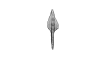
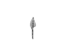
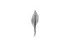
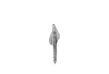
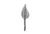
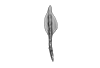
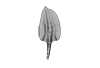
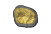
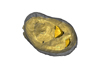
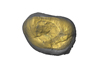
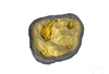
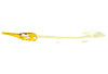
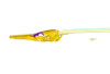
This project presents the 3D models of two isolated petrosals from the Oligocene locality of Pech de Fraysse (Quercy, France) here attributed to the genus Prodremotherium Filhol, 1877. Our aim is to describe the petrosal morphology of this Oligocene “early ruminant” as only few data are available in the literature for Oligocene taxa.
Prodremotherium sp. UM PFY 4053 View specimen

|
M3#7Labelled 3D model of right isolated petrosal of Prodremotherium sp. from Pech de Fraysse (Quercy, MP 28) Type: "3D_surfaces"doi: 10.18563/m3.sf7 state:published |
Download 3D surface file |
Prodremotherium sp. UM PFY 4054 View specimen

|
M3#8Labelled 3D model of right isolated petrosal of Prodremotherium sp. from Pech de Fraysse (Quercy, MP 28) Type: "3D_surfaces"doi: 10.18563/m3.sf8 state:published |
Download 3D surface file |

This contribution contains the 3D models of the set of Famennian conodont elements belonging to the species Polygnathus glaber and Polygnathus communis analyzed in the following publication: Renaud et al. 2021: Patterns of bilateral asymmetry and allometry in Late Devonian Polygnathus. Palaeontology. https://doi.org/10.1111/pala.12513
Polygnathus glaber UM BUS 001 View specimen

|
M3#574right P1 element Type: "3D_surfaces"doi: 10.18563/m3.sf.574 state:published |
Download 3D surface file |
Polygnathus glaber UM BUS 002 View specimen
|
M3#575right P1 element Type: "3D_surfaces"doi: 10.18563/m3.sf.575 state:published |
Download 3D surface file |
Polygnathus glaber UM BUS 003 View specimen

|
M3#576right P1 element Type: "3D_surfaces"doi: 10.18563/m3.sf.576 state:published |
Download 3D surface file |
Polygnathus glaber UM BUS 004 View specimen

|
M3#577left P1 element Type: "3D_surfaces"doi: 10.18563/m3.sf.577 state:published |
Download 3D surface file |
Polygnathus glaber UM BUS 005 View specimen

|
M3#578left P1 element Type: "3D_surfaces"doi: 10.18563/m3.sf.578 state:published |
Download 3D surface file |
Polygnathus glaber UM BUS 006 View specimen
|
M3#579right P1 element Type: "3D_surfaces"doi: 10.18563/m3.sf.579 state:published |
Download 3D surface file |
Polygnathus glaber UM BUS 007 View specimen

|
M3#580right P1 element Type: "3D_surfaces"doi: 10.18563/m3.sf.580 state:published |
Download 3D surface file |
Polygnathus glaber UM BUS 008 View specimen
|
M3#581left P1 element Type: "3D_surfaces"doi: 10.18563/m3.sf.581 state:published |
Download 3D surface file |
Polygnathus glaber UM BUS 009 View specimen
|
M3#582left P1 element Type: "3D_surfaces"doi: 10.18563/m3.sf.582 state:published |
Download 3D surface file |
Polygnathus glaber UM BUS 010 View specimen

|
M3#583right P1 element Type: "3D_surfaces"doi: 10.18563/m3.sf.583 state:published |
Download 3D surface file |
Polygnathus glaber UM BUS 011 View specimen

|
M3#584right P1 element Type: "3D_surfaces"doi: 10.18563/m3.sf.584 state:published |
Download 3D surface file |
Polygnathus glaber UM BUS 012 View specimen
|
M3#585right P1 element Type: "3D_surfaces"doi: 10.18563/m3.sf.585 state:published |
Download 3D surface file |
Polygnathus glaber UM BUS 013 View specimen

|
M3#586left P1 element Type: "3D_surfaces"doi: 10.18563/m3.sf.586 state:published |
Download 3D surface file |
Polygnathus glaber UM BUS 014 View specimen

|
M3#587left P1 element Type: "3D_surfaces"doi: 10.18563/m3.sf.587 state:published |
Download 3D surface file |
Polygnathus glaber UM BUS 015 View specimen

|
M3#588left P1 element Type: "3D_surfaces"doi: 10.18563/m3.sf.588 state:published |
Download 3D surface file |
Polygnathus glaber UM BUS 016 View specimen
|
M3#589right P1 element Type: "3D_surfaces"doi: 10.18563/m3.sf.589 state:published |
Download 3D surface file |
Polygnathus glaber UM BUS 017 View specimen

|
M3#590left P1 element Type: "3D_surfaces"doi: 10.18563/m3.sf.590 state:published |
Download 3D surface file |
Polygnathus glaber UM BUS 018 View specimen

|
M3#591left P1 element Type: "3D_surfaces"doi: 10.18563/m3.sf.591 state:published |
Download 3D surface file |
Polygnathus glaber UM BUS 019 View specimen
|
M3#592left P1 element Type: "3D_surfaces"doi: 10.18563/m3.sf.592 state:published |
Download 3D surface file |
Polygnathus glaber UM BUS 020 View specimen

|
M3#593left P1 element Type: "3D_surfaces"doi: 10.18563/m3.sf.593 state:published |
Download 3D surface file |
Polygnathus glaber UM BUS 021 View specimen

|
M3#594right P1 element Type: "3D_surfaces"doi: 10.18563/m3.sf.594 state:published |
Download 3D surface file |
Polygnathus glaber UM BUS 022 View specimen
|
M3#595left P1 element Type: "3D_surfaces"doi: 10.18563/m3.sf.595 state:published |
Download 3D surface file |
Polygnathus glaber UM BUS 023 View specimen

|
M3#596left P1 element Type: "3D_surfaces"doi: 10.18563/m3.sf.596 state:published |
Download 3D surface file |
Polygnathus glaber UM BUS 024 View specimen

|
M3#597left P1 element Type: "3D_surfaces"doi: 10.18563/m3.sf.597 state:published |
Download 3D surface file |
Polygnathus glaber UM BUS 025 View specimen

|
M3#598left P1 element Type: "3D_surfaces"doi: 10.18563/m3.sf.598 state:published |
Download 3D surface file |
Polygnathus glaber UM BUS 026 View specimen

|
M3#599left P1 element Type: "3D_surfaces"doi: 10.18563/m3.sf.599 state:published |
Download 3D surface file |
Polygnathus glaber UM BUS 027 View specimen

|
M3#600right P1 element Type: "3D_surfaces"doi: 10.18563/m3.sf.600 state:published |
Download 3D surface file |
Polygnathus glaber UM BUS 028 View specimen
|
M3#601right P1 element Type: "3D_surfaces"doi: 10.18563/m3.sf.601 state:published |
Download 3D surface file |
Polygnathus glaber UM BUS 029 View specimen

|
M3#602right P1 element Type: "3D_surfaces"doi: 10.18563/m3.sf.602 state:published |
Download 3D surface file |
Polygnathus glaber UM BUS 030 View specimen

|
M3#603right P1 element Type: "3D_surfaces"doi: 10.18563/m3.sf.603 state:published |
Download 3D surface file |
Polygnathus communis UM CTB 001 View specimen
|
M3#604right P1 element Type: "3D_surfaces"doi: 10.18563/m3.sf.604 state:published |
Download 3D surface file |
Polygnathus communis UM CTB 002 View specimen

|
M3#605right P1 element Type: "3D_surfaces"doi: 10.18563/m3.sf.605 state:published |
Download 3D surface file |
Polygnathus communis UM CTB 003 View specimen

|
M3#606right P1 element Type: "3D_surfaces"doi: 10.18563/m3.sf.606 state:published |
Download 3D surface file |
Polygnathus communis UM CTB 004 View specimen

|
M3#607right P1 element Type: "3D_surfaces"doi: 10.18563/m3.sf.607 state:published |
Download 3D surface file |
Polygnathus communis UM CTB 005 View specimen

|
M3#608left P1 element Type: "3D_surfaces"doi: 10.18563/m3.sf.608 state:published |
Download 3D surface file |
Polygnathus communis UM CTB 006 View specimen

|
M3#609left P1 element Type: "3D_surfaces"doi: 10.18563/m3.sf.609 state:published |
Download 3D surface file |
Polygnathus communis UM CTB 007 View specimen

|
M3#610left P1 element Type: "3D_surfaces"doi: 10.18563/m3.sf.610 state:published |
Download 3D surface file |
Polygnathus communis UM CTB 008 View specimen

|
M3#611left P1 element Type: "3D_surfaces"doi: 10.18563/m3.sf.611 state:published |
Download 3D surface file |
Polygnathus communis UM CTB 009 View specimen

|
M3#612right P1 element Type: "3D_surfaces"doi: 10.18563/m3.sf.612 state:published |
Download 3D surface file |
Polygnathus communis UM CTB 010 View specimen

|
M3#613left P1 element Type: "3D_surfaces"doi: 10.18563/m3.sf.613 state:published |
Download 3D surface file |
Polygnathus communis UM CTB 011 View specimen

|
M3#614right P1 element Type: "3D_surfaces"doi: 10.18563/m3.sf.614 state:published |
Download 3D surface file |
Polygnathus communis UM CTB 012 View specimen

|
M3#615right P1 element Type: "3D_surfaces"doi: 10.18563/m3.sf.615 state:published |
Download 3D surface file |
Polygnathus communis UM CTB 013 View specimen

|
M3#616right P1 element Type: "3D_surfaces"doi: 10.18563/m3.sf.616 state:published |
Download 3D surface file |
Polygnathus communis UM CTB 014 View specimen

|
M3#617right P1 element Type: "3D_surfaces"doi: 10.18563/m3.sf.617 state:published |
Download 3D surface file |
Polygnathus communis UM CTB 015 View specimen

|
M3#618right P1 element Type: "3D_surfaces"doi: 10.18563/m3.sf.618 state:published |
Download 3D surface file |
Polygnathus communis UM CTB 016 View specimen

|
M3#619left P1 element Type: "3D_surfaces"doi: 10.18563/m3.sf.619 state:published |
Download 3D surface file |
Polygnathus communis UM CTB 017 View specimen

|
M3#620right P1 element Type: "3D_surfaces"doi: 10.18563/m3.sf.620 state:published |
Download 3D surface file |
Polygnathus communis UM CTB 018 View specimen

|
M3#621right P1 element Type: "3D_surfaces"doi: 10.18563/m3.sf.621 state:published |
Download 3D surface file |
Polygnathus communis UM CTB 019 View specimen

|
M3#622right P1 element Type: "3D_surfaces"doi: 10.18563/m3.sf.622 state:published |
Download 3D surface file |
Polygnathus communis UM CTB 020 View specimen

|
M3#623right P1 element Type: "3D_surfaces"doi: 10.18563/m3.sf.623 state:published |
Download 3D surface file |
Polygnathus communis UM CTB 021 View specimen

|
M3#624left P1 element Type: "3D_surfaces"doi: 10.18563/m3.sf.624 state:published |
Download 3D surface file |
Polygnathus communis UM CTB 022 View specimen

|
M3#625left element Type: "3D_surfaces"doi: 10.18563/m3.sf.625 state:published |
Download 3D surface file |
Polygnathus communis UM CTB 023 View specimen

|
M3#626left P1 element Type: "3D_surfaces"doi: 10.18563/m3.sf.626 state:published |
Download 3D surface file |
Polygnathus communis UM CTB 024 View specimen

|
M3#627left P1 element Type: "3D_surfaces"doi: 10.18563/m3.sf.627 state:published |
Download 3D surface file |
Polygnathus communis UM CTB 025 View specimen

|
M3#628left P1 element Type: "3D_surfaces"doi: 10.18563/m3.sf.628 state:published |
Download 3D surface file |
Polygnathus communis UM CTB 026 View specimen

|
M3#629left P1 element Type: "3D_surfaces"doi: 10.18563/m3.sf.629 state:published |
Download 3D surface file |
Polygnathus communis UM CTB 027 View specimen

|
M3#630left P1 element Type: "3D_surfaces"doi: 10.18563/m3.sf.630 state:published |
Download 3D surface file |
Polygnathus communis UM CTB 028 View specimen

|
M3#631left P1 element Type: "3D_surfaces"doi: 10.18563/m3.sf.631 state:published |
Download 3D surface file |
Polygnathus communis UM CTB 029 View specimen

|
M3#632left P1 element Type: "3D_surfaces"doi: 10.18563/m3.sf.632 state:published |
Download 3D surface file |
Polygnathus communis UM CTB 030 View specimen

|
M3#633left P1 element Type: "3D_surfaces"doi: 10.18563/m3.sf.633 state:published |
Download 3D surface file |
Polygnathus communis UM CTB 031 View specimen
|
M3#634left P1 element Type: "3D_surfaces"doi: 10.18563/m3.sf.634 state:published |
Download 3D surface file |
Polygnathus communis UM CTB 032 View specimen

|
M3#635left P1 element Type: "3D_surfaces"doi: 10.18563/m3.sf.635 state:published |
Download 3D surface file |
Polygnathus communis UM CTB 033 View specimen

|
M3#636left P1 element Type: "3D_surfaces"doi: 10.18563/m3.sf.636 state:published |
Download 3D surface file |
Polygnathus communis UM CTB 034 View specimen

|
M3#637right P1 element Type: "3D_surfaces"doi: 10.18563/m3.sf.637 state:published |
Download 3D surface file |
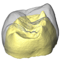
The present 3D Dataset contains the 3D models of external and internal aspects of human upper permanent second molars from the Neolithic necropolis analyzed in the following publication: Le Luyer M., Coquerelle M., Rottier S., Bayle P. (2016): Internal tooth structure and burial practices: insights into the Neolithic necropolis of Gurgy (France, 5100-4000 cal. BC). Plos One 11(7): e0159688. doi: 10.1371/journal.pone.0159688.
Homo sapiens GLN04-201-ULM2 View specimen
|
M3#74Outer enamel surface (OES) and enamel-dentine junction (EDJ) of Neolithic upper permanent left second molar Type: "3D_surfaces"doi: 10.18563/m3.sf.74 state:published |
Download 3D surface file |
Homo sapiens GLN04-206-ULM2 View specimen
|
M3#75Outer enamel surface (OES) and enamel-dentine junction (EDJ) of Neolithic upper permanent left second molar Type: "3D_surfaces"doi: 10.18563/m3.sf.75 state:published |
Download 3D surface file |
Homo sapiens GLN05-213-URM2 View specimen
|
M3#76Outer enamel surface (OES) and enamel-dentine junction (EDJ) of Neolithic upper permanent right second molar Type: "3D_surfaces"doi: 10.18563/m3.sf.76 state:published |
Download 3D surface file |
Homo sapiens GLN05-215A-URM2 View specimen
|
M3#77Outer enamel surface (OES) and enamel-dentine junction (EDJ) of Neolithic upper permanent right second molar Type: "3D_surfaces"doi: 10.18563/m3.sf.77 state:published |
Download 3D surface file |
Homo sapiens GLN06-215B-URM2 View specimen
|
M3#78Outer enamel surface (OES) and enamel-dentine junction (EDJ) of Neolithic upper permanent right second molar Type: "3D_surfaces"doi: 10.18563/m3.sf.78 state:published |
Download 3D surface file |
Homo sapiens GLN06-223-URM2 View specimen
|
M3#79Outer enamel surface (OES) and enamel-dentine junction (EDJ) of Neolithic upper permanent right second molar Type: "3D_surfaces"doi: 10.18563/m3.sf.79 state:published |
Download 3D surface file |
Homo sapiens GLN04-229-URM2 View specimen
|
M3#80Outer enamel surface (OES) and enamel-dentine junction (EDJ) of Neolithic upper permanent right second molar Type: "3D_surfaces"doi: 10.18563/m3.sf.80 state:published |
Download 3D surface file |
Homo sapiens GLN05-243B-ULM2 View specimen
|
M3#81Outer enamel surface (OES) and enamel-dentine junction (EDJ) with reconstructed dentine horn tip of Neolithic upper permanent left second molar Type: "3D_surfaces"doi: 10.18563/m3.sf.81 state:published |
Download 3D surface file |
Homo sapiens GLN04-248-ULM2 View specimen
|
M3#82Outer enamel surface (OES) and enamel-dentine junction (EDJ) with reconstructed dentine horn tip of Neolithic upper permanent left second molar Type: "3D_surfaces"doi: 10.18563/m3.sf.82 state:published |
Download 3D surface file |
Homo sapiens GLN04-252-ULM2 View specimen
|
M3#83Outer enamel surface (OES) and enamel-dentine junction (EDJ) of Neolithic upper permanent left second molar Type: "3D_surfaces"doi: 10.18563/m3.sf.83 state:published |
Download 3D surface file |
Homo sapiens GLN04-253-ULM2 View specimen
|
M3#84Outer enamel surface (OES) and enamel-dentine junction (EDJ) of Neolithic upper permanent left second molar Type: "3D_surfaces"doi: 10.18563/m3.sf.84 state:published |
Download 3D surface file |
Homo sapiens GLN05-257-URM2 View specimen
|
M3#85Outer enamel surface (OES) and enamel-dentine junction (EDJ) with reconstructed dentine horn tip of Neolithic upper permanent right second molar Type: "3D_surfaces"doi: 10.18563/m3.sf.85 state:published |
Download 3D surface file |
Homo sapiens GLN04-264-ULM2 View specimen
|
M3#86Outer enamel surface (OES) and enamel-dentine junction (EDJ) of Neolithic upper permanent left second molar Type: "3D_surfaces"doi: 10.18563/m3.sf.86 state:published |
Download 3D surface file |
Homo sapiens GLN04-277-URM2 View specimen
|
M3#87Outer enamel surface (OES) and enamel-dentine junction (EDJ) of Neolithic upper permanent right second molar Type: "3D_surfaces"doi: 10.18563/m3.sf.87 state:published |
Download 3D surface file |
Homo sapiens GLN04-289B-URM2 View specimen
|
M3#88Outer enamel surface (OES) and enamel-dentine junction (EDJ) of Neolithic upper permanent right second molar Type: "3D_surfaces"doi: 10.18563/m3.sf.88 state:published |
Download 3D surface file |
Homo sapiens GLN06-291-URM2 View specimen
|
M3#89Outer enamel surface (OES) and enamel-dentine junction (EDJ) with reconstructed dentine horn tip of Neolithic upper permanent right second molar Type: "3D_surfaces"doi: 10.18563/m3.sf.89 state:published |
Download 3D surface file |
Homo sapiens GLN05-292-URM2 View specimen
|
M3#90Outer enamel surface (OES) and enamel-dentine junction (EDJ) of Neolithic upper permanent right second molar Type: "3D_surfaces"doi: 10.18563/m3.sf.90 state:published |
Download 3D surface file |
Homo sapiens GLN05-294-ULM2 View specimen
|
M3#91Outer enamel surface (OES) and enamel-dentine junction (EDJ) with reconstructed dentine horn tip of Neolithic upper permanent left second molar Type: "3D_surfaces"doi: 10.18563/m3.sf.91 state:published |
Download 3D surface file |
Homo sapiens GLN05-308-URM2 View specimen
|
M3#93Outer enamel surface (OES) and enamel-dentine junction (EDJ) of Neolithic upper permanent right second molar Type: "3D_surfaces"doi: 10.18563/m3.sf.93 state:published |
Download 3D surface file |
Homo sapiens GLN05-301-ULM2 View specimen
|
M3#92Outer enamel surface (OES) and enamel-dentine junction (EDJ) of Neolithic upper permanent left second molar Type: "3D_surfaces"doi: 10.18563/m3.sf.92 state:published |
Download 3D surface file |
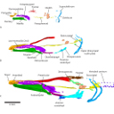
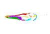
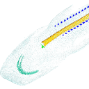
Using X-ray microtomography, we describe the ossification events during the larval development of a non-teleost actinopterygian species: the Cuban gar Atractosteus tristoechus from the order Lepisosteiformes. We provide a detailed developmental series for each anatomical structure, covering a large sequence of mineralization events going from an early stage (13 days post-hatching, 21mm total length) to an almost fully ossified larval stage (118dph or 87mm in standard length). With this work, we expect to bring new developmental data to be used in further comparative studies with other lineages of bony vertebrates. We also hope that the on-line publication of these twelve successive 3D reconstructions, fully labelled and flagged, will be an educational tool for all students in comparative anatomy.
Atractosteus tristoechus At1-13dph View specimen

|
M3#94At1-13dph : 13 dph larvae, 21 mm TL Type: "3D_surfaces"doi: 10.18563/m3.sf.94 state:published |
Download 3D surface file |
Atractosteus tristoechus At2-16dph View specimen

|
M3#95Atractosteus tristoechus larva, 16 dph, 26mm SL. Type: "3D_surfaces"doi: 10.18563/m3.sf.95 state:published |
Download 3D surface file |
Atractosteus tristoechus At3-19dph View specimen

|
M3#96Atractosteus tristoechus larva, 19 dph, 27mm SL. Type: "3D_surfaces"doi: 10.18563/m3.sf.96 state:published |
Download 3D surface file |
Atractosteus tristoechus At4-22dph View specimen
|
M3#97Atractosteus tristoechus larva, 22dph, 30mm SL. Type: "3D_surfaces"doi: 10.18563/m3.sf.97 state:published |
Download 3D surface file |
Atractosteus tristoechus At5-26dph View specimen
|
M3#98Atractosteus tristoechus larva, 26 dph, 32mm SL. Type: "3D_surfaces"doi: 10.18563/m3.sf.98 state:published |
Download 3D surface file |
Atractosteus tristoechus At6-31dph View specimen
|
M3#99Atractosteus tristoechus larva, 31 dph, 39mm SL. Type: "3D_surfaces"doi: 10.18563/m3.sf.99 state:published |
Download 3D surface file |
Atractosteus tristoechus At7-37dph View specimen
|
M3#100Atractosteus tristoechus larva, 37 dph, 43mm SL. Type: "3D_surfaces"doi: 10.18563/m3.sf.100 state:published |
Download 3D surface file |
Atractosteus tristoechus At8-52dph View specimen
|
M3#101Atractosteus tristoechus larva, 52 dph, 46mm SL. Type: "3D_surfaces"doi: 10.18563/m3.sf.101 state:published |
Download 3D surface file |
Atractosteus tristoechus At9-74dph View specimen
|
M3#102Atractosteus tristoechus larva, 74 dph, 61mm SL. Not all structures are colored, only newly ossified ones. Type: "3D_surfaces"doi: 10.18563/m3.sf.102 state:published |
Download 3D surface file |
Atractosteus tristoechus At10-89dph View specimen
|
M3#103Atractosteus tristoechus larva, 89 dph, 63mm SL. Not all structures are colored, only newly ossified ones. You may find the tag file in the At1-13dph reconstruction data. Type: "3D_surfaces"doi: 10.18563/m3.sf.103 state:published |
Download 3D surface file |
Atractosteus tristoechus At11-104dph View specimen
|
M3#104Atractosteus tristoechus larva, 104 dph, 70mm SL. Not all structures are colored, only newly ossified ones. Type: "3D_surfaces"doi: 10.18563/m3.sf.104 state:published |
Download 3D surface file |
Atractosteus tristoechus At12-118dph View specimen
|
M3#105Atractosteus tristoechus larva, 118 dph, 87mm SL. Type: "3D_surfaces"doi: 10.18563/m3.sf.105 state:published |
Download 3D surface file |

This contribution contains the 3D models analyzed in Müller et al. (2021) “Pushing the boundary? Testing the ‘functional elongation hypothesis’ of the giraffe’s neck”.
Aepyceros melampus ZFMK 2001.278 View specimen

|
M3#643Vertebrae C7, T1 Type: "3D_surfaces"doi: 10.18563/m3.sf.643 state:published |
Download 3D surface file |
Giraffa camelopardalis ZMB 66393 View specimen

|
M3#644Vertebrae Type: "3D_surfaces"doi: 10.18563/m3.sf.644 state:published |
Download 3D surface file |
Giraffa camelopardalis ZSM 1967/17 View specimen

|
M3#645Vertebrae Type: "3D_surfaces"doi: 10.18563/m3.sf.645 state:published |
Download 3D surface file |
Giraffa camelopardalis ZSM 1981/19 View specimen

|
M3#646C3, C4, C5, C6, C7, T1, T2 Type: "3D_surfaces"doi: 10.18563/m3.sf.646 state:published |
Download 3D surface file |
Giraffa camelopardalis KMDA M-10861 View specimen

|
M3#647C3, C4, C5, C6, C7, T1, T2. Acquired via laser scanner. Type: "3D_surfaces"doi: 10.18563/m3.sf.647 state:published |
Download 3D surface file |
Giraffa camelopardalis SMF 84214 View specimen

|
M3#648C7, T1. Warning : photogrammetric models (unit scale is CM, not MM). Type: "3D_surfaces"doi: 10.18563/m3.sf.648 state:published |
Download 3D surface file |
Giraffa camelopardalis SMF 78299 View specimen

|
M3#649C7, T1. Warning : unscaled photogrammetric 3D models (unknown size). Type: "3D_surfaces"doi: 10.18563/m3.sf.649 state:published |
Download 3D surface file |
Giraffa camelopardalis SMF o. N View specimen

|
M3#650C7, T1. Warning : unscaled photogrammetric 3D models (unknown size). Type: "3D_surfaces"doi: 10.18563/m3.sf.650 state:published |
Download 3D surface file |
Giraffa camelopardalis SMNS 19138 View specimen

|
M3#671C7, T1. Warning : unscaled photogrammetric 3D models (unknown size). Type: "3D_surfaces"doi: 10.18563/m3.sf.671 state:published |
Download 3D surface file |
Okapia johnstoni ZMB 62086 View specimen

|
M3#651C3, C4, C5, C6, C7, T1, T2 Type: "3D_surfaces"doi: 10.18563/m3.sf.651 state:published |
Download 3D surface file |
Okapia johnstoni ZMB 70325 View specimen

|
M3#652C3, C4, C5, C6, C7, T1, T2 Type: "3D_surfaces"doi: 10.18563/m3.sf.652 state:published |
Download 3D surface file |
Sivatherium giganteum NHMUK 15707 View specimen
|
M3#653C7. Warning : unscaled photogrammetric 3D model (unknown size). Type: "3D_surfaces"doi: 10.18563/m3.sf.653 state:published |
Download 3D surface file |
Sivatherium giganteum NHMUK 15297 View specimen

|
M3#654T1. Warning : unscaled photogrammetric 3D model (unknown size). Type: "3D_surfaces"doi: 10.18563/m3.sf.654 state:published |
Download 3D surface file |
Cervus elaphus ZMB 47502 View specimen

|
M3#655C3, C4, C5, C6, C7, T1, T2 Type: "3D_surfaces"doi: 10.18563/m3.sf.655 state:published |
Download 3D surface file |
Axis axis SMF 1450 View specimen

|
M3#656C7, T1 Type: "3D_surfaces"doi: 10.18563/m3.sf.656 state:published |
Download 3D surface file |
Cervus nippon SMF 4368 View specimen

|
M3#657C7, T1 Type: "3D_surfaces"doi: 10.18563/m3.sf.657 state:published |
Download 3D surface file |
Capreolus capreolus SMF 79852 View specimen

|
M3#658C7, T1 Type: "3D_surfaces"doi: 10.18563/m3.sf.658 state:published |
Download 3D surface file |
Capreolus capreolus ZFMK 67.237 View specimen

|
M3#659C7, T1 Type: "3D_surfaces"doi: 10.18563/m3.sf.659 state:published |
Download 3D surface file |
Muntiacus reevesi SMF 92954 View specimen

|
M3#660C7, T1 Type: "3D_surfaces"doi: 10.18563/m3.sf.660 state:published |
Download 3D surface file |
Muntiacus reevesi SMF 92332 View specimen

|
M3#661C7, T1 Type: "3D_surfaces"doi: 10.18563/m3.sf.661 state:published |
Download 3D surface file |
Alces alces SMF 35549 View specimen

|
M3#662C7, T1 Type: "3D_surfaces"doi: 10.18563/m3.sf.662 state:published |
Download 3D surface file |
Dama dama ZFMK 86.125 View specimen

|
M3#663C7, T1 Type: "3D_surfaces"doi: 10.18563/m3.sf.663 state:published |
Download 3D surface file |
Antilope cervicapra ZMB 78829 View specimen

|
M3#664C3, C4, C5, C6, C7, T1, T2 Type: "3D_surfaces"doi: 10.18563/m3.sf.664 state:published |
Download 3D surface file |
Bison bonasus SMNS 2998 View specimen

|
M3#665C7, T1. Warning : unscaled photogrammetric 3D models (unknown size). Type: "3D_surfaces"doi: 10.18563/m3.sf.665 state:published |
Download 3D surface file |
Nanger dama SMF 74435 View specimen

|
M3#666C7, T1 Type: "3D_surfaces"doi: 10.18563/m3.sf.666 state:published |
Download 3D surface file |
Litocranius walleri SMF 23747 View specimen

|
M3#667C7, T1 Type: "3D_surfaces"doi: 10.18563/m3.sf.667 state:published |
Download 3D surface file |
Litocranius walleri SMF 23749 View specimen

|
M3#669C7, T1 Type: "3D_surfaces"doi: 10.18563/m3.sf.669 state:published |
Download 3D surface file |
Tragelaphus eurycerus SMF 95875 View specimen

|
M3#670C7, T1 Type: "3D_surfaces"doi: 10.18563/m3.sf.670 state:published |
Download 3D surface file |
Bos javanicus SMF 64934 View specimen

|
M3#672C7, T1 Type: "3D_surfaces"doi: 10.18563/m3.sf.672 state:published |
Download 3D surface file |
Ovis aries ZFMK 1982.338 View specimen

|
M3#673C7, T1 Type: "3D_surfaces"doi: 10.18563/m3.sf.673 state:published |
Download 3D surface file |
Rupicapra rupicapra ZFMK 72.367 View specimen

|
M3#674C7, T1 Type: "3D_surfaces"doi: 10.18563/m3.sf.674 state:published |
Download 3D surface file |
Kobus ellipsiprymnus SMNS 4443 View specimen

|
M3#675C7, T1 Type: "3D_surfaces"doi: 10.18563/m3.sf.675 state:published |
Download 3D surface file |
Sylvicapra grimmia SMNS 15292 View specimen

|
M3#676C7, T1 Type: "3D_surfaces"doi: 10.18563/m3.sf.676 state:published |
Download 3D surface file |
Syncerus caffer SMNS 7347 View specimen

|
M3#677C7, T1. Warning : unscaled photogrammetric 3D models (unknown size). Type: "3D_surfaces"doi: 10.18563/m3.sf.677 state:published |
Download 3D surface file |
Procapra gutturosa SMNS 5796 View specimen

|
M3#678C7, T1 Type: "3D_surfaces"doi: 10.18563/m3.sf.678 state:published |
Download 3D surface file |
Damaliscus pygargus SMNS 21617 View specimen

|
M3#679C7, T1 Type: "3D_surfaces"doi: 10.18563/m3.sf.679 state:published |
Download 3D surface file |
Madoqua kirkii SMNS 4432 View specimen

|
M3#680C7, T1 Type: "3D_surfaces"doi: 10.18563/m3.sf.680 state:published |
Download 3D surface file |
Bubalus mindorensis SMNS 2054 View specimen

|
M3#681C7, T1. Warning : unscaled photogrammetric 3D models (unknown size). Type: "3D_surfaces"doi: 10.18563/m3.sf.681 state:published |
Download 3D surface file |
Capra hircus SMNS 51328 View specimen

|
M3#682C7, T1 Type: "3D_surfaces"doi: 10.18563/m3.sf.682 state:published |
Download 3D surface file |
Connochaetes taurinus SMNS 4442 View specimen

|
M3#683C7, T1. Warning : unscaled photogrammetric 3D models (unknown size). Type: "3D_surfaces"doi: 10.18563/m3.sf.683 state:published |
Download 3D surface file |
Antilocapra americana ZSM 1964/218 View specimen

|
M3#684C3, C4, C5, C6, C7, T1, T2 Type: "3D_surfaces"doi: 10.18563/m3.sf.684 state:published |
Download 3D surface file |
Antilocapra americana ZMB 77281 View specimen

|
M3#685C7, T1 Type: "3D_surfaces"doi: 10.18563/m3.sf.685 state:published |
Download 3D surface file |
Moschus moschiferus ZMB 62080 View specimen

|
M3#686C3, C4, C5, C6, C7, T1, T2 Type: "3D_surfaces"doi: 10.18563/m3.sf.686 state:published |
Download 3D surface file |
Moschus moschiferus ZMB 60367 View specimen

|
M3#687C7, T1 Type: "3D_surfaces"doi: 10.18563/m3.sf.687 state:published |
Download 3D surface file |
Moschus moschiferus ZMB 51830 View specimen

|
M3#688C7, T1 Type: "3D_surfaces"doi: 10.18563/m3.sf.688 state:published |
Download 3D surface file |
Tragulus javanicus SMF 82179 View specimen

|
M3#689C7, T1 Type: "3D_surfaces"doi: 10.18563/m3.sf.689 state:published |
Download 3D surface file |
Tragulus javanicus ZMB 86222 View specimen

|
M3#690C7, T1 Type: "3D_surfaces"doi: 10.18563/m3.sf.690 state:published |
Download 3D surface file |
Tragulus sp. ZMB o. N. View specimen

|
M3#691C7, T1 Type: "3D_surfaces"doi: 10.18563/m3.sf.691 state:published |
Download 3D surface file |
Hyemoschus aquaticus ZMB 71071 View specimen

|
M3#692C7, T1 Type: "3D_surfaces"doi: 10.18563/m3.sf.692 state:published |
Download 3D surface file |
Hyemoschus aquaticus ZMB 103235 View specimen

|
M3#693C7, T1 Type: "3D_surfaces"doi: 10.18563/m3.sf.693 state:published |
Download 3D surface file |
Vicugna vicugna SMF 94752 View specimen

|
M3#694C7, T1 Type: "3D_surfaces"doi: 10.18563/m3.sf.694 state:published |
Download 3D surface file |
Camelus dromedarius SMF 70473 View specimen

|
M3#695C7, T1. Warning : unscaled photogrammetric 3D models (unknown size). Type: "3D_surfaces"doi: 10.18563/m3.sf.695 state:published |
Download 3D surface file |
Camelus bactrianus SMF 25542 View specimen

|
M3#696C7, T1. Warning : unscaled photogrammetric 3D models (unknown size). Type: "3D_surfaces"doi: 10.18563/m3.sf.696 state:published |
Download 3D surface file |
Lama glama SMNS 31175 View specimen

|
M3#697C7, T1 Type: "3D_surfaces"doi: 10.18563/m3.sf.697 state:published |
Download 3D surface file |
Vicugna pacos SMNS 46255 View specimen

|
M3#698C7, T1 Type: "3D_surfaces"doi: 10.18563/m3.sf.698 state:published |
Download 3D surface file |
Vicugna pacos SMNS 7349 View specimen

|
M3#699C7, T1 Type: "3D_surfaces"doi: 10.18563/m3.sf.699 state:published |
Download 3D surface file |
This contribution contains the 3D models described and figured in the following publication: Shiraishi N et al. Morphology and morphometry of the human embryonic brain: A three-dimensional analysis NeuroImage 115, 2015, 96-103, DOI: 10.1016/j.neuroimage.2015.04.044.
Homo sapiens KC-CS13BRN50455 View specimen

|
M3#24Computationally reconstructed cerebral parenchyma and ventricle of the human embryo at Carnegie Stage 13. Type: "3D_surfaces"doi: 10.18563/m3.sf24 state:published |
Download 3D surface file |
Homo sapiens KC-CS14BRN18834 View specimen

|
M3#25Computationally reconstructed cerebral parenchyma and ventricle of the human embryo at Carnegie Stage 14. Type: "3D_surfaces"doi: 10.18563/m3.sf25 state:published |
Download 3D surface file |
Homo sapiens KC-CS15BRN19975 View specimen

|
M3#26Computationally reconstructed cerebral parenchyma and ventricle of the human embryo at Carnegie Stage 15. Type: "3D_surfaces"doi: 10.18563/m3.sf26 state:published |
Download 3D surface file |
Homo sapiens KC-CS16BRN7870 View specimen

|
M3#27Computationally reconstructed cerebral parenchyma and ventricle of the human embryo at Carnegie Stage 16. Type: "3D_surfaces"doi: 10.18563/m3.sf27 state:published |
Download 3D surface file |
Homo sapiens KC-CS17BRN26702 View specimen

|
M3#28Computationally reconstructed cerebral parenchyma and ventricle of the human embryo at Carnegie Stage 17. Type: "3D_surfaces"doi: 10.18563/m3.sf28 state:published |
Download 3D surface file |
Homo sapiens KC-CS18BRN25914 View specimen

|
M3#29Computationally reconstructed cerebral parenchyma and ventricle of the human embryo at Carnegie Stage 18. Type: "3D_surfaces"doi: 10.18563/m3.sf29 state:published |
Download 3D surface file |
Homo sapiens KC-CS19BRN16508 View specimen

|
M3#30Computationally reconstructed cerebral parenchyma and ventricle of the human embryo at Carnegie Stage 19. Type: "3D_surfaces"doi: 10.18563/m3.sf30 state:published |
Download 3D surface file |
Homo sapiens KC-CS20BRN26581 View specimen

|
M3#31Computationally reconstructed cerebral parenchyma and ventricle of the human embryo at Carnegie Stage 20. Type: "3D_surfaces"doi: 10.18563/m3.sf31 state:published |
Download 3D surface file |
Homo sapiens KC-CS21BRN33434 View specimen

|
M3#32Computationally reconstructed cerebral parenchyma and ventricle of the human embryo at Carnegie Stage 21. Type: "3D_surfaces"doi: 10.18563/m3.sf32 state:published |
Download 3D surface file |
Homo sapiens KC-CS22BRN27960 View specimen

|
M3#33Computationally reconstructed cerebral parenchyma and ventricle of the human embryo at Carnegie Stage 22. Type: "3D_surfaces"doi: 10.18563/m3.sf33 state:published |
Download 3D surface file |
Homo sapiens KC-CS23BRN28189 View specimen

|
M3#34Computationally reconstructed cerebral parenchyma and ventricle of the human embryo at Carnegie Stage 23. Type: "3D_surfaces"doi: 10.18563/m3.sf34 state:published |
Download 3D surface file |

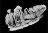
This project presents a µCT dataset and an associated 3D surface model of the holotype of Donrussellia magna (UM PAT 17; Primates, Adapiformes). UM PAT17 is the only known specimen for the species and consists of a well-preserved left lower jaw with p4-m3. It documents one of the oldest European primates, eventually dated near the Paleocene Eocene Thermal Maximum.
Donrussellia magna UM PAT 17 View specimen

|
M3#173D surface file model of UM PAT 17 (type specimen of Donrussellia magna), which is a well preserved left lower jaw with p4-m3. The teeth (and roots) were manually segmented. Type: "3D_surfaces"doi: 10.18563/m3.sf17 state:published |
Download 3D surface file |
|
M3#18CT Scan Data of Donrussellia magna UM PAT 17. Voxel size (in µm): 36µm (isotropic voxels). Dimensions in x,y,z : 594 pixels, 294 pixels, 1038 pixels. Image type : 8-bit voxels. Image format : raw data format (no header). Type: "3D_CT"doi: 10.18563/m3.sf18 state:published |
Download CT data |
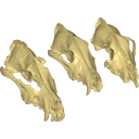
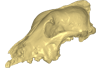
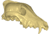
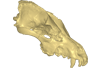
Archaeozoological studies are increasingly using new methods and approaches to explore questions about domestication. Here, we provide 3D models of three archaeological Canis lupus skulls from Belgium originating from the sites of Goyet (31,680±250BP; 31,890+240/-220BP), Trou des Nutons (21,810±90BP) and Trou Balleux (postglacial). Since their identification as either wolves or early dogs is still debated, we present these models as additional tools for further investigating their evolutionary history and the history of dog domestication.
Canis lupus Goyet 2860 View specimen
|
M3#213D surface model of the cranium of the Late Pleistocene Canis lupus "Goyet 2860" from the Royal Belgian Institute of Natural Sciences. Type: "3D_surfaces"doi: 10.18563/m3.sf21 state:published |
Download 3D surface file |
Canis lupus Trou Balleux no-nr View specimen
|
M3#223D surface model of the cranium of the Late Pleistocene Canis lupus "Trou Balleux no-nr" from the University of Liège, Belgium Type: "3D_surfaces"doi: 10.18563/m3.sf22 state:published |
Download 3D surface file |
Canis lupus Trou des Nutons 2559-1 View specimen
|
M3#233D surface model of the cranium of the Late Pleistocene Canis lupus "Trou des Nutons 2559-1" from the Royal Belgian Institute of Natural Sciences. Type: "3D_surfaces"doi: 10.18563/m3.sf23 state:published |
Download 3D surface file |
Current knowledge on the skeletogenesis of Chondrichthyes is scarce compared with their extant sister group, the bony fishes. Most of the previously described developmental tables in Chondrichthyes have focused on embryonic external morphology only. Due to its small body size and relative simplicity to raise eggs in laboratory conditions, the small-spotted catshark Scyliorhinus canicula has emerged as a reference species to describe developmental mechanisms in the Chondrichthyes lineage. Here we investigate the dynamic of mineralization in a set of six embryonic specimens using X-ray microtomography and describe the developing units of both the dermal skeleton (teeth and dermal scales) and endoskeleton (vertebral axis). This preliminary data on skeletogenesis in the catshark sets the first bases to a more complete investigation of the skeletal developmental in Chondrichthyes. It should provide comparison points with data known in osteichthyans and could thus be used in the broader context of gnathostome skeletal evolution.
Scyliorhinus canicula SC6_2_2015_03_20 View specimen

|
M3#50Mineralized skeleton of a 6,2 cm long embryo of Scyliorhinus canicula Type: "3D_surfaces"doi: 10.18563/m3.sf.50 state:published |
Download 3D surface file |
Scyliorhinus canicula SC6_7_2015_03_20 View specimen

|
M3#51Mineralized skeleton of a 6,7 cm long embryo of Scyliorhinus canicula Type: "3D_surfaces"doi: 10.18563/m3.sf.51 state:published |
Download 3D surface file |
Scyliorhinus canicula SC7_1_2015_04_03 View specimen

|
M3#52Mineralized skeleton of a 7,1 cm long embryo of Scyliorhinus canicula Type: "3D_surfaces"doi: 10.18563/m3.sf.52 state:published |
Download 3D surface file |
Scyliorhinus canicula SC7_5_2015_03_13 View specimen

|
M3#53Mineralized skeleton of a 7,5 cm long embryo of Scyliorhinus canicula Type: "3D_surfaces"doi: 10.18563/m3.sf.53 state:published |
Download 3D surface file |
Scyliorhinus canicula SC8_2015_03_20 View specimen

|
M3#54Mineralized skeleton of a 8 cm long embryo of Scyliorhinus canicula Type: "3D_surfaces"doi: 10.18563/m3.sf.54 state:published |
Download 3D surface file |
Scyliorhinus canicula SC10_2015_02_27 View specimen

|
M3#55Mineralized skeleton of a 10 cm long embryo of Scyliorhinus canicula Type: "3D_surfaces"doi: 10.18563/m3.sf.55 state:published |
Download 3D surface file |
The present 3D Dataset contains the 3D models analyzed in: Toyoda S et al., 2015, Morphogenesis of the inner ear at different stages of normal human development. The Anatomical Record. doi : 10.1002/ar.23268
Homo sapiens KC-CS17IER29248 View specimen

|
M3#36Computationally reconstructed membranous labyrinth of a human embryo (KC-CS17IER29248) at Carnegie Stage 17 (Crown Rump Length= 7mm). Type: "3D_surfaces"doi: 10.18563/m3.sf36 state:published |
Download 3D surface file |
Homo sapiens KC-CS18IER17746 View specimen

|
M3#37Computationally reconstructed membranous labyrinth of a human embryo (KC-CS18IER17746) at Carnegie Stage 18 (Crown Rump Length= 12mm). Type: "3D_surfaces"doi: 10.18563/m3.sf37 state:published |
Download 3D surface file |
Homo sapiens KC-CS19IER16127 View specimen

|
M3#38Computationally reconstructed membranous labyrinth of a human embryo (KC-CS19IER16127) at Carnegie Stage 19 (Crown Rump Length= 13mm). Type: "3D_surfaces"doi: 10.18563/m3.sf38 state:published |
Download 3D surface file |
Homo sapiens KC-CS20IER20268 View specimen

|
M3#39Computationally reconstructed membranous labyrinth of a human embryo (KC-CS20IER20268) at Carnegie Stage 20 (Crown Rump Length= 13.7mm). Type: "3D_surfaces"doi: 10.18563/m3.sf39 state:published |
Download 3D surface file |
Homo sapiens KC-CS21IER28066 View specimen

|
M3#40Computationally reconstructed membranous labyrinth of a human embryo (KC-CS21IER28066) at Carnegie Stage 21 (Crown Rump Length= 16.7mm). Type: "3D_surfaces"doi: 10.18563/m3.sf40 state:published |
Download 3D surface file |
Homo sapiens KC-CS22IER35233 View specimen

|
M3#41Computationally reconstructed membranous labyrinth of a human embryo (KC-CS22IER35233) at Carnegie Stage 22 (Crown Rump Length= 22mm). Type: "3D_surfaces"doi: 10.18563/m3.sf41 state:published |
Download 3D surface file |
Homo sapiens KC-CS23IER15919 View specimen

|
M3#42Computationally reconstructed membranous labyrinth of a human embryo (KC-CS23IER15919) at Carnegie Stage 23 (Crown Rump Length= 32.3mm). Type: "3D_surfaces"doi: 10.18563/m3.sf42 state:published |
Download 3D surface file |
Homo sapiens KC-FIER52730 View specimen

|
M3#43Computationally reconstructed human membranous labyrinth in post embryonic phase (KC-FIER52730). Crown Rump Length: 43.5mm. Type: "3D_surfaces"doi: 10.18563/m3.sf43 state:published |
Download 3D surface file |
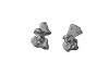
Considerable morphological variations are found in the middle ear among mammals. Here I present a three-dimensional atlas of the middle ear ossicles of eulipotyphlan mammals. This group has radiated into various environments as terrestrial, aquatic, and subterranean habitats independently in multiple lineages. Therefore, eulipotyphlans are an ideal group to explore the form-function relationship of the middle ear ossicles. This comparative atlas of hedgehogs, true shrews, water shrews, mole shrews, true moles, and shrew moles encourages future studies of the middle ear morphology of this diverse group.
Erinaceus europaeus DK2331 View specimen
|
M3#151Left middle ear ossicles Type: "3D_surfaces"doi: 10.18563/m3.sf.151 state:published |
Download 3D surface file |
Anourosorex yamashinai SIK_yamashinai View specimen
|
M3#152Left middle ear ossicles Type: "3D_surfaces"doi: 10.18563/m3.sf.152 state:published |
Download 3D surface file |
Blarina brevicauda M8003 View specimen
|
M3#153Right middle ear ossicles Type: "3D_surfaces"doi: 10.18563/m3.sf.153 state:published |
Download 3D surface file |
Chimarrogale platycephala DK5481 View specimen
|
M3#162Left middle ear ossicles Type: "3D_surfaces"doi: 10.18563/m3.sf.162 state:published |
Download 3D surface file |
Suncus murinus DK1227 View specimen
|
M3#155Left middle ear ossicles Type: "3D_surfaces"doi: 10.18563/m3.sf.155 state:published |
Download 3D surface file |
Condylura cristata SIK0050 View specimen
|
M3#156Right middle ear ossicles Type: "3D_surfaces"doi: 10.18563/m3.sf.156 state:published |
Download 3D surface file |
Euroscaptor klossi SIK0673 View specimen
|
M3#163Left middle ear ossicles Type: "3D_surfaces"doi: 10.18563/m3.sf.163 state:published |
Download 3D surface file |
Euroscaptor malayana SIK_malayana View specimen
|
M3#164Left middle ear ossicles Type: "3D_surfaces"doi: 10.18563/m3.sf.164 state:published |
Download 3D surface file |
Mogera wogura DK2551 View specimen
|
M3#159Left middle ear ossicles Type: "3D_surfaces"doi: 10.18563/m3.sf.159 state:published |
Download 3D surface file |
Talpa altaica SIK_altaica View specimen
|
M3#161Right middle ear ossicles Type: "3D_surfaces"doi: 10.18563/m3.sf.161 state:published |
Download 3D surface file |
Urotrichus talpoides DK0887 View specimen
|
M3#165Left middle ear ossicles Type: "3D_surfaces"doi: 10.18563/m3.sf.165 state:published |
Download 3D surface file |
Oreoscaptor mizura DK6545 View specimen
|
M3#166Left middle ear ossicles Type: "3D_surfaces"doi: 10.18563/m3.sf.166 state:published |
Download 3D surface file |
Scalopus aquaticus SIK_aquaticus View specimen
|
M3#167Left middle ear ossicles Type: "3D_surfaces"doi: 10.18563/m3.sf.167 state:published |
Download 3D surface file |
Scapanus orarius SIK_orarius View specimen

|
M3#168Left middle ear ossicles Type: "3D_surfaces"doi: 10.18563/m3.sf.168 state:published |
Download 3D surface file |
Neurotrichus gibbsii SIK_gibbsii View specimen
|
M3#169Left middle ear ossicles Type: "3D_surfaces"doi: 10.18563/m3.sf.169 state:published |
Download 3D surface file |
The present 3D Dataset contains the 3D models analyzed in: Hirose, A., Nakashima, T., Yamada, S., Uwabe, C., Kose, K., Takakuwa, T. 2012. Embryonic liver morphology and morphometry by magnetic resonance microscopic imaging. Anat Rec (Hoboken) 295, 51-59. doi: 10.1002/ar.21496
Homo sapiens KC-CS14LIV1387 View specimen
|
M3#64Human liver at Carnegie Stage (CS) 14 Type: "3D_surfaces"doi: 10.18563/m3.sf.64 state:published |
Download 3D surface file |
Homo sapiens KC-CS15LIV5074 View specimen
|
M3#65Human liver at Carnegie Stage (CS) 15 Type: "3D_surfaces"doi: 10.18563/m3.sf.65 state:published |
Download 3D surface file |
Homo sapiens KC-CS16LIV2578 View specimen

|
M3#66Human liver at Carnegie Stage (CS) 16 Type: "3D_surfaces"doi: 10.18563/m3.sf.66 state:published |
Download 3D surface file |
Homo sapiens KC-CS17LIV17832 View specimen
|
M3#67Human liver at Carnegie Stage (CS) 17 Type: "3D_surfaces"doi: 10.18563/m3.sf.67 state:published |
Download 3D surface file |
Homo sapiens KC-CS18LIV21124 View specimen
|
M3#68Human liver at Carnegie Stage (CS) 18 Type: "3D_surfaces"doi: 10.18563/m3.sf.68 state:published |
Download 3D surface file |
Homo sapiens KC-CS19LIV14353 View specimen

|
M3#69Human liver at Carnegie Stage (CS) 19 Type: "3D_surfaces"doi: 10.18563/m3.sf.69 state:published |
Download 3D surface file |
Homo sapiens KC-CS20LIV20701 View specimen

|
M3#70Human liver at Carnegie Stage (CS) 20 Type: "3D_surfaces"doi: 10.18563/m3.sf.70 state:published |
Download 3D surface file |
Homo sapiens KC-CS21LIV25858 View specimen
|
M3#71Human liver at Carnegie Stage (CS) 21 Type: "3D_surfaces"doi: 10.18563/m3.sf.71 state:published |
Download 3D surface file |
Homo sapiens KC-CS22LIV22226 View specimen
|
M3#72Human liver at Carnegie Stage (CS) 22 Type: "3D_surfaces"doi: 10.18563/m3.sf.72 state:published |
Download 3D surface file |
Homo sapiens KC-CS23LIV25704 View specimen
|
M3#73Human liver at Carnegie Stage (CS) 23 Type: "3D_surfaces"doi: 10.18563/m3.sf.73 state:published |
Download 3D surface file |
This contribution contains the 3D reconstruction of Canariomys bravoi, described and figured in the following publication: Michaux J., Hautier L., Hutterer R., Lebrun R., Guy F., García-Talavera F., 2012 : Body shape and life style of the extinct rodent Canariomys bravoi (Mammalia, Murinae) from Tenerife, Canary Islands (Spain). Comptes Rendus Palevol 11 (7), 485-494. DOI: 10.1016/j.crpv.2012.06.004
Canariomys bravoi TFMCV872-873 View specimen

|
M3#6This file contains the 3D reconstruction of Canariomys bravoi, described and figured in the following publication: Michaux J., Hautier L., Hutterer R., Lebrun R., Guy F., García-Talavera F., 2012 : Body shape and life style of the extinct rodent Canariomys bravoi (Mammalia, Murinae) from Tenerife, Canary Islands (Spain). Comptes Rendus Palevol 11 (7), 485-494. Type: "3D_surfaces"doi: 10.18563/m3.sf6 state:published |
Download 3D surface file |
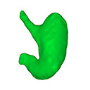
The present 3D Dataset contains the 3D models analyzed in: Kaigai N et al. Morphogenesis and three-dimensional movement of the stomach during the human embryonic period, Anat Rec (Hoboken). 2014 May;297(5):791-797. doi: 10.1002/ar.22833.
Homo sapiens KC-CS16STM27159 View specimen
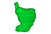
|
M3#56computationally reconstructed stomach of the human embryo (M3#56_KC-CS16STM27159) at Carnegie Stage 16 (Crown Rump Length= 9.9mm). Type: "3D_surfaces"doi: 10.18563/m3.sf56 state:published |
Download 3D surface file |
Homo sapiens KC-CS17STM20383 View specimen
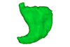
|
M3#57computationally reconstructed stomach of the human embryo (M3#57_KC-CS17STM20383) at Carnegie Stage 17 (Crown Rump Length= 12.3mm). Type: "3D_surfaces"doi: 10.18563/m3.sf57 state:published |
Download 3D surface file |
Homo sapiens KC-CS18STM21807 View specimen
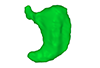
|
M3#58computationally reconstructed stomach of the human embryo (M3#58_KC-CS18STM21807) at Carnegie Stage 18 (Crown Rump Length= 14.7mm). Type: "3D_surfaces"doi: 10.18563/m3.sf58 state:published |
Download 3D surface file |
Homo sapiens KC-CS19STM17998 View specimen
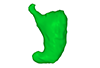
|
M3#59computationally reconstructed stomach of the human embryo (M3#59_KC-CS19STM17998) at Carnegie Stage 19 (Crown Rump Length was unmeasured ). Type: "3D_surfaces"doi: 10.18563/m3.sf59 state:published |
Download 3D surface file |
Homo sapiens KC-CS20STM20785 View specimen
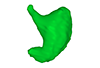
|
M3#60computationally reconstructed stomach of the human embryo (M3#60_KC-CS20STM20785) at Carnegie Stage 20 (Crown Rump Length= 18.7 mm). Type: "3D_surfaces"doi: 10.18563/m3.sf60 state:published |
Download 3D surface file |
Homo sapiens KC-CS21STM24728 View specimen
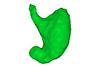
|
M3#61computationally reconstructed stomach of the human embryo (M3#61_KC-CS21STM24728) at Carnegie Stage 21 (Crown Rump Length= 20.9 mm). Type: "3D_surfaces"doi: 10.18563/m3.sf61 state:published |
Download 3D surface file |
Homo sapiens KC-CS22STM26438 View specimen

|
M3#62computationally reconstructed stomach of the human embryo (M3#62_KC-CS22STM26438) at Carnegie Stage 22 (Crown Rump Length= 21.5 mm). Type: "3D_surfaces"doi: 10.18563/m3.sf62 state:published |
Download 3D surface file |
Homo sapiens KC-CS23STM20018 View specimen
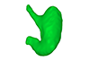
|
M3#63computationally reconstructed stomach of the human embryo (M3#63_KC-CS23STM20018) at Carnegie Stage 23 (Crown Rump Length= 23.1 mm). Type: "3D_surfaces"doi: 10.18563/m3.sf63 state:published |
Download 3D surface file |
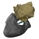
This contribution includes the 3D models of the reconstructed ossicular chain of the cainotheriid Caenomeryx filholi from the late Oligocene locality of Pech Desse (MP28, Quercy, France) described and figured in the publication of Assemat et al. (2020). It represents the oldest ossicular chain reconstruction for a Paleogene terrestrial artiodactyl species.
Caenomeryx filholi UM PDS 3353 View specimen
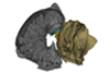
|
M3#508reconstruction of the middle ear with petrosal, bulla, stapes, incus, malleus Type: "3D_surfaces"doi: 10.18563/m3.sf.508 state:published |
Download 3D surface file |

This contribution contains the 3D models described and figured in the following publication: Molnar, JL, Pierce, SE, Bhullar, B-A, Turner, AH, Hutchinson, JR (accepted). Morphological and functional changes in the crocodylomorph vertebral column with increasing aquatic adaptation. Royal Society Open Science.
Protosuchus richardsoni AMNH-VP 3024 View specimen

|
M3#448th and 9th dorsal vertebrae, 1st and 2nd lumbar vertebrae, and 5th lumbar and sacral vertebrae. Type: "3D_surfaces"doi: 10.18563/m3.sf44 state:published |
Download 3D surface file |
Terrestrisuchus gracilis NHM-PV R 7562 View specimen

|
M3#451st and 2nd lumbar vertebrae, and 5th lumbar and sacral vertebrae Type: "3D_surfaces"doi: 10.18563/m3.sf45 state:published |
Download 3D surface file |
Pelagosaurus typus NHM-PV OR 32598 View specimen

|
M3#467th and 8th dorsal vertebrae, 11th and 12th dorsal vertebrae, 15th dorsal vertebra and sacral vertebra. Type: "3D_surfaces"doi: 10.18563/m3.sf46 state:published |
Download 3D surface file |
Metriorhynchus superciliosus NHM-PV R 2054 View specimen

|
M3#476th and 7th dorsal vertebrae, 10th and 11th dorsal vertebrae, 17th dorsal vertebra and sacral vertebra Type: "3D_surfaces"doi: 10.18563/m3.sf47 state:published |
Download 3D surface file |
Crocodylus niloticus FNC0 View specimen

|
M3#487th and 8th dorsal vertebrae, 1st and 2nd lumbar vertebrae, 5th lumbar vertebra and sacral vertebra. Type: "3D_surfaces"doi: 10.18563/m3.sf48 state:published |
Download 3D surface file |
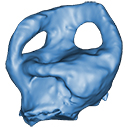
The present 3D Dataset contains the 3D models analyzed in "Neenan, J. M., Reich, T., Evers, S., Druckenmiller, P. S., Voeten, D. F. A. E., Choiniere, J. N., Barrett, P. M., Pierce, S. E. and Benson, R. B. J. Evolution of the sauropterygian labyrinth with increasingly pelagic lifestyles. Current Biology, 27." https://doi.org/10.1016/j.cub.2017.10.069
Amblyrhynchus cristatus OUMNH 11616 View specimen
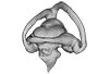
|
M3#322Right labyrinth of Amblyrhynchus cristatus (OUMNH 11616). Type: "3D_surfaces"doi: 10.18563/m3.sf.322 state:published |
Download 3D surface file |
Augustasaurus hagdorni FMNH PR 1974 View specimen
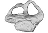
|
M3#333Right labyrinth model of Augustasaurus FMNH PR 1974 Type: "3D_surfaces"doi: 10.18563/m3.sf.333 state:published |
Download 3D surface file |
Callawayasaurus colombiensis UCMP V-38349 / UCMP V-125328 View specimen
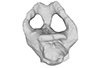
|
M3#331Composite left labyrinth of Callawayasaurus. The majority of the model is from the holotype (UCMP V-38349), but the anterior portion is formed from the right labyrinth (reflected) from the paratype (UCMP V-125328). Type: "3D_surfaces"doi: 10.18563/m3.sf.331 state:published |
Download 3D surface file |
Lepidochelys olivacea SMNS 11070 View specimen
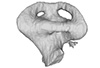
|
M3#330Left labyrinth model of Lepidochelys SMNS 11070 Type: "3D_surfaces"doi: 10.18563/m3.sf.330 state:published |
Download 3D surface file |
Macrochelys temminckii FMNH 22111 View specimen
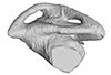
|
M3#334Left labyrinth model of Macrochelys FMNH 22111 Type: "3D_surfaces"doi: 10.18563/m3.sf.334 state:published |
Download 3D surface file |
Macroplata tenuiceps NHMUK R 5488 View specimen
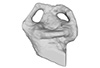
|
M3#328Left labyrinth of Macroplata NHMUK R 5488 Type: "3D_surfaces"doi: 10.18563/m3.sf.328 state:published |
Download 3D surface file |
Microcleidus homalospondylus NHMUK 36184 View specimen
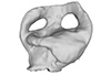
|
M3#327Right labyrinth model of Microcleidus NHMUK 36184 Type: "3D_surfaces"doi: 10.18563/m3.sf.327 state:published |
Download 3D surface file |
Nothosaurus sp. NME 16/4 View specimen
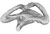
|
M3#326Right labyrinth model of Nothosaurus sp. NME 16/4 Type: "3D_surfaces"doi: 10.18563/m3.sf.326 state:published |
Download 3D surface file |
Peloneustes philarchus NHMUK R 3803 View specimen
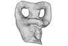
|
M3#325Left labyrinth model of Peloneustes philarchus NHMUK R 3803 Type: "3D_surfaces"doi: 10.18563/m3.sf.325 state:published |
Download 3D surface file |
Placodus gigas UMO BT 13 View specimen
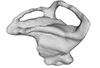
|
M3#324Right labyrinth model of Placodus gigas UMO BT 13 Type: "3D_surfaces"doi: 10.18563/m3.sf.324 state:published |
Download 3D surface file |
Puppigerus camperi NHMUK R 38955 View specimen
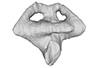
|
M3#323Left labyrinth model of Puppigerus NHMUK R 38955 Type: "3D_surfaces"doi: 10.18563/m3.sf.323 state:published |
Download 3D surface file |
Simosaurus gaillardoti GPIT RE/09313 View specimen
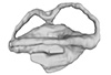
|
M3#332Right labyrinth model of Simosaurus GPIT RE/09313 Type: "3D_surfaces"doi: 10.18563/m3.sf.332 state:published |
Download 3D surface file |
Libonectes morgani SMUSMP 69120 View specimen
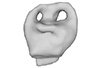
|
M3#335Right labyrinth model of Libonected morgani (SMUSMP 69120) Type: "3D_surfaces"doi: 10.18563/m3.sf.335 state:published |
Download 3D surface file |


