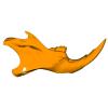
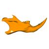
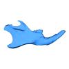
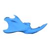
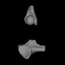
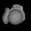
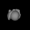
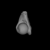
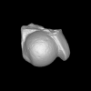
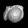
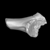
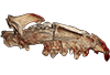
3D models of Kalakocetus, the earliest Cetacea
3D models related to the publication: Hidden diversity of Palaeogene metatherians: a new family of polydolopimorphian marsupials from Peruvian Amazonia
Explodable 3D Dog Skull for Veterinary Education
3D GM dataset of bird skeletal variation
Skeletal embryonic development in the catshark
Bony connexions of the petrosal bone of extant hippos
bony labyrinth (14) , inner ear (11) , Eocene (11) , geometric morphometrics (10) , CT-scan (10) , Oligocene (9) , Micro-CT (9)
Maëva Judith Orliac (24) , Lionel Hautier (24) , Laurent Marivaux (18) , Renaud Lebrun (15) , Rodolphe Tabuce (14) , Pierre-Olivier Antoine (13) , Bastien Mennecart (13)
|
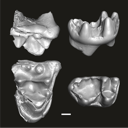
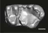
3D models related to the publication: “Trophic differentiation between the endemic Cypriot mouse and the house mouse: a study coupling stable isotopes and morphometrics”
|
|
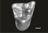
M3#15843D model of the right mandible Type: "3D_surfaces"doi: 10.18563/m3.sf.1584 state:published |
Download 3D surface file |
Mus cypriacus Cypriacus_BET2 View specimen

|
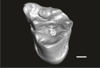
M3#15853D model of the right mandible Type: "3D_surfaces"doi: 10.18563/m3.sf.1585 state:published |
Download 3D surface file |
Mus cypriacus Cypriacus_FON1 View specimen
|
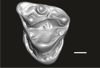
M3#15863D model of the right mandible Type: "3D_surfaces"doi: 10.18563/m3.sf.1586 state:published |
Download 3D surface file |
Mus cypriacus Cypriacus_FON2 View specimen
|
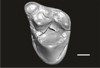
M3#15873D model of the right mandible Type: "3D_surfaces"doi: 10.18563/m3.sf.1587 state:published |
Download 3D surface file |
Mus cypriacus Cypriacus_KOU1 View specimen
|
M3#15883D model of the right mandible Type: "3D_surfaces"doi: 10.18563/m3.sf.1588 state:published |
Download 3D surface file |
Mus musculus Cyprus_dom_KOF1 View specimen
|
M3#15893D model of the right mandible Type: "3D_surfaces"doi: 10.18563/m3.sf.1589 state:published |
Download 3D surface file |
Mus musculus Cyprus_dom_LEF1 View specimen
|
M3#15903D model of the right mandible Type: "3D_surfaces"doi: 10.18563/m3.sf.1590 state:published |
Download 3D surface file |
Mus musculus Cyprus_dom_MEN1 View specimen

|
M3#15913D model of the right mandible Type: "3D_surfaces"doi: 10.18563/m3.sf.1591 state:published |
Download 3D surface file |
Mus musculus Cyprus_dom_TSE2 View specimen
|
M3#15923D model of the mirrored left mandible Type: "3D_surfaces"doi: 10.18563/m3.sf.1592 state:published |
Download 3D surface file |
Mus musculus Cyprus_dom_XYL5 View specimen
|
M3#15933D model of the right mandible Type: "3D_surfaces"doi: 10.18563/m3.sf.1593 state:published |
Download 3D surface file |
This contribution contains the three-dimensional digital models of eleven isolated fossil teeth of a merialine paroxyclaenid (Welcommoides gurki), discovered from lower Oligocene deposits of the Bugti Hills (Balochistan, Pakistan). These fossils were described, figured and discussed in the following publication: Solé et al. (2024), An unexpected late paroxyclaenid (Mammalia, Cimolesta) out of Europe: dental evidence from the Oligocene of the Bugti Hills, Pakistan. Papers in Palaeontology. https://doi.org/10.1002/spp2.1599
Welcommoides gurki UM-DBC 2225 View specimen
|
M3#1083Left m3 Type: "3D_surfaces"doi: 10.18563/m3.sf.1083 state:published |
Download 3D surface file |
Welcommoides gurki UM-DBC 2226 View specimen
|
M3#1084Right m3 Type: "3D_surfaces"doi: 10.18563/m3.sf.1084 state:published |
Download 3D surface file |
Welcommoides gurki UM-DBC 2227 View specimen
|
M3#1085Trigonid of a right lower molar Type: "3D_surfaces"doi: 10.18563/m3.sf.1085 state:published |
Download 3D surface file |
Welcommoides gurki UM-DBC 2230 View specimen
|
M3#1086Right DP4 Type: "3D_surfaces"doi: 10.18563/m3.sf.1086 state:published |
Download 3D surface file |
Welcommoides gurki UM-DBC 2228 View specimen
|
M3#1093Right M1 Type: "3D_surfaces"doi: 10.18563/m3.sf.1093 state:published |
Download 3D surface file |
Welcommoides gurki UM-DBC 2229 View specimen
|
M3#1087Right M2 Type: "3D_surfaces"doi: 10.18563/m3.sf.1087 state:published |
Download 3D surface file |
Welcommoides gurki UM-DBC 2236 View specimen
|
M3#1088Left M2 Type: "3D_surfaces"doi: 10.18563/m3.sf.1088 state:published |
Download 3D surface file |
Welcommoides gurki UM-DBC 2231 View specimen
|
M3#1089Left M3 Type: "3D_surfaces"doi: 10.18563/m3.sf.1089 state:published |
Download 3D surface file |
Welcommoides gurki UM-DBC 2232 View specimen
|
M3#1090Left M3 Type: "3D_surfaces"doi: 10.18563/m3.sf.1090 state:published |
Download 3D surface file |
Welcommoides gurki UM-DBC 2234 View specimen
|
M3#1091Left M3 Type: "3D_surfaces"doi: 10.18563/m3.sf.1091 state:published |
Download 3D surface file |
Welcommoides gurki UM-DBC 2233 View specimen
|
M3#1092Left M3 Type: "3D_surfaces"doi: 10.18563/m3.sf.1092 state:published |
Download 3D surface file |
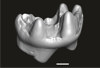
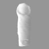
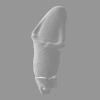
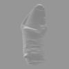
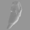
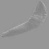
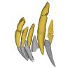
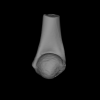
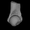
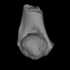
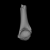
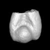
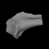
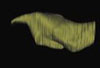
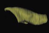
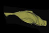
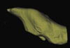
The present 3D dataset contains 3D models of new material from the middle Eocene of the Upper Subathu Formation in the Kalakot area (India), documenting the anterior dentition of the raoellid Indohyus indirae. Raoellidae are closely related to stem cetaceans and bring crucial information to understand the earliest phase of land to water transition in Cetacea.
Indohyus indirae GU/RJ/31 View specimen
|
M3#1505Right i1 Type: "3D_surfaces"doi: 10.18563/m3.sf.1505 state:published |
Download 3D surface file |
Indohyus indirae GU/RJ/32 View specimen
|
M3#1506Right i1 Type: "3D_surfaces"doi: 10.18563/m3.sf.1506 state:published |
Download 3D surface file |
Indohyus indirae GU/RJ/16 View specimen
|
M3#1507Left I3 Type: "3D_surfaces"doi: 10.18563/m3.sf.1507 state:published |
Download 3D surface file |
Indohyus indirae GU/RJ/23 View specimen
|
M3#1508Left I2 Type: "3D_surfaces"doi: 10.18563/m3.sf.1508 state:published |
Download 3D surface file |
Indohyus indirae GU/RJ/25 View specimen
|
M3#1509Left I1 Type: "3D_surfaces"doi: 10.18563/m3.sf.1509 state:published |
Download 3D surface file |
Indohyus indirae GU/RJ/26 View specimen
|
M3#1510Right I3 Type: "3D_surfaces"doi: 10.18563/m3.sf.1510 state:published |
Download 3D surface file |
Indohyus indirae GU/RJ/57 View specimen
|
M3#1511Left I1 Type: "3D_surfaces"doi: 10.18563/m3.sf.1511 state:published |
Download 3D surface file |
Indohyus indirae GU/RJ/61 View specimen
|
M3#1512Right upper canine Type: "3D_surfaces"doi: 10.18563/m3.sf.1512 state:published |
Download 3D surface file |
Indohyus indirae GU/RJ/63 View specimen

|
M3#1513Left upper canine Type: "3D_surfaces"doi: 10.18563/m3.sf.1513 state:published |
Download 3D surface file |
Indohyus indirae GU/RJ/74 View specimen
|
M3#1514Left upper canine Type: "3D_surfaces"doi: 10.18563/m3.sf.1514 state:published |
Download 3D surface file |
Indohyus indirae GU/RJ/439 View specimen
|
M3#1515Left upper canine Type: "3D_surfaces"doi: 10.18563/m3.sf.1515 state:published |
Download 3D surface file |
Indohyus indirae GU/RJ/457 View specimen
|
M3#1516Left upper canine Type: "3D_surfaces"doi: 10.18563/m3.sf.1516 state:published |
Download 3D surface file |
Indohyus indirae GU/RJ/846 View specimen
|
M3#1517Left upper canine Type: "3D_surfaces"doi: 10.18563/m3.sf.1517 state:published |
Download 3D surface file |
Indohyus indirae GU/RJ/822 View specimen
|
M3#1518Right fragmentary maxillary with decidual canine and I3 Type: "3D_surfaces"doi: 10.18563/m3.sf.1518 state:published |
Download 3D surface file |
Indohyus indirae GU/RJ/824 View specimen
|
M3#1519Right fragmentary mandible with lower canine and small part of the i3 Type: "3D_surfaces"doi: 10.18563/m3.sf.1519 state:published |
Download 3D surface file |
Indohyus indirae GU/RJ/838 View specimen
|
M3#1520Right fragmentary mandible with permanent i2, i3 and canine and small part of the root of the decidual i3 Type: "3D_surfaces"doi: 10.18563/m3.sf.1520 state:published |
Download 3D surface file |
Indohyus indirae GU/RJ/842 View specimen
|
M3#1521Left fragmentary mandible with decidual and permanent canine Type: "3D_surfaces"doi: 10.18563/m3.sf.1521 state:published |
Download 3D surface file |
Indohyus indirae GU/RJ/26,32,56,57,457,822,838,842 : Composite Anterior dentition View specimen
|
M3#15293D composite reconstruction of the anterior dentition of Indohyus indirae with GU/RJ/57 (I1), 56 (I2), 26 (I3), 457 (Upper canine), 822 (P1), 32 (i1), 838 (i2, i3 and lower canine) and 842 (p1) Type: "3D_surfaces"doi: 10.18563/m3.sf.1529 state:published |
Download 3D surface file |
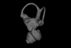
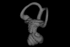
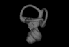
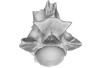
This contribution contains the three-dimensional models of the inner ear of the hetaxodontid rodents Amblyrhiza, Clidomys and Elasmodontomys from the West Indies. These specimens were analyzed and discussed in : The inner ear of caviomorph rodents: phylogenetic implications and application to extinct West Indian taxa.
Amblyrhiza inundata 11842 View specimen
|
M3#11543D surface of the left-oriented inner ear of Amblyrhiza. Type: "3D_surfaces"doi: 10.18563/m3.sf.1154 state:published |
Download 3D surface file |
Clidomys sp NA View specimen
|
M3#11553D surface of the left-oriented inner ear of Clidomys sp. Type: "3D_surfaces"doi: 10.18563/m3.sf.1155 state:published |
Download 3D surface file |
Elasmodontomys obliquus 17127 View specimen
|
M3#11563D surface of the left-oriented inner ear of Elasmodontomys obliquus. Type: "3D_surfaces"doi: 10.18563/m3.sf.1156 state:published |
Download 3D surface file |
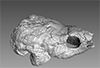
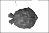
The present 3D Dataset contains the 3D model analyzed in the following publication: Paulina-Carabajal, A., Sterli, J., Werneburg, I., 2019. The endocranial anatomy of the stem turtle Naomichelys speciosa from the Early Cretaceous of North America. Acta Palaeontologica Polonica, https://doi.org/10.4202/app.00606.2019
Naomichelys speciosa FMNH PR273 View specimen
|
M3#428FMNH_PR273_1 - Naomichlys speciosa - skull Type: "3D_surfaces"doi: 10.18563/m3.sf.428 state:published |
Download 3D surface file |
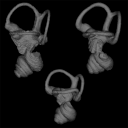
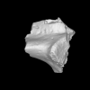
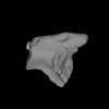
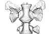
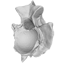
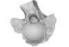
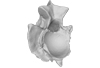
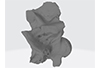
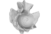
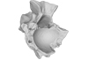
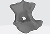
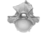
The present 3D Dataset contains the 3D models analyzed in Mennecart, B., Duranthon, F., & Costeur, L. 2024. Systematic contribution of the auditory region to the knowledge of the oldest European Bovidae (Mammalia, Ruminantia). Journal of Anatomy XXX. https://doi.org/10.1111/joa.14132
Pusillutragus montrealensis MHNT.PAL.2015.0.2261.4 View specimen
|
M3#1522Right petrosal, bony labyrinth, stapes Type: "3D_surfaces"doi: 10.18563/m3.sf.1522 state:published |
Download 3D surface file |
Pusillutragus montrealensis MHNT.PAL.2015.0.2261.9 View specimen
|
M3#1523Left petrosal and left bony labyrinth Type: "3D_surfaces"doi: 10.18563/m3.sf.1523 state:published |
Download 3D surface file |
Eotragus artenensis SMNS-P-41625 View specimen
|
M3#1524Petrosal (right), bony labyrinth (left) Type: "3D_surfaces"doi: 10.18563/m3.sf.1524 state:published |
Download 3D surface file |
Eotragus clavatus NMB San.15056 View specimen
|
M3#1528Right petrosal and right bony labyrinth Type: "3D_surfaces"doi: 10.18563/m3.sf.1528 state:published |
Download 3D surface file |
Eotragus clavatus NMB San.15055 View specimen
|
M3#1526Left Petrosal Type: "3D_surfaces"doi: 10.18563/m3.sf.1526 state:published |
Download 3D surface file |
This contribution contains the 3D models of the fossil teeth of two chinchilloid caviomorph rodents (Borikenomys praecursor and Chinchilloidea gen. et sp. indet.) discovered from lower Oligocene deposits of Puerto Rico, San Sebastian Formation (locality LACM Loc. 8060). These fossils were described and figured in the following publication: Marivaux et al. (2020), Early Oligocene chinchilloid caviomorphs from Puerto Rico and the initial rodent colonization of the West Indies. Proceedings of the Royal Society B. http://dx.doi.org/10.1098/rspb.2019.2806
Borikenomys praecursor LACM 162447 View specimen
|
M3#638Right lower m3. This isolated tooth was scanned with a resolution of 6 µm using a μ-CT-scanning station EasyTom 150 / Rx Solutions (Montpellier RIO Imaging, ISE-M, Montpellier, France). AVIZO 7.1 (Visualization Sciences Group) software was used for visualization, segmentation, and 3D rendering. The specimen was prepared within a “labelfield” module of AVIZO, using the segmentation threshold selection tool. Type: "3D_surfaces"doi: 10.18563/m3.sf.638 state:published |
Download 3D surface file |
Borikenomys praecursor LACM 162446 View specimen
|
M3#639Fragment of lower molar (most of the mesial part). This isolated broken tooth was scanned with a resolution of 6 µm using a μ-CT-scanning station EasyTom 150 / Rx Solutions (Montpellier RIO Imaging, ISE-M, Montpellier, France). AVIZO 7.1 (Visualization Sciences Group) software was used for visualization, segmentation, and 3D rendering. The specimen was prepared within a “labelfield” module of AVIZO, using the segmentation threshold selection tool. Type: "3D_surfaces"doi: 10.18563/m3.sf.639 state:published |
Download 3D surface file |
indet indet LACM 162448 View specimen
|
M3#640Fragment of either an upper tooth (mesial laminae) or a lower tooth (distal laminae). The specimen was scanned with a resolution of 6 µm using a μ-CT-scanning station EasyTom 150 / Rx Solutions (Montpellier RIO Imaging, ISE-M, Montpellier, France). AVIZO 7.1 (Visualization Sciences Group) software was used for visualization, segmentation, and 3D rendering. This fragment of tooth was prepared within a “labelfield” module of AVIZO, using the segmentation threshold selection tool. Type: "3D_surfaces"doi: 10.18563/m3.sf.640 state:published |
Download 3D surface file |
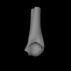
The present contribution contains the 3D models of fossil humeri and ilia of anurans from various Eocene-Miocene deposits of Peruvian Amazonia. These fossils were described and figured in the following publication: Jansen et al. (2023), First Eocene–Miocene anuran fossils from Peruvian Amazonia: insights into Neotropical frog evolution and diversity. Papers in Palaeontology, The Palaeontological Association.
Indet. indet. MUSM 4746 View specimen
|
M3#1231Humeral fragment (distal end) Type: "3D_surfaces"doi: 10.18563/m3.sf.1231 state:published |
Download 3D surface file |
Indet. indet. MUSM 4747 View specimen
|
M3#1232Humeral fragment (distal end) Type: "3D_surfaces"doi: 10.18563/m3.sf.1232 state:published |
Download 3D surface file |
Indet. indet. MUSM 4748 View specimen
|
M3#1233Humeral fragment (distal end) Type: "3D_surfaces"doi: 10.18563/m3.sf.1233 state:published |
Download 3D surface file |
Indet. indet. MUSM 4755 View specimen
|
M3#1234Humeral fragment (distal end) Type: "3D_surfaces"doi: 10.18563/m3.sf.1234 state:published |
Download 3D surface file |
Indet. indet. MUSM 4756 View specimen
|
M3#1235Humeral fragment (distal end) Type: "3D_surfaces"doi: 10.18563/m3.sf.1235 state:published |
Download 3D surface file |
Indet. indet. MUSM 4757 View specimen

|
M3#1236Humeral fragment (distal end) Type: "3D_surfaces"doi: 10.18563/m3.sf.1236 state:published |
Download 3D surface file |
Indet. indet. MUSM 4761 View specimen
|
M3#1237Humeral fragment (distal end) Type: "3D_surfaces"doi: 10.18563/m3.sf.1237 state:published |
Download 3D surface file |
Indet. indet. MUSM 4763 View specimen
|
M3#1238Humeral fragment (distal end) Type: "3D_surfaces"doi: 10.18563/m3.sf.1238 state:published |
Download 3D surface file |
Indet. indet. MUSM 4765 View specimen
|
M3#1239Humeral fragment (distal end) Type: "3D_surfaces"doi: 10.18563/m3.sf.1239 state:published |
Download 3D surface file |
Indet. indet. MUSM 4766 View specimen
|
M3#1240Humeral fragment (distal end) Type: "3D_surfaces"doi: 10.18563/m3.sf.1240 state:published |
Download 3D surface file |
Indet. indet. MUSM 4775 View specimen
|
M3#1241Humeral fragment (distal end) Type: "3D_surfaces"doi: 10.18563/m3.sf.1241 state:published |
Download 3D surface file |
cf. Pipa sp. MUSM 4776 View specimen
|
M3#1242Humeral fragment (distal end) Type: "3D_surfaces"doi: 10.18563/m3.sf.1242 state:published |
Download 3D surface file |
Indet. indet. MUSM 4788 View specimen
|
M3#1243Ilial fragment Type: "3D_surfaces"doi: 10.18563/m3.sf.1243 state:published |
Download 3D surface file |
Indet. indet. MUSM 4789 View specimen
|
M3#1244Ilial fragment Type: "3D_surfaces"doi: 10.18563/m3.sf.1244 state:published |
Download 3D surface file |
Indet. indet. MUSM 4790 View specimen
|
M3#1245Ilial fragment Type: "3D_surfaces"doi: 10.18563/m3.sf.1245 state:published |
Download 3D surface file |
Indet. indet. MUSM 4792 View specimen
|
M3#1246Ilial fragment Type: "3D_surfaces"doi: 10.18563/m3.sf.1246 state:published |
Download 3D surface file |
Indet. indet. MUSM 4793 View specimen
|
M3#1247Ilial fragment Type: "3D_surfaces"doi: 10.18563/m3.sf.1247 state:published |
Download 3D surface file |
Indet. indet. MUSM 4794 View specimen
|
M3#1249Ilial fragment Type: "3D_surfaces"doi: 10.18563/m3.sf.1249 state:published |
Download 3D surface file |
Indet. indet. MUSM 4795 View specimen
|
M3#1250Ilial fragment Type: "3D_surfaces"doi: 10.18563/m3.sf.1250 state:published |
Download 3D surface file |
cf. Pipa sp. MUSM 4796 View specimen
|
M3#1251Ilial fragment Type: "3D_surfaces"doi: 10.18563/m3.sf.1251 state:published |
Download 3D surface file |
cf. Pipa sp. MUSM 4797 View specimen
|
M3#1252Ilial fragment Type: "3D_surfaces"doi: 10.18563/m3.sf.1252 state:published |
Download 3D surface file |
The present 3D Dataset contains 26 3D models analyzed in the study: On the “cartilaginous rider” in the endocasts of turtle brain cavities, published by the authors in the journal Vertebrate Zoology.
Annemys sp. IVPP-V-18106 View specimen
|
M3#7723D surface(s) file to specimen IVPP-V-18106 Type: "3D_surfaces"doi: 10.18563/m3.sf.772 state:published |
Download 3D surface file |
Apalone spinifera FMNH 22178 View specimen

|
M3#7733D surface(s) file to specimen FMNH 22178 Type: "3D_surfaces"doi: 10.18563/m3.sf.773 state:published |
Download 3D surface file |
Caretta caretta NHMUK1940.3.15.1 View specimen
|
M3#7863D surface(s) file to specimen NHMUK1940.3.15.1 Type: "3D_surfaces"doi: 10.18563/m3.sf.786 state:published |
Download 3D surface file |
Chelodina reimanni ZMB 49659 View specimen
|
M3#7743D surface(s) file to specimen ZMB 49659 Type: "3D_surfaces"doi: 10.18563/m3.sf.774 state:published |
Download 3D surface file |
Chelonia mydas ZMB-37416MS View specimen

|
M3#7753D surface(s) file to specimen ZMB-37416MS Type: "3D_surfaces"doi: 10.18563/m3.sf.775 state:published |
Download 3D surface file |
Cuora amboinensis NHMUK69.42.145_4 View specimen
|
M3#7763D surface(s) file to specimen NHMUK69.42.145_4 Type: "3D_surfaces"doi: 10.18563/m3.sf.776 state:published |
Download 3D surface file |
Emydura subglobosa IW92 View specimen
|
M3#7773D surface(s) file to specimen IW92 Type: "3D_surfaces"doi: 10.18563/m3.sf.777 state:published |
Download 3D surface file |
Eubaena cephalica DMNH 96004 View specimen
|
M3#7783D surface(s) file to specimen DMNH 96004 Type: "3D_surfaces"doi: 10.18563/m3.sf.778 state:published |
Download 3D surface file |
Gopherus berlandieri AMNH-73816 View specimen
|
M3#7793D surface(s) file to specimen AMNH-73816 Type: "3D_surfaces"doi: 10.18563/m3.sf.779 state:published |
Download 3D surface file |
Kinixys belliana AMNH-10028 View specimen
|
M3#7803D surface(s) file to specimen AMNH-10028 Type: "3D_surfaces"doi: 10.18563/m3.sf.780 state:published |
Download 3D surface file |
Macrochelys temminckii GPIT-PV-79430 View specimen
|
M3#7813D surface(s) file to specimen GPIT-PV-79430 Type: "3D_surfaces"doi: 10.18563/m3.sf.781 state:published |
Download 3D surface file |
Malacochersus tornieri SMF-58702 View specimen
|
M3#7873D surface(s) file to specimen SMF-58702 Type: "3D_surfaces"doi: 10.18563/m3.sf.787 state:published |
Download 3D surface file |
Naomichelys speciosa FMNH-PR-273 View specimen
|
M3#7823D surface(s) file to specimen FMNH-PR-273 Type: "3D_surfaces"doi: 10.18563/m3.sf.782 state:published |
Download 3D surface file |
Pelodiscus sinensis IW576-2 View specimen
|
M3#7833D surface(s) file to specimen IW576-2 Type: "3D_surfaces"doi: 10.18563/m3.sf.783 state:published |
Download 3D surface file |
Platysternon megacephalum SMF-69684 View specimen
|
M3#7843D surface(s) file to specimen SMF-69684 Type: "3D_surfaces"doi: 10.18563/m3.sf.784 state:published |
Download 3D surface file |
Podocnemis unifilis SMF-55470 View specimen
|
M3#7853D surface(s) file to specimen SMF-55470 Type: "3D_surfaces"doi: 10.18563/m3.sf.785 state:published |
Download 3D surface file |
Proganochelys quenstedtii MB 1910.45.2 View specimen
|
M3#7883D surface(s) file to specimen MB 1910.45.2 Type: "3D_surfaces"doi: 10.18563/m3.sf.788 state:published |
Download 3D surface file |
Proganochelys quenstedtii SMNS 16980 View specimen
|
M3#7893D surface(s) file to specimen SMNS 16980 Type: "3D_surfaces"doi: 10.18563/m3.sf.789 state:published |
Download 3D surface file |
Rhinochelys pulchriceps CAMSM_B55775 View specimen
|
M3#7903D surface(s) file to specimen CAMSM_B55775 Type: "3D_surfaces"doi: 10.18563/m3.sf.790 state:published |
Download 3D surface file |
Rhinoclemmys funereal YPM12174 View specimen
|
M3#7913D surface(s) file to specimen YPM12174 Type: "3D_surfaces"doi: 10.18563/m3.sf.791 state:published |
Download 3D surface file |
Sandownia harrisi MIWG3480 View specimen
|
M3#7923D surface(s) file to specimen MIWG3480 Type: "3D_surfaces"doi: 10.18563/m3.sf.792 state:published |
Download 3D surface file |
Testudo graeca YPM14342 View specimen
|
M3#7933D surface(s) file to specimen YPM14342 Type: "3D_surfaces"doi: 10.18563/m3.sf.793 state:published |
Download 3D surface file |
Testudo hermanni AMNH134518 View specimen
|
M3#7943D surface(s) file to specimen AMNH134518 Type: "3D_surfaces"doi: 10.18563/m3.sf.794 state:published |
Download 3D surface file |
Trachemys scripta NN View specimen
|
M3#7953D surface(s) file to specimen Trachemys scripta Type: "3D_surfaces"doi: 10.18563/m3.sf.795 state:published |
Download 3D surface file |
Xinjiangchelys radiplicatoides IVPP V9539 View specimen
|
M3#7963D surface(s) file to specimen IVPP V9539 Type: "3D_surfaces"doi: 10.18563/m3.sf.796 state:published |
Download 3D surface file |
Chelydra serpentina UFR VP1 View specimen

|
M3#801Brain endocast Type: "3D_surfaces"doi: 10.18563/m3.sf.801 state:published |
Download 3D surface file |
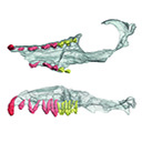
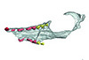
This contribution contains 3D models of the cranial skeleton and muscles in an elephantfish (Callorhinchus milii) and a catshark (Scyliorhinus canicula), based on synchrotron tomographic scans. These datasets were analyzed and described in Dearden et al. (2021) “The morphology and evolution of chondrichthyan cranial muscles: a digital dissection of the elephantfish Callorhinchus milii and the catshark Scyliorhinus canicula.” Journal of Anatomy.
Callorhinchus milii 001 View specimen
|
M3#7083D models of the cranial skeleton and muscles of Callorhinchus milii, created using Mimics. Type: "3D_surfaces"doi: 10.18563/m3.sf.708 state:published |
Download 3D surface file |
Scyliorhinus canicula 002 View specimen
|
M3#7093D models of the cranial skeleton and muscles of Scyliorhinus canicula, created using Mimics. Type: "3D_surfaces"doi: 10.18563/m3.sf.709 state:published |
Download 3D surface file |
This contribution contains the 3D models described and figured in the following publication: Paulina-Carabajal, A. and Nieto, M. N. In press. Brief comment on the brain and inner ear of Giganotosaurus carolinii (Dinosauria: Theropoda) based on CT scans. Ameghiniana. https://doi.org/10.5710/AMGH.25.10.2019.3237
Giganotosaurus carolinii MUCPv-CH-1 View specimen
|
M3#504The current file contents 3D models of the braincase, brain, left and right inner ears Type: "3D_surfaces"doi: 10.18563/m3.sf.504 state:published |
Download 3D surface file |
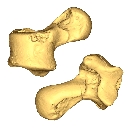
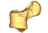
The present 3D Dataset contains the 3D models of the sacral vertebrae analyzed in “Sacral co-ossification in dinosaurs: The oldest record of fused sacral vertebrae in Dinosauria and the diversity of sacral co-ossification patterns in the group”.
Buriolestes schultzi CAPPA/UFSM 0035 View specimen
|
M3#705Sacral vertebrae of Buriolestes schultzi Type: "3D_surfaces"doi: 10.18563/m3.sf.705 state:published |
Download 3D surface file |
indet indet CAPPA/UFSM 0228 View specimen
|
M3#706Sacral vertebrae of a saurischian dinosaur indet. Type: "3D_surfaces"doi: 10.18563/m3.sf.706 state:published |
Download 3D surface file |
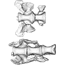
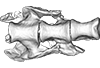
This contribution contains the 3D models described and figured in the following publication: Georgalis, G.L., G. Guinot, K.E. Kassegne, Y.Z. Amoudji, A.K.C. Johnson, H. Cappetta and L. Hautier. 2021. An assemblage of giant aquatic snakes (Serpentes, Palaeophiidae) from the Eocene of Togo. Swiss Journal of Palaeontology 140, https://doi.org/10.1186/s13358-021-00236-w
Palaeophis africanus UM KPO 21 View specimen
|
M3#821Trunk vertebra UM KPO 21 of Palaeophis africanus Type: "3D_surfaces"doi: 10.18563/m3.sf.821 state:published |
Download 3D surface file |
Palaeophis africanus UM KPO 22 View specimen
|
M3#822Trunk vertebra UM KPO 22 of Palaeophis africanus from the Eocene of Togo Type: "3D_surfaces"doi: 10.18563/m3.sf.822 state:published |
Download 3D surface file |
Palaeophis africanus UM KPO 23 View specimen
|
M3#823Trunk vertebra UM KPO 23 of Palaeophis africanus Type: "3D_surfaces"doi: 10.18563/m3.sf.823 state:published |
Download 3D surface file |
Palaeophis africanus UM KPO 24 View specimen
|
M3#824Trunk vertebra UM KPO 24 of Palaeophis africanus Type: "3D_surfaces"doi: 10.18563/m3.sf.824 state:published |
Download 3D surface file |
Palaeophis africanus UM KPO 25 View specimen
|
M3#825Trunk vertebra UM KPO 25 of Palaeophis africanus Type: "3D_surfaces"doi: 10.18563/m3.sf.825 state:published |
Download 3D surface file |
Palaeophis africanus UM KPO 26 View specimen
|
M3#826Trunk vertebra UM KPO 26 of Palaeophis africanus Type: "3D_surfaces"doi: 10.18563/m3.sf.826 state:published |
Download 3D surface file |
Palaeophis africanus UM KPO 27 View specimen
|
M3#827Trunk vertebra UM KPO 27 of Palaeophis africanus Type: "3D_surfaces"doi: 10.18563/m3.sf.827 state:published |
Download 3D surface file |
Palaeophis africanus UM KPO 28 View specimen
|
M3#828Trunk vertebra UM KPO 28 of Palaeophis africanus Type: "3D_surfaces"doi: 10.18563/m3.sf.828 state:published |
Download 3D surface file |
Palaeophis africanus UM KPO 29 View specimen
|
M3#829Trunk vertebra UM KPO 29 of Palaeophis africanus Type: "3D_surfaces"doi: 10.18563/m3.sf.829 state:published |
Download 3D surface file |
Palaeophis africanus UM KPO 30 View specimen
|
M3#830Trunk vertebra UM KPO 30 of Palaeophis africanus Type: "3D_surfaces"doi: 10.18563/m3.sf.830 state:published |
Download 3D surface file |
Palaeophis africanus UM KPO 31 View specimen
|
M3#831Trunk vertebra UM KPO 28 of Palaeophis africanus Type: "3D_surfaces"doi: 10.18563/m3.sf.831 state:published |
Download 3D surface file |
Palaeophis africanus UM KPO 32 View specimen
|
M3#832Trunk vertebra UM KPO 32 of Palaeophis africanus Type: "3D_surfaces"doi: 10.18563/m3.sf.832 state:published |
Download 3D surface file |
Palaeophis africanus UM KPO 33 View specimen
|
M3#833Trunk vertebra UM KPO 33 of Palaeophis africanus Type: "3D_surfaces"doi: 10.18563/m3.sf.833 state:published |
Download 3D surface file |
Palaeophis africanus UM KPO 34 View specimen

|
M3#839Trunk vertebra UM KPO 34 of Palaeophis africanus Type: "3D_surfaces"doi: 10.18563/m3.sf.839 state:published |
Download 3D surface file |
Palaeophis africanus UM KPO 35 View specimen
|
M3#840Trunk vertebra UM KPO 35 of Palaeophis africanus Type: "3D_surfaces"doi: 10.18563/m3.sf.840 state:published |
Download 3D surface file |
Palaeophis africanus UM KPO 36 View specimen
|
M3#841Trunk vertebra UM KPO 36 of Palaeophis africanus Type: "3D_surfaces"doi: 10.18563/m3.sf.841 state:published |
Download 3D surface file |
Palaeophis africanus UM KPO 37 View specimen
|
M3#842Trunk vertebra UM KPO 37 of Palaeophis africanus Type: "3D_surfaces"doi: 10.18563/m3.sf.842 state:published |
Download 3D surface file |
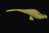
The present 3D Dataset contains the 3D models of the brain endocast analyzed in “Virtual brain endocast of Antifer (Mammalia: Cervidae), an extinct large cervid from South America”.
Antifer ensenadensis U-4922 View specimen
|
M3#550Brain endocast Type: "3D_surfaces"doi: 10.18563/m3.sf.550 state:published |
Download 3D surface file |
Antifer ensenadensis MCN-PV 943 View specimen
|
M3#551Brain endocast Type: "3D_surfaces"doi: 10.18563/m3.sf.551 state:published |
Download 3D surface file |
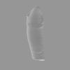
This contribution contains the 3D model of the fossil talus of a small-bodied anthropoid primate (Platyrrhini, Cebidae, Cebinae) discovered from lower Miocene deposits of Peruvian Amazonia (MD-61 locality, Upper Madre de Dios Basin). This fossil was described and figured in the following publication: Marivaux et al. (2012), A platyrrhine talus from the early Miocene of Peru (Amazonian Madre de Dios Sub-Andean Zone). Journal of Human Evolution. http://dx.doi.org/10.1016/j.jhevol.2012.07.005
Cebinae indet. sp. MUSM-2024 View specimen
|
M3#380Right talus 3D surface of a Miocene Cebinae indet. primate Type: "3D_surfaces"doi: 10.18563/m3.sf.380 state:published |
Download 3D surface file |
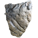
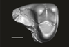
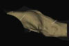
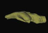
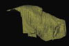
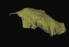
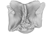
The present 3D Dataset contains the 3D model analyzed in Hendrickx, C. and Bell, P. R. 2021. The scaly skin of the abelisaurid Carnotaurus sastrei (Theropoda: Ceratosauria) from the Upper Cretaceous of Patagonia. Cretaceous Research. https://doi.org/10.1016/j.cretres.2021.104994
Carnotaurus sastrei MACN 894 View specimen
|
M3#8023D reconstruction of the biggest patch of skin (~1200 cm2) from the anterior tail region of the holotype of Carnotaurus, which is the largest single patch of squamous integument available for any saurischian. The skin consists of medium to large (up to 65 mm in diameter) conical feature scales surrounded by a network of low and small (< 14 mm) irregular basement scales separated by narrow interstitial tissue. Type: "3D_surfaces"doi: 10.18563/m3.sf.802 state:published |
Download 3D surface file |
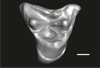
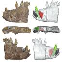
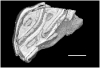
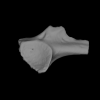
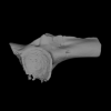
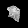
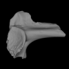
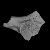
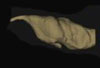
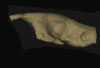
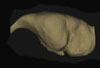
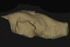
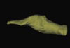
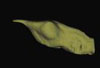
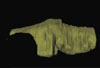
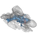
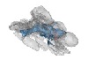
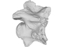
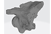
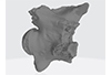
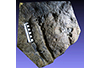
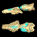
This contribution contains the 3D models described and figured in the following publication: Kassegne K. E., Mourlam M. J., Guinot G., Amoudji Y. Z., Martin J. E., Togbe K. A., Johnson A. K., Hautier L. 2021. First partial cranium of Togocetus from Kpogamé (Togo) and the protocetid diversity in the Togolese phosphate basin. Annales de Paléontologie, Issue 2, April–June 2021, 102488. https://doi.org/10.1016/j.annpal.2021.102488
Togocetus cf. traversei ULDG-KPO1 View specimen
|
M3#768The specimen consists of a partial cranium prepared out of a calcareous phosphate matrix. The partial cranium lacks the anterior part of the rostrum, the cranial roof, and most of the basicranium apart from the left zygomatic process of the squamosal. The maxilla, nasal, palatine, pterygoid, alisphenoid, and squamosal bones are preserved, as well as two incomplete dental rows described hereafter. Type: "3D_surfaces"doi: 10.18563/m3.sf.768 state:published |
Download 3D surface file |
|
M3#770µCT . Resolution: 0.3156mm. This scan can easily be opened with Fiji, MorphoDig, 3DSlicer, or any software that reads .MHD file format. Also, the .RAW file can be opened easily with other software such as Avizo/Amira when providing the correct dimensions (which are enclosed within the file name) Type: "3D_CT"doi: 10.18563/m3.sf.770 state:published |
Download CT data |
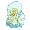
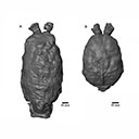
The present 3D Dataset contains the 3D models of brain endocast of traversodontid cynodonts studied in: Pavanatto et al. 2019. Virtual reconstruction of cranial endocasts of traversodontid cynodonts (Eucynodontia: Gomphodontia) from the upper Triassic of Southern Brazil. Journal of Morphology. https://doi.org/10.1002/jmor.21029
Siriusgnathus niemeyerorum CAPPA/UFSM 0032 View specimen
|
M3#4253D model of the brain endocast Type: "3D_surfaces"doi: 10.18563/m3.sf.425 state:published |
Download 3D surface file |
Exaeretodon riograndensis CAPPA/UFSM 0030 View specimen

|
M3#4263D model of the brain endocast Type: "3D_surfaces"doi: 10.18563/m3.sf.426 state:published |
Download 3D surface file |
Exaeretodon riograndensis CAPPA/UFSM 0227 View specimen
|
M3#4273D model of the brain endocast Type: "3D_surfaces"doi: 10.18563/m3.sf.427 state:published |
Download 3D surface file |
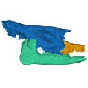
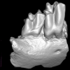
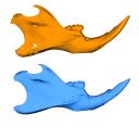
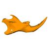
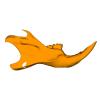
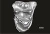
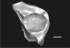
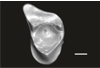
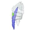
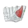
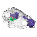
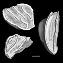
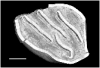
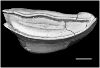
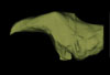
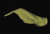
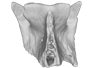
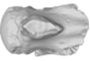
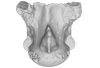
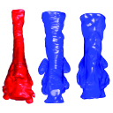
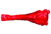
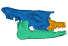
The present 3D Dataset contains two 3D models described in Tissier et al. (https://doi.org/10.1098/rsos.200633): the only known complete mandible of the early-branching rhinocerotoid Epiaceratherium magnum Uhlig, 1999, and a hypothetical reconstruction of the complete archetypic skull of Epiaceratherium Heissig, 1969, created by merging three cranial parts from three distinct Epiaceratherium species.
Epiaceratherium magnum NMB.O.B.928 View specimen
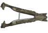
|
M3#5343D surface model of the mandible NMB.O.B.928 of Epiaceratherium magnum, with texture file. Type: "3D_surfaces"doi: 10.18563/m3.sf.534 state:published |
Download 3D surface file |
Epiaceratherium magnum NMB.O.B.928 + MJSN POI007–245 + NMB.I.O.43 View specimen
|
M3#535Archetypal reconstruction of the skull of Epiaceratherium, generated by 3D virtual association of the cranium of E. delemontense (MJSN POI007–245, in blue), mandible of E. magnum (NMB.O.B.928, green) and snout of E. bolcense (NMB.I.O.43, in orange). Type: "3D_surfaces"doi: 10.18563/m3.sf.535 state:published |
Download 3D surface file |
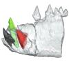
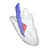
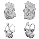
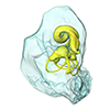
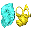
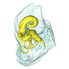
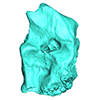
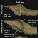
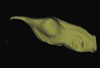
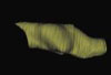
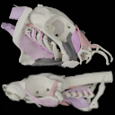
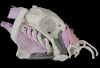
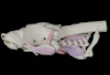
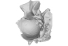
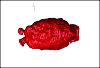
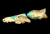
This contribution contains the 3D model described and figured in the following publication: Dubied, M., Mennecart, B. and Solé, F. 2019. The cranium of Proviverra typica (Mammalia, Hyaenodonta) and its impact on hyaenodont phylogeny and endocranial evolution. Palaeontology. https://doi.org/10.1111/pala.12437
Proviverra typica NMB Em18 View specimen
|
M3#355The file contain the cranium (yellow) and the endocast (blue) of the facial part and the brain case part of the type specimen of Proviverra typica (NMB Em18). Type: "3D_surfaces"doi: 10.18563/m3.sf.355 state:published |
Download 3D surface file |