3D models of Kalakocetus, the earliest Cetacea
The specimens of Speothos pacivorus
3D models related to the publication: Hidden diversity of Palaeogene metatherians: a new family of polydolopimorphian marsupials from Peruvian Amazonia
3D GM dataset of bird skeletal variation
Skeletal embryonic development in the catshark
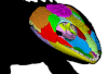
Bony connexions of the petrosal bone of extant hippos
bony labyrinth (14) , inner ear (11) , Eocene (11) , geometric morphometrics (10) , CT-scan (10) , Oligocene (9) , Micro-CT (9)
Lionel Hautier (25) , Maëva Judith Orliac (24) , Laurent Marivaux (18) , Renaud Lebrun (15) , Rodolphe Tabuce (14) , Pierre-Olivier Antoine (13) , Bastien Mennecart (13)
|
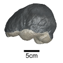
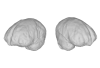
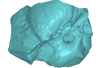
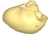
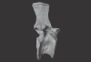
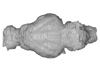
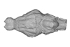
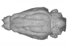
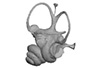
The endocranial cast of a 10 ka intentionally deformed human cranium from ChinaYin Qiyu
Published online: 27/07/2022 |
|
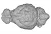
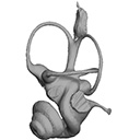
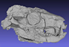
M3#972The frontal region of the endocast is flattened, probably formed by the constant pressure on the frontal bone during growth. There is a well-developed frontal crest on the endocranial surface. The endocast widens posteriorly from the frontal lobe. The widest point of the endocast is at the lateral border of the parietal lobe. The lower parietal areas display a marked lateral expansion. The overall shape of the endocast is asymmetrical, with the left side of the parietal lobe being more laterally expanded than the right side. Like the frontal lobe, the occipital lobe is also anteroposteriorly flattened. Type: "3D_surfaces"doi: 10.18563/m3.sf.972 state:published |
Download 3D surface file |
|
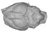
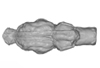
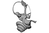
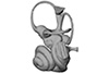
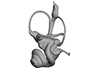
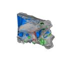
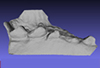
M3#976The original endocranial cast model (with texture) of IVPP-PA1616. It shows the original structures of the specimen, and was not altered in any way. Type: "3D_surfaces"doi: 10.18563/m3.sf.976 state:published |
Download 3D surface file |
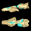
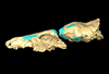
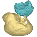
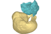
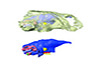
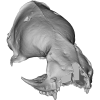
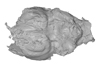
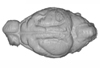
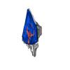
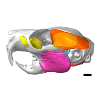
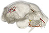
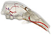
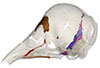
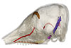
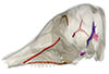
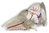
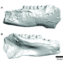
This contribution contains the 3D model described and figured in the following publication: Dubied, M., Mennecart, B. and Solé, F. 2019. The cranium of Proviverra typica (Mammalia, Hyaenodonta) and its impact on hyaenodont phylogeny and endocranial evolution. Palaeontology. https://doi.org/10.1111/pala.12437
Proviverra typica NMB Em18 View specimen
|
M3#355The file contain the cranium (yellow) and the endocast (blue) of the facial part and the brain case part of the type specimen of Proviverra typica (NMB Em18). Type: "3D_surfaces"doi: 10.18563/m3.sf.355 state:published |
Download 3D surface file |
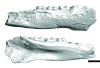
This contribution contains the 3D models described and figured in the following publication: Mourlam, M., Orliac, M. J. (2017), Protocetid (Cetacea, Artiodactyla) bullae and petrosals from the Middle Eocene locality of Kpogamé, Togo: new insights into the early history of cetacean hearing. Journal of Systematic Palaeontology https://doi.org/10.1080/14772019.2017.1328378
?Carolinacetus indet. UM KPG-M 164 View specimen
|
M3#132left petrosal of ?Carolinacetus sp. from the locality of Kpogamé, Togo Type: "3D_surfaces"doi: 10.18563/m3.sf.132 state:published |
Download 3D surface file |
indet. indet. UM KPG-M 73 View specimen

|
M3#133labelled surface of the left petrosal Type: "3D_surfaces"doi: 10.18563/m3.sf.133 state:published |
Download 3D surface file |
|
M3#134left bullaof Protocetidae indeterminate from Kpogamé, Togo Type: "3D_surfaces"doi: 10.18563/m3.sf.134 state:published |
Download 3D surface file |
|
M3#135petrotympanic complex of Protocetidae indeterminate from Kpogamé, Togo Type: "3D_surfaces"doi: 10.18563/m3.sf.135 state:published |
Download 3D surface file |
?Carolinacetus indet. UM KPG-M 33 View specimen
|
M3#136left auditory bulla of a juvenile specimen of ?Carolinacetus sp. from Kpogamé, Togo Type: "3D_surfaces"doi: 10.18563/m3.sf.136 state:published |
Download 3D surface file |
Togocetus traversei UM KPG-M 80 View specimen

|
M3#137fragmentary right auditory bulla of Togocetus traversei from Kpogamé, Togo Type: "3D_surfaces"doi: 10.18563/m3.sf.137 state:published |
Download 3D surface file |
This contribution contains the 3D reconstruction of Canariomys bravoi, described and figured in the following publication: Michaux J., Hautier L., Hutterer R., Lebrun R., Guy F., García-Talavera F., 2012 : Body shape and life style of the extinct rodent Canariomys bravoi (Mammalia, Murinae) from Tenerife, Canary Islands (Spain). Comptes Rendus Palevol 11 (7), 485-494. DOI: 10.1016/j.crpv.2012.06.004
Canariomys bravoi TFMCV872-873 View specimen

|
M3#6This file contains the 3D reconstruction of Canariomys bravoi, described and figured in the following publication: Michaux J., Hautier L., Hutterer R., Lebrun R., Guy F., García-Talavera F., 2012 : Body shape and life style of the extinct rodent Canariomys bravoi (Mammalia, Murinae) from Tenerife, Canary Islands (Spain). Comptes Rendus Palevol 11 (7), 485-494. Type: "3D_surfaces"doi: 10.18563/m3.sf6 state:published |
Download 3D surface file |
This contribution contains the 3D model described and figured in the following publication: New turtles from the Late Cretaceous of Monte Alto-SP, Brazil, including cranial osteology, neuroanatomy and phylogenetic position of a new taxon. PalZ. https://doi.org/10.1007/s12542-017-0397-x
Yuraramirim montealtensis 04-0008/89 View specimen
|
M3#2783D surfaces related to specimen MPMA 04-0008/89. Type: "3D_surfaces"doi: 10.18563/m3.sf.278 state:published |
Download 3D surface file |
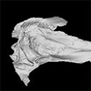
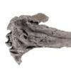
The present 3D Dataset contains the 3D model analyzed in The largest freshwater odontocete: a South Asian river dolphin relative from the Proto-Amazonia.
Pebanista yacuruna MUSM 4017 View specimen
|
M3#1394Holotype skull of Pebanista yacuruna MUSM 4017 Type: "3D_surfaces"doi: 10.18563/m3.sf.1394 state:published |
Download 3D surface file |
The present 3D Dataset contains 3D models of the cranium surface and of the bony labyrinth endocast of the stem bat Vielasia sigei. They are used by (Hand et al., 2023) to explore the phylogenetic position of this species, to infer its laryngeal echolocating capabilities, and to eventually discuss chiropteran evolution before the crown clade diversification.
Vielasia sigei UM VIE-250 View specimen
|
M3#1269External surface of the cranium Type: "3D_surfaces"doi: 10.18563/m3.sf.1269 state:published |
Download 3D surface file |
|
M3#1270Virtual endocast of the right bony labyrinth Type: "3D_surfaces"doi: 10.18563/m3.sf.1270 state:published |
Download 3D surface file |
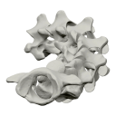
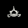
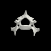
The present 3D Dataset contains the 3D models analyzed in Merten, L.J.F, Manafzadeh, A.R., Herbst, E.C., Amson, E., Tambusso, P.S., Arnold, P., Nyakatura, J.A., 2023. The functional significance of aberrant cervical counts in sloths: insights from automated exhaustive analysis of cervical range of motion. Proceedings of the Royal Society B. doi: 10.1098/rspb.2023.1592
Ailurus fulgens PMJ_Mam_6639 View specimen
|
M3#1260cervical vertebral series (7 vertebrae) Type: "3D_surfaces"doi: 10.18563/m3.sf.1260 state:published |
Download 3D surface file |
Bradypus variegatus ZMB_Mam_91345 View specimen
|
M3#1261cervical vertebral series (8 vertebrae) + first thoracic vertebra Type: "3D_surfaces"doi: 10.18563/m3.sf.1261 state:published |
Download 3D surface file |
Bradypus variegatus ZMB_Mam_35824 View specimen

|
M3#1262cervical vertebral series (8 vertebrae) + first & second thoracic vertebra Type: "3D_surfaces"doi: 10.18563/m3.sf.1262 state:published |
Download 3D surface file |
Choloepus didactylus ZMB_Mam_38388 View specimen
|
M3#1263cervical vertebral series (7 vertebrae) Type: "3D_surfaces"doi: 10.18563/m3.sf.1263 state:published |
Download 3D surface file |
Choloepus didactylus ZMB_Mam_102634 View specimen
|
M3#1264cervical vertebral series (6 vertebrae) + first thoracic vertebra Type: "3D_surfaces"doi: 10.18563/m3.sf.1264 state:published |
Download 3D surface file |
Tamandua tetradactyla ZMB_Mam_91288 View specimen
|
M3#1266cervical vertebral series (7 vertebrae) + first thoracic vertebra Type: "3D_surfaces"doi: 10.18563/m3.sf.1266 state:published |
Download 3D surface file |
Glossotherium robustum MNHN_n/n View specimen
|
M3#1267cervical vertebral series (7 vertebrae) + first thoracic vertebra Type: "3D_surfaces"doi: 10.18563/m3.sf.1267 state:published |
Download 3D surface file |
This contribution contains the three-dimensional models of the inner ear of the hetaxodontid rodents Amblyrhiza, Clidomys and Elasmodontomys from the West Indies. These specimens were analyzed and discussed in : The inner ear of caviomorph rodents: phylogenetic implications and application to extinct West Indian taxa.
Amblyrhiza inundata 11842 View specimen
|
M3#11543D surface of the left-oriented inner ear of Amblyrhiza. Type: "3D_surfaces"doi: 10.18563/m3.sf.1154 state:published |
Download 3D surface file |
Clidomys sp NA View specimen
|
M3#11553D surface of the left-oriented inner ear of Clidomys sp. Type: "3D_surfaces"doi: 10.18563/m3.sf.1155 state:published |
Download 3D surface file |
Elasmodontomys obliquus 17127 View specimen
|
M3#11563D surface of the left-oriented inner ear of Elasmodontomys obliquus. Type: "3D_surfaces"doi: 10.18563/m3.sf.1156 state:published |
Download 3D surface file |
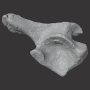
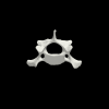
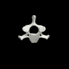
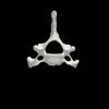
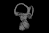
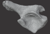
The present 3D Dataset contains the 3D models of an ilium, a vertebra, and a partial scapula of Prestosuchus sp. that were analyzed in “New Loricata remains from the Pinheiros-Chiniquá Sequence (Middle-Upper Triassic), southern Brazil”.
Prestosuchus sp. UFSM11603 View specimen
|
M3#1080Surface scan of a right ilium of Prestosuchus sp. with a 0.4 mm resolution. Type: "3D_surfaces"doi: 10.18563/m3.sf.1080 state:published |
Download 3D surface file |
Prestosuchus sp. UFSM11233 View specimen
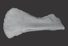
|
M3#1081Surface scan of a partial right scapula of Prestosuchus sp. with a 0.4mm resolution. Type: "3D_surfaces"doi: 10.18563/m3.sf.1081 state:published |
Download 3D surface file |
Prestosuchus sp. UFSM11602a View specimen
|
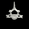
M3#1082Surface scan of a anterior dorsal vertebra of Prestosuchus sp. with a 0.2 mm resolution. Type: "3D_surfaces"doi: 10.18563/m3.sf.1082 state:published |
Download 3D surface file |
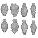
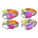
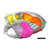
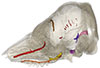
The present 3D Dataset contains the 3D models illustrated and described in the chapter “Paleoneurology of Artiodactyla, an overview of the evolution of the artiodactyl brain” (Orliac et al. 2022) published in "Paleoneurology of amniotes: new directions in the study of fossil endocasts", edited by Dozo, Paulina-Carabajal, Macrini and Walsh.
Homacodon vagans AMNH 12695 View specimen
|
M3#1063Endocranial cast Type: "3D_surfaces"doi: 10.18563/m3.sf.1063 state:published |
Download 3D surface file |
Helohyus sp. AMNH 13079 View specimen
|
M3#1064Endocranial cast Type: "3D_surfaces"doi: 10.18563/m3.sf.1064 state:published |
Download 3D surface file |
Leptauchenia sp. AMNH 45508 View specimen
|
M3#1065endocranial cast Type: "3D_surfaces"doi: 10.18563/m3.sf.1065 state:published |
Download 3D surface file |
Agriochoerus sp. AMNH 95330 View specimen
|
M3#1067endocranial cast Type: "3D_surfaces"doi: 10.18563/m3.sf.1067 state:published |
Download 3D surface file |
Mouillacitherium elegans UM ACQ 6625 View specimen
|
M3#1068endocranial cast Type: "3D_surfaces"doi: 10.18563/m3.sf.1068 state:published |
Download 3D surface file |
Caenomeryx filholi UM PDS 2570 View specimen
|
M3#1069endocranial cast Type: "3D_surfaces"doi: 10.18563/m3.sf.1069 state:published |
Download 3D surface file |
Dichobune leporina MNHN.F.QU16586 View specimen
|
M3#1070endocranial cast Type: "3D_surfaces"doi: 10.18563/m3.sf.1070 state:published |
Download 3D surface file |
Anoplotherium sp. not numbered View specimen
|
M3#1071endocranial cast Type: "3D_surfaces"doi: 10.18563/m3.sf.1071 state:published |
Download 3D surface file |
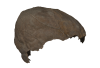
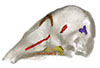
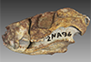
This contribution contains the 3D model of the holotype of Chambius kasserinensis, the basalmost ‘elephant-shrew’ figured in the following publication: New remains of Chambius kasserinensis from the Eocene of Tunisia and evaluation of proposed affinities for Macroscelidea (Mammalia, Afrotheria). https://doi.org/10.1080/08912963.2017.1297433
Chambius kasserinensis CBI-1-06 View specimen
|
M3#1463D model of the holotype maxilla of Chambius kasserinensis. The 3D surface was extracted manually from the limestone matrix within AVIZO 9.2 Type: "3D_surfaces"doi: 10.18563/m3.sf.146 state:published |
Download 3D surface file |
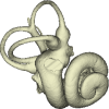
The present 3D Dataset contains the 3D models analyzed in the article Mennecart, B., and L. Costeur. 2016. A Dorcatherium (Mammalia, Ruminantia, Middle Miocene) petrosal bone and the tragulid ear region. Journal of Vertebrate Paleontology 36(6), 1211665(1)-1211665(7). DOI: 10.1080/02724634.2016.1211665.
Tragulus javanicus 10028 View specimen
|
M3#1193D surface of the left bony labyrinth of Tragulus javanicus NMB 10028 Type: "3D_surfaces"doi: 10.18563/m3.sf.119 state:published |
Download 3D surface file |
Moschiola meminna C.2453 View specimen
|
M3#1203D surface of the left bony labyrinth of Moschiola meminna NMB C.2453 Type: "3D_surfaces"doi: 10.18563/m3.sf.120 state:published |
Download 3D surface file |
Hyemoschus aquaticus C.1930 View specimen
|
M3#1223D surface of the right bony labyrinth of Hyemoschus aquaticus NMB C.1930 Type: "3D_surfaces"doi: 10.18563/m3.sf.122 state:published |
Download 3D surface file |
Dorcatherium crassum San.15053 View specimen
|
M3#1233D surface of the right bony labyrinth of Dorcatherium crassum NMB San.15053 Type: "3D_surfaces"doi: 10.18563/m3.sf.123 state:published |
Download 3D surface file |
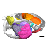
The present 3D Dataset contains the 3D models analyzed in the following publication: Paulina-Carabajal, A., and Porfiri, J.D. 2026. Novel information on the braincase of Megaraptor namunhuaiquii (Dinosauria: Theropoda) using X-ray tomography: pneumaticity, paleoneurology and their paleobiological implications. Ameghiniana 63(1), 16-32
Megaraptor namunhuaiquii MUC-PV 595 View specimen
|
M3#18733D models of the braincase, lateral wall of the braincase, brain, right and left inner ears Type: "3D_surfaces"doi: 10.18563/m3.sf.1873 state:in_press |
Download 3D surface file |
This contribution contains the 3D model(s) described and figured in the following publication: Da Cunha, L., Fabre, P.-H. & Hautier, L. (2024) Springhares, flying and flightless scaly-tailed squirrels (Anomaluromorpha, Rodentia) are the squirrely mouse: comparative anatomy of the masticatory musculature and its implications on the evolution of hystricomorphy in rodents. Journal of Anatomy, 244, 900–928.
Anomalurus derbianus 21804 View specimen
|
M3#1493Masticatory apparatus of Anomalurus Type: "3D_surfaces"doi: 10.18563/m3.sf.1493 state:published |
Download 3D surface file |
Idiurus macrotis 29335 View specimen
|
M3#1492Masticatory apparatus of Idiurus Type: "3D_surfaces"doi: 10.18563/m3.sf.1492 state:published |
Download 3D surface file |
Zenkerella insignis 5.5.23.27 View specimen
|
M3#1490Masticatory apparatus of Zenkerella Type: "3D_surfaces"doi: 10.18563/m3.sf.1490 state:published |
Download 3D surface file |
Pedetes capensis NA View specimen

|
M3#1491Masticatory apparatus of Pedetes Type: "3D_surfaces"doi: 10.18563/m3.sf.1491 state:published |
Download 3D surface file |
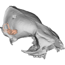
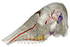
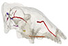
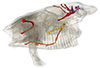
The present 3D Dataset contains the 3D models analyzed in the following publication: Le Verger K., González Ruiz L.R., Billet G. 2021. Comparative anatomy and phylogenetic contribution of intracranial osseous canals and cavities in armadillos and glyptodonts (Xenarthra, Cingulata). Journal of Anatomy 00: 1-30 p. https://doi.org/10.1111/joa.13512
Bradypus tridactylus MNHN ZM-MO-1999-1065 View specimen
|
M3#844Bradypus tridactylus MNHN ZM-MO-1999-1065: cranium, cranial canals & alveolar cavities. Type: "3D_surfaces"doi: 10.18563/m3.sf.844 state:published |
Download 3D surface file |
Tamandua tetradactyla NHMUK ZD-1903.7.7.135 View specimen
|
M3#845Tamandua tetradactyla NHMUK ZD-1903.7.7.135: cranium, cranial canals & alveolar cavities. Type: "3D_surfaces"doi: 10.18563/m3.sf.845 state:published |
Download 3D surface file |
Dasypus novemcinctus AMNH 33150 View specimen
|
M3#846Dasypus novemcinctus AMNH 33150: cranium, cranial canals & alveolar cavities. Type: "3D_surfaces"doi: 10.18563/m3.sf.846 state:published |
Download 3D surface file |
Dasypus novemcinctus AMNH 133261 View specimen
|
M3#847Dasypus novemcinctus AMNH 133261: cranium, cranial canals & alveolar cavities. Type: "3D_surfaces"doi: 10.18563/m3.sf.847 state:published |
Download 3D surface file |
Dasypus novemcinctus AMNH 133328 View specimen
|
M3#848Dasypus novemcinctus AMNH 133328: cranium, cranial canals & alveolar cavities. Type: "3D_surfaces"doi: 10.18563/m3.sf.848 state:published |
Download 3D surface file |
Zaedyus pichiy ZMB-MAM-49039 View specimen
|
M3#849Zaedyus pichiy ZMB-MAM-49039: cranium, cranial canals & alveolar cavities. Type: "3D_surfaces"doi: 10.18563/m3.sf.849 state:published |
Download 3D surface file |
Zaedyus pichiy MHNG 1627.053 View specimen

|
M3#850Zaedyus pichiy MHNG 1627.053: cranium, cranial canals & alveolar cavities. Type: "3D_surfaces"doi: 10.18563/m3.sf.850 state:published |
Download 3D surface file |
Zaedyus pichiy MHNG 1276.076 View specimen
|
M3#851Zaedyus pichiy MHNG 1276.076: cranium, cranial canals & alveolar cavities. Type: "3D_surfaces"doi: 10.18563/m3.sf.851 state:published |
Download 3D surface file |
Cabassous unicinctus NBC_ZMA.MAM.26326.a View specimen
|
M3#852Cabassous unicinctus NBC ZMA.MAM.26326.a: cranium, cranial canals & alveolar cavities. Type: "3D_surfaces"doi: 10.18563/m3.sf.852 state:published |
Download 3D surface file |
Cabassous unicinctus MNHN-CG-1999-1044 View specimen
|
M3#853Cabassous unicinctus MNHN-CG-1999-1044: cranium, cranial canals & alveolar cavities. Type: "3D_surfaces"doi: 10.18563/m3.sf.853 state:published |
Download 3D surface file |
Vassallia maxima FMNH P14424 View specimen
|
M3#854Vassallia maxima FMNH P14424: cranium, cranial canals & alveolar cavities. Type: "3D_surfaces"doi: 10.18563/m3.sf.854 state:published |
Download 3D surface file |
Glyptodon sp. MNHN-F-PAM-759 View specimen
|
M3#855Glyptodon sp. MNHN-F-PAM-759: cranium, cranial canals & alveolar cavities. Type: "3D_surfaces"doi: 10.18563/m3.sf.855 state:published |
Download 3D surface file |
Glyptodon sp. MNHN-F-PAM-760 View specimen

|
M3#856Glyptodon sp. MNHN-F-PAM-760: cranium, cranial canals & alveolar cavities. Type: "3D_surfaces"doi: 10.18563/m3.sf.856 state:published |
Download 3D surface file |
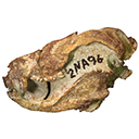
The present 3D Dataset contains the 3D models analyzed in Hendrickx, C., Gaetano, L. C., Choiniere, J., Mocke, H. and Abdala, F. in press. A new traversodontid cynodont with a peculiar postcanine dentition from the Middle/Late Triassic of Namibia and dental evolution in basal gomphodonts. Journal of Systematic Palaeontology.
Etjoia dentitransitus GSN F1591 View specimen
|
M3#557Surface model derived from µCT data of the holotype of Etjoia dentitransitus Type: "3D_surfaces"doi: 10.18563/m3.sf.557 state:published |
Download 3D surface file |
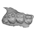
|
M3#558Photogrammetric 3D surface model of the postcanines of the Holotype of Etjoia dentitransitus Type: "3D_surfaces"doi: 10.18563/m3.sf.558 state:published |
Download 3D surface file |
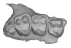
|
M3#559Photogrammetric 3D surface model of the Holotype of Etjoia dentitransitus Type: "3D_surfaces"doi: 10.18563/m3.sf.559 state:published |
Download 3D surface file |
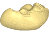
The present 3D Dataset contains the 3D model of a left dentary with m1-m3 analyzed in “A new fossil of Tayassuidae (Mammalia: Certartiodactyla) from the Pleistocene of northern Brazil”. The 3D model was generated using a laser scanning.
cf. Pecari tajacu UFSM 11606 View specimen
|
M3#498Left dentary with m1-m3 Type: "3D_surfaces"doi: 10.18563/m3.sf.498 state:published |
Download 3D surface file |
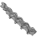
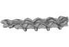
This contribution contains the 3D model described and figured in the following publication: Albino, A., Carrillo-Briceño, J. D. & Neenan, J. M. 2016. An enigmatic aquatic snake from the Cenomanian of northern South America. PeerJ 4:e2027 http://dx.doi.org/10.7717/peerj.2027
Lunaophis aquaticus MCNC-1827-F View specimen
|
M3#116Articulated precloacal vertebrae of Lunaophis aquaticus Type: "3D_surfaces"doi: 10.18563/m3.sf.116 state:published |
Download 3D surface file |
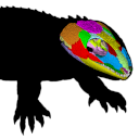
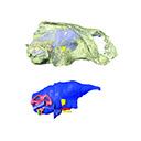
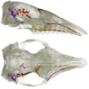
The present 3D Dataset contains the 3D models analyzed in: Abel P., Pommery Y., Ford D. P., Koyabu D., Werneburg I. 2022. Skull sutures and cranial mechanics in the Permian reptile Captorhinus aguti and the evolution of the temporal region in early amniotes. Frontiers in Ecology and Evolution. https://doi.org/10.3389/fevo.2022.841784
Captorhinus aguti OMNH 44816 View specimen
|
M3#965Segmented cranial bone surfaces of OMNH 44816 Type: "3D_surfaces"doi: 10.18563/m3.sf.965 state:published |
Download 3D surface file |


