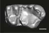
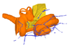
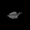
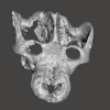
3D models of Kalakocetus, the earliest Cetacea
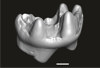
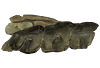
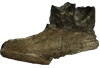
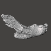
The specimens of Speothos pacivorus
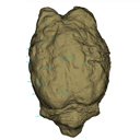
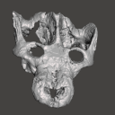
3D models related to the publication: Hidden diversity of Palaeogene metatherians: a new family of polydolopimorphian marsupials from Peruvian Amazonia
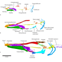
3D GM dataset of bird skeletal variation
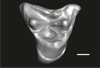
Skeletal embryonic development in the catshark
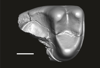
Bony connexions of the petrosal bone of extant hippos
bony labyrinth (14) , inner ear (11) , Eocene (11) , geometric morphometrics (10) , CT-scan (10) , Oligocene (9) , Micro-CT (9)
Lionel Hautier (25) , Maëva Judith Orliac (24) , Laurent Marivaux (18) , Renaud Lebrun (15) , Rodolphe Tabuce (14) , Pierre-Olivier Antoine (13) , Bastien Mennecart (13)
|
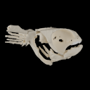
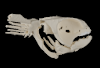
3D models related to the publication: Evidence for high-performance suction feeding in the Pennsylvanian stem-group holocephalan Iniopera.Richard Dearden
Published online: 18/01/2023 |
|
M3#1034plys of the head endoskeleton of Iniopera sp. Type: "3D_surfaces"doi: 10.18563/m3.sf.1034 state:published |
Download 3D surface file |
The present 3D Dataset contains the 3D model of the brain endocast of Neoepiblema acreensis analyzed in “Small within the largest: Brain size and anatomy of the extinct Neoepiblema acreensis, a giant rodent from the Neotropics”. The 3D model was generated using CT-Scanning and techniques of virtual reconstruction.
Neoepiblema acreensis UFAC 4515 View specimen
|
M3#502Brain endocast of Neoepiblema acreensis Type: "3D_surfaces"doi: 10.18563/m3.sf.502 state:published |
Download 3D surface file |
The present 3D dataset contains the 3D models analyzed in the publication: Rosa, R. M., Salvador, R. B., & Cavallari, D. C. (2025). The disappearing act of the magician tree snail: anatomy, distribution, and phylogenetic relationships of Drymaeus magus (Gastropoda: Bulimulidae), a long-lost species hidden in plain sight. Zoological Journal of the Linnean Society.
Drymaeus magus CMRP 1049 View specimen
|
M3#1597Internal organs of Drymaeus magus Type: "3D_surfaces"doi: 10.18563/m3.sf.1597 state:published |
Download 3D surface file |
|
M3#1598External surface of Drymaeus magus Type: "3D_surfaces"doi: 10.18563/m3.sf.1598 state:published |
Download 3D surface file |
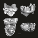
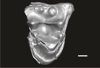
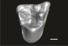
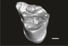
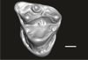
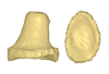
This contribution contains the three-dimensional digital models of eleven isolated fossil teeth of a merialine paroxyclaenid (Welcommoides gurki), discovered from lower Oligocene deposits of the Bugti Hills (Balochistan, Pakistan). These fossils were described, figured and discussed in the following publication: Solé et al. (2024), An unexpected late paroxyclaenid (Mammalia, Cimolesta) out of Europe: dental evidence from the Oligocene of the Bugti Hills, Pakistan. Papers in Palaeontology. https://doi.org/10.1002/spp2.1599
Welcommoides gurki UM-DBC 2225 View specimen
|
M3#1083Left m3 Type: "3D_surfaces"doi: 10.18563/m3.sf.1083 state:published |
Download 3D surface file |
Welcommoides gurki UM-DBC 2226 View specimen
|
M3#1084Right m3 Type: "3D_surfaces"doi: 10.18563/m3.sf.1084 state:published |
Download 3D surface file |
Welcommoides gurki UM-DBC 2227 View specimen
|
M3#1085Trigonid of a right lower molar Type: "3D_surfaces"doi: 10.18563/m3.sf.1085 state:published |
Download 3D surface file |
Welcommoides gurki UM-DBC 2230 View specimen
|
M3#1086Right DP4 Type: "3D_surfaces"doi: 10.18563/m3.sf.1086 state:published |
Download 3D surface file |
Welcommoides gurki UM-DBC 2228 View specimen
|
M3#1093Right M1 Type: "3D_surfaces"doi: 10.18563/m3.sf.1093 state:published |
Download 3D surface file |
Welcommoides gurki UM-DBC 2229 View specimen
|
M3#1087Right M2 Type: "3D_surfaces"doi: 10.18563/m3.sf.1087 state:published |
Download 3D surface file |
Welcommoides gurki UM-DBC 2236 View specimen
|
M3#1088Left M2 Type: "3D_surfaces"doi: 10.18563/m3.sf.1088 state:published |
Download 3D surface file |
Welcommoides gurki UM-DBC 2231 View specimen
|
M3#1089Left M3 Type: "3D_surfaces"doi: 10.18563/m3.sf.1089 state:published |
Download 3D surface file |
Welcommoides gurki UM-DBC 2232 View specimen
|
M3#1090Left M3 Type: "3D_surfaces"doi: 10.18563/m3.sf.1090 state:published |
Download 3D surface file |
Welcommoides gurki UM-DBC 2234 View specimen
|
M3#1091Left M3 Type: "3D_surfaces"doi: 10.18563/m3.sf.1091 state:published |
Download 3D surface file |
Welcommoides gurki UM-DBC 2233 View specimen
|
M3#1092Left M3 Type: "3D_surfaces"doi: 10.18563/m3.sf.1092 state:published |
Download 3D surface file |
The present 3D Dataset contains the 3D models analyzed in Keppeler, H., Schultz, J. A., Ruf, I., & Martin, T., 2023. Cranial anatomy of Hypisodus minimus (Artiodactyla: Ruminantia) from the Oligocene Brule Formation of North America. Palaeontographica Abteilung A.
Hypisodus minimus SMNK-PAL 27212 View specimen
|
M3#1031CT image stack of a skull of Hypisodus minimus. Also includes a lumbar vertebra and a probable proximal phalanx of digit III or IV. Type: "3D_CT"doi: 10.18563/m3.sf.1031 state:published |
Download CT data |
|
M3#10363D surface models of a skull of Hypisodus minimus (SMNK-PAL27212). The data includes a surface model for: basisphenoid, tympanic bullae, ethmoid (lamina perpendicularis), frontals, jugal (left), jugal (right), lacrimals, lower dentition, mandibles, mastoid processes, maxillaries, maxilloturbinals, nasals, occipital, palatine, parietals, petrosals, presphenoid, squamosals, turbinates, upper dentition, and the vomer. Type: "3D_surfaces"doi: 10.18563/m3.sf.1036 state:published |
Download 3D surface file |
Hypisodus minimus SMNK-PAL 27213 View specimen
|
M3#1033CT image stack of a skull of Hypisodus minimus. Also shows numerous postcranial material including an atlas articulated with the occipital bone, the distal part of a left humerus articulated to radius and ulna, a part of a femur, a part of a tibia and fibula, unidentifiable tarsal bones, parts of the metatarsals II, III, IV and V and their phalanges, a proximal phalanx of digit III or IV, a middle phalanx of digit III or IV, a possible patella and calcaneus, as well as numerous unidentifiable broken bony fragments. Type: "3D_CT"doi: 10.18563/m3.sf.1033 state:published |
Download CT data |
|
M3#10353D surface models of a skull of Hypisodus minimus (SMNK-PAL27213). The data includes a surface model for: atlas, basisphenoid, tympanic bullae, nasals, occipital, the petrosals, and the inner ear. Type: "3D_surfaces"doi: 10.18563/m3.sf.1035 state:published |
Download 3D surface file |
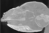
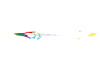
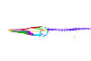
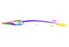
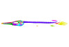
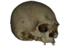
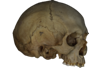
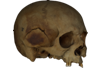
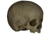
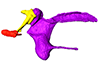
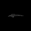
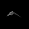
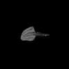
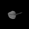
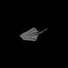
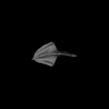
In this contribution, we describe the external and internal morphology of a delphinid petrosal bone collected from Ahu Tahai, a burial site located on the Southwestern coast of Easter Island, at Hangaroa. We discuss the taxonomic attribution of this archaeological item and describe its internal structures based on µCT data, including the bony labyrinth and the nerve and vein patterns. Identification of the nerves exists lead us to relocate the identification of the foramen singulare in delphinid petrosals.
indet. indet. AT1 View specimen
|
M3#420Stapes Type: "3D_surfaces"doi: 10.18563/m3.sf.420 state:published |
Download 3D surface file |
|
M3#421petrosal bone Type: "3D_surfaces"doi: 10.18563/m3.sf.421 state:published |
Download 3D surface file |
|
M3#422in situ bony labyrinth Type: "3D_surfaces"doi: 10.18563/m3.sf.422 state:published |
Download 3D surface file |
|
M3#423bony labyrinth and associated nerves and blood vessels Type: "3D_surfaces"doi: 10.18563/m3.sf.423 state:published |
Download 3D surface file |
Using X-ray microtomography, we describe the ossification events during the larval development of a non-teleost actinopterygian species: the Cuban gar Atractosteus tristoechus from the order Lepisosteiformes. We provide a detailed developmental series for each anatomical structure, covering a large sequence of mineralization events going from an early stage (13 days post-hatching, 21mm total length) to an almost fully ossified larval stage (118dph or 87mm in standard length). With this work, we expect to bring new developmental data to be used in further comparative studies with other lineages of bony vertebrates. We also hope that the on-line publication of these twelve successive 3D reconstructions, fully labelled and flagged, will be an educational tool for all students in comparative anatomy.
Atractosteus tristoechus At1-13dph View specimen

|
M3#94At1-13dph : 13 dph larvae, 21 mm TL Type: "3D_surfaces"doi: 10.18563/m3.sf.94 state:published |
Download 3D surface file |
Atractosteus tristoechus At2-16dph View specimen
|
M3#95Atractosteus tristoechus larva, 16 dph, 26mm SL. Type: "3D_surfaces"doi: 10.18563/m3.sf.95 state:published |
Download 3D surface file |
Atractosteus tristoechus At3-19dph View specimen

|
M3#96Atractosteus tristoechus larva, 19 dph, 27mm SL. Type: "3D_surfaces"doi: 10.18563/m3.sf.96 state:published |
Download 3D surface file |
Atractosteus tristoechus At4-22dph View specimen
|
M3#97Atractosteus tristoechus larva, 22dph, 30mm SL. Type: "3D_surfaces"doi: 10.18563/m3.sf.97 state:published |
Download 3D surface file |
Atractosteus tristoechus At5-26dph View specimen
|
M3#98Atractosteus tristoechus larva, 26 dph, 32mm SL. Type: "3D_surfaces"doi: 10.18563/m3.sf.98 state:published |
Download 3D surface file |
Atractosteus tristoechus At6-31dph View specimen
|
M3#99Atractosteus tristoechus larva, 31 dph, 39mm SL. Type: "3D_surfaces"doi: 10.18563/m3.sf.99 state:published |
Download 3D surface file |
Atractosteus tristoechus At7-37dph View specimen
|
M3#100Atractosteus tristoechus larva, 37 dph, 43mm SL. Type: "3D_surfaces"doi: 10.18563/m3.sf.100 state:published |
Download 3D surface file |
Atractosteus tristoechus At8-52dph View specimen
|
M3#101Atractosteus tristoechus larva, 52 dph, 46mm SL. Type: "3D_surfaces"doi: 10.18563/m3.sf.101 state:published |
Download 3D surface file |
Atractosteus tristoechus At9-74dph View specimen
|
M3#102Atractosteus tristoechus larva, 74 dph, 61mm SL. Not all structures are colored, only newly ossified ones. Type: "3D_surfaces"doi: 10.18563/m3.sf.102 state:published |
Download 3D surface file |
Atractosteus tristoechus At10-89dph View specimen
|
M3#103Atractosteus tristoechus larva, 89 dph, 63mm SL. Not all structures are colored, only newly ossified ones. You may find the tag file in the At1-13dph reconstruction data. Type: "3D_surfaces"doi: 10.18563/m3.sf.103 state:published |
Download 3D surface file |
Atractosteus tristoechus At11-104dph View specimen
|
M3#104Atractosteus tristoechus larva, 104 dph, 70mm SL. Not all structures are colored, only newly ossified ones. Type: "3D_surfaces"doi: 10.18563/m3.sf.104 state:published |
Download 3D surface file |
Atractosteus tristoechus At12-118dph View specimen
|
M3#105Atractosteus tristoechus larva, 118 dph, 87mm SL. Type: "3D_surfaces"doi: 10.18563/m3.sf.105 state:published |
Download 3D surface file |
The present 3D Dataset contains the 3D models analyzed in: Hirose, A., Nakashima, T., Yamada, S., Uwabe, C., Kose, K., Takakuwa, T. 2012. Embryonic liver morphology and morphometry by magnetic resonance microscopic imaging. Anat Rec (Hoboken) 295, 51-59. doi: 10.1002/ar.21496
Homo sapiens KC-CS14LIV1387 View specimen
|
M3#64Human liver at Carnegie Stage (CS) 14 Type: "3D_surfaces"doi: 10.18563/m3.sf.64 state:published |
Download 3D surface file |
Homo sapiens KC-CS15LIV5074 View specimen
|
M3#65Human liver at Carnegie Stage (CS) 15 Type: "3D_surfaces"doi: 10.18563/m3.sf.65 state:published |
Download 3D surface file |
Homo sapiens KC-CS16LIV2578 View specimen
|
M3#66Human liver at Carnegie Stage (CS) 16 Type: "3D_surfaces"doi: 10.18563/m3.sf.66 state:published |
Download 3D surface file |
Homo sapiens KC-CS17LIV17832 View specimen
|
M3#67Human liver at Carnegie Stage (CS) 17 Type: "3D_surfaces"doi: 10.18563/m3.sf.67 state:published |
Download 3D surface file |
Homo sapiens KC-CS18LIV21124 View specimen
|
M3#68Human liver at Carnegie Stage (CS) 18 Type: "3D_surfaces"doi: 10.18563/m3.sf.68 state:published |
Download 3D surface file |
Homo sapiens KC-CS19LIV14353 View specimen

|
M3#69Human liver at Carnegie Stage (CS) 19 Type: "3D_surfaces"doi: 10.18563/m3.sf.69 state:published |
Download 3D surface file |
Homo sapiens KC-CS20LIV20701 View specimen

|
M3#70Human liver at Carnegie Stage (CS) 20 Type: "3D_surfaces"doi: 10.18563/m3.sf.70 state:published |
Download 3D surface file |
Homo sapiens KC-CS21LIV25858 View specimen
|
M3#71Human liver at Carnegie Stage (CS) 21 Type: "3D_surfaces"doi: 10.18563/m3.sf.71 state:published |
Download 3D surface file |
Homo sapiens KC-CS22LIV22226 View specimen
|
M3#72Human liver at Carnegie Stage (CS) 22 Type: "3D_surfaces"doi: 10.18563/m3.sf.72 state:published |
Download 3D surface file |
Homo sapiens KC-CS23LIV25704 View specimen
|
M3#73Human liver at Carnegie Stage (CS) 23 Type: "3D_surfaces"doi: 10.18563/m3.sf.73 state:published |
Download 3D surface file |

This project presents a µCT dataset and an associated 3D surface model of the holotype of Donrussellia magna (UM PAT 17; Primates, Adapiformes). UM PAT17 is the only known specimen for the species and consists of a well-preserved left lower jaw with p4-m3. It documents one of the oldest European primates, eventually dated near the Paleocene Eocene Thermal Maximum.
Donrussellia magna UM PAT 17 View specimen

|
M3#173D surface file model of UM PAT 17 (type specimen of Donrussellia magna), which is a well preserved left lower jaw with p4-m3. The teeth (and roots) were manually segmented. Type: "3D_surfaces"doi: 10.18563/m3.sf17 state:published |
Download 3D surface file |
|
M3#18CT Scan Data of Donrussellia magna UM PAT 17. Voxel size (in µm): 36µm (isotropic voxels). Dimensions in x,y,z : 594 pixels, 294 pixels, 1038 pixels. Image type : 8-bit voxels. Image format : raw data format (no header). Type: "3D_CT"doi: 10.18563/m3.sf18 state:published |
Download CT data |
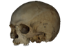
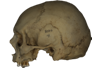
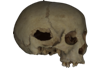
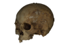
This contribution contains the 3D models described and figured in the following publications:
- Marini E., Lussu P., 2020. A virtual physical anthropology lab. Teaching in the time of coronavirus, in prep.;
- Lussu P., Bratzu D., Marini E., 2020. Cloud-based ultra close-range digital photogrammetry: validation of an approach for the effective virtual reconstruction of skeletal remains, in prep.
Homo sapiens MSAE 59 View specimen

|
M3#509MSAE 59 Type: "3D_surfaces"doi: 10.18563/m3.sf.509 state:published |
Download 3D surface file |
Homo sapiens MSAE 62 View specimen
|
M3#510MSAE 62 Type: "3D_surfaces"doi: 10.18563/m3.sf.510 state:published |
Download 3D surface file |
Homo sapiens MSAE 63 View specimen
|
M3#512MSAE 63 Type: "3D_surfaces"doi: 10.18563/m3.sf.512 state:published |
Download 3D surface file |
Homo sapiens MSAE 78 View specimen
|
M3#514MSAE 78 Type: "3D_surfaces"doi: 10.18563/m3.sf.514 state:published |
Download 3D surface file |
Homo sapiens MSAE 95 View specimen
|
M3#515MSAE 95 Type: "3D_surfaces"doi: 10.18563/m3.sf.515 state:published |
Download 3D surface file |
Homo sapiens MSAE 1852 View specimen
|
M3#516MSAE 1852 Type: "3D_surfaces"doi: 10.18563/m3.sf.516 state:published |
Download 3D surface file |
Homo sapiens MSAE 6426 View specimen
|
M3#517MSAE 6426 Type: "3D_surfaces"doi: 10.18563/m3.sf.517 state:published |
Download 3D surface file |
Homo sapiens MSAE 6428 View specimen
|
M3#518MSAE 6428 Type: "3D_surfaces"doi: 10.18563/m3.sf.518 state:published |
Download 3D surface file |
Homo sapiens MSAE 6992 View specimen
|
M3#519MSAE 6992 Type: "3D_surfaces"doi: 10.18563/m3.sf.519 state:published |
Download 3D surface file |
Homo sapiens MSAE 7688 View specimen
|
M3#520MSAE 7688 Type: "3D_surfaces"doi: 10.18563/m3.sf.520 state:published |
Download 3D surface file |
This contribution contains the 3D models described and figured in the following publication: Paulina-Carabajal, A. and Nieto, M. N. In press. Brief comment on the brain and inner ear of Giganotosaurus carolinii (Dinosauria: Theropoda) based on CT scans. Ameghiniana. https://doi.org/10.5710/AMGH.25.10.2019.3237
Giganotosaurus carolinii MUCPv-CH-1 View specimen
|
M3#504The current file contents 3D models of the braincase, brain, left and right inner ears Type: "3D_surfaces"doi: 10.18563/m3.sf.504 state:published |
Download 3D surface file |
This contribution contains the 3D models described and figured in the following publication: Tissier et al. (in prep.).
Sellamynodon zimborensis UBB MPS 15795 View specimen
|
M3#297Incomplete skull with left M3. Type: "3D_surfaces"doi: 10.18563/m3.sf.297 state:published |
Download 3D surface file |
Sellamynodon zimborensis UBB MPS 15795 View specimen

|
M3#298Mandible with complete molar and premolar rows, lacking symphysis. Type: "3D_surfaces"doi: 10.18563/m3.sf.298 state:published |
Download 3D surface file |
Amynodontopsis aff. bodei UBB MPS V545 View specimen
|
M3#299Maxillary fragment with M1-3. Type: "3D_surfaces"doi: 10.18563/m3.sf.299 state:published |
Download 3D surface file |
Amynodontopsis aff. bodei UBB MPS V546 View specimen
|
M3#300Unworn m1/2 on mandible fragment. Type: "3D_surfaces"doi: 10.18563/m3.sf.300 state:published |
Download 3D surface file |
Considerable morphological variations are found in the middle ear among mammals. Here I present a three-dimensional atlas of the middle ear ossicles of eulipotyphlan mammals. This group has radiated into various environments as terrestrial, aquatic, and subterranean habitats independently in multiple lineages. Therefore, eulipotyphlans are an ideal group to explore the form-function relationship of the middle ear ossicles. This comparative atlas of hedgehogs, true shrews, water shrews, mole shrews, true moles, and shrew moles encourages future studies of the middle ear morphology of this diverse group.
Erinaceus europaeus DK2331 View specimen
|
M3#151Left middle ear ossicles Type: "3D_surfaces"doi: 10.18563/m3.sf.151 state:published |
Download 3D surface file |
Anourosorex yamashinai SIK_yamashinai View specimen
|
M3#152Left middle ear ossicles Type: "3D_surfaces"doi: 10.18563/m3.sf.152 state:published |
Download 3D surface file |
Blarina brevicauda M8003 View specimen
|
M3#153Right middle ear ossicles Type: "3D_surfaces"doi: 10.18563/m3.sf.153 state:published |
Download 3D surface file |
Chimarrogale platycephala DK5481 View specimen
|
M3#162Left middle ear ossicles Type: "3D_surfaces"doi: 10.18563/m3.sf.162 state:published |
Download 3D surface file |
Suncus murinus DK1227 View specimen
|
M3#155Left middle ear ossicles Type: "3D_surfaces"doi: 10.18563/m3.sf.155 state:published |
Download 3D surface file |
Condylura cristata SIK0050 View specimen
|
M3#156Right middle ear ossicles Type: "3D_surfaces"doi: 10.18563/m3.sf.156 state:published |
Download 3D surface file |
Euroscaptor klossi SIK0673 View specimen
|
M3#163Left middle ear ossicles Type: "3D_surfaces"doi: 10.18563/m3.sf.163 state:published |
Download 3D surface file |
Euroscaptor malayana SIK_malayana View specimen
|
M3#164Left middle ear ossicles Type: "3D_surfaces"doi: 10.18563/m3.sf.164 state:published |
Download 3D surface file |
Mogera wogura DK2551 View specimen
|
M3#159Left middle ear ossicles Type: "3D_surfaces"doi: 10.18563/m3.sf.159 state:published |
Download 3D surface file |
Talpa altaica SIK_altaica View specimen
|
M3#161Right middle ear ossicles Type: "3D_surfaces"doi: 10.18563/m3.sf.161 state:published |
Download 3D surface file |
Urotrichus talpoides DK0887 View specimen
|
M3#165Left middle ear ossicles Type: "3D_surfaces"doi: 10.18563/m3.sf.165 state:published |
Download 3D surface file |
Oreoscaptor mizura DK6545 View specimen
|
M3#166Left middle ear ossicles Type: "3D_surfaces"doi: 10.18563/m3.sf.166 state:published |
Download 3D surface file |
Scalopus aquaticus SIK_aquaticus View specimen
|
M3#167Left middle ear ossicles Type: "3D_surfaces"doi: 10.18563/m3.sf.167 state:published |
Download 3D surface file |
Scapanus orarius SIK_orarius View specimen

|
M3#168Left middle ear ossicles Type: "3D_surfaces"doi: 10.18563/m3.sf.168 state:published |
Download 3D surface file |
Neurotrichus gibbsii SIK_gibbsii View specimen
|
M3#169Left middle ear ossicles Type: "3D_surfaces"doi: 10.18563/m3.sf.169 state:published |
Download 3D surface file |
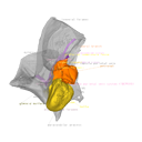
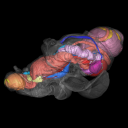
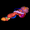
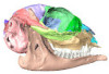
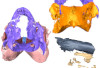
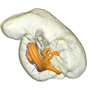
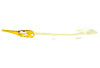
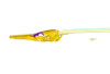
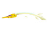
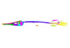
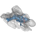
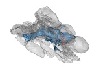
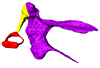
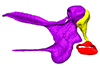
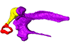
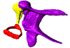
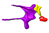
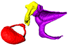
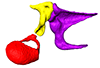
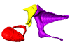
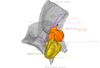
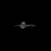
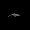
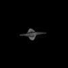
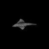
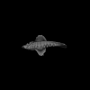
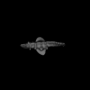
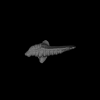
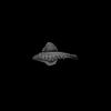
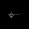
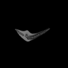
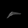
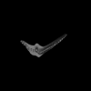
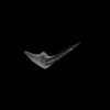
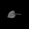
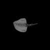
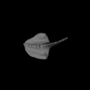
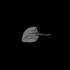
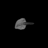
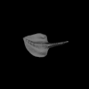
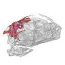
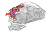
This project presents the osteological connexions of the petrosal bone of the extant Hippopotamidae Hippopotamus amphibius and Choeropsis liberiensis by a virtual osteological dissection of the ear region. The petrosal, the bulla, the sinuses and the major morphological features surrounding the petrosal bone are labelled, both in situ and in an exploded model presenting disassembly views. The directional underwater hearing mode of Hippopotamidae is discussed based on the new observations.
Choeropsis liberiensis UPPal-M09-5-005a View specimen
|
M3#1Labelled compact model of the right ear region of Choeropsis liberiensis (UPPal-M09-5-005a) Type: "3D_surfaces"doi: 10.18563/m3.sf1 state:published |
Download 3D surface file |

|
M3#2Labelled exploded model of the right ear region of Choeropsis liberiensis (UPPal-M09-5-005a) Type: "3D_surfaces"doi: 10.18563/m3.sf2 state:published |
Download 3D surface file |
Hippopotamus amphibius UM N179 View specimen

|
M3#3Labelled compact model of the right ear region of Hippopotamus amphibius (UM N 179) Type: "3D_surfaces"doi: 10.18563/m3.sf3 state:published |
Download 3D surface file |

|
M3#4Labelled exploded model of the right ear region of Hippopotamus amphibius (UM N 179) Type: "3D_surfaces"doi: 10.18563/m3.sf4 state:published |
Download 3D surface file |
The present 3D Dataset contains the 3D models analyzed in Assemat et al. 2023: Shape diversity in conodont elements, a quantitative study using 3D topography. Marine Micropaleontology 184. https://doi.org/10.1016/j.marmicro.2023.102292
P1 elements represent dental components of the conodont apparatus that perform the final stage of food processing before ingestion. Consequently, quantifying the shape of P1 elements across the topographic indices of different conodont species becomes crucial for deciphering the diversity in feeding behavior within this group.
Bispathodus aculeatus UM CTB 082 View specimen
|
M3#1404P element Type: "3D_surfaces"doi: 10.18563/m3.sf.1404 state:published |
Download 3D surface file |
Bispathodus aculeatus UM CTB 083 View specimen

|
M3#1405P element Type: "3D_surfaces"doi: 10.18563/m3.sf.1405 state:published |
Download 3D surface file |
Bispathodus aculeatus UM CTB 086 View specimen
|
M3#1406P element Type: "3D_surfaces"doi: 10.18563/m3.sf.1406 state:published |
Download 3D surface file |
Bispathodus ultimus UM CTB 088 View specimen

|
M3#1407P element Type: "3D_surfaces"doi: 10.18563/m3.sf.1407 state:published |
Download 3D surface file |
Bispathodus aculeatus UM CTB 089 View specimen
|
M3#1408P element Type: "3D_surfaces"doi: 10.18563/m3.sf.1408 state:published |
Download 3D surface file |
Bispathodus costatus UM CTB 090 View specimen
|
M3#1409P element Type: "3D_surfaces"doi: 10.18563/m3.sf.1409 state:published |
Download 3D surface file |
Bispathodus ultimus UM CTB 092 View specimen
|
M3#1410P element Type: "3D_surfaces"doi: 10.18563/m3.sf.1410 state:published |
Download 3D surface file |
Bispathodus costatus UM CTB 093 View specimen
|
M3#1411P element Type: "3D_surfaces"doi: 10.18563/m3.sf.1411 state:published |
Download 3D surface file |
Bispathodus spinulicostatus UM CTB 094 View specimen
|
M3#1412P element Type: "3D_surfaces"doi: 10.18563/m3.sf.1412 state:published |
Download 3D surface file |
Bispathodus aculeatus UM CTB 096 View specimen

|
M3#1413P element Type: "3D_surfaces"doi: 10.18563/m3.sf.1413 state:published |
Download 3D surface file |
Bispathodus ultimus UM CTB 098 View specimen

|
M3#1414P element Type: "3D_surfaces"doi: 10.18563/m3.sf.1414 state:published |
Download 3D surface file |
Bispathodus costatus UM CTB 060 View specimen
|
M3#1415P element Type: "3D_surfaces"doi: 10.18563/m3.sf.1415 state:published |
Download 3D surface file |
Bispathodus spinulicostatus UM CTB 073 View specimen

|
M3#1416P element Type: "3D_surfaces"doi: 10.18563/m3.sf.1416 state:published |
Download 3D surface file |
Branmehla suprema UM CTB 049 View specimen

|
M3#1417P element Type: "3D_surfaces"doi: 10.18563/m3.sf.1417 state:published |
Download 3D surface file |
Branmehla inornata UM CTB 100 View specimen
|
M3#1418P element Type: "3D_surfaces"doi: 10.18563/m3.sf.1418 state:published |
Download 3D surface file |
Bispathodus stabilis (morphe 1) UM CTB 101 View specimen

|
M3#1419P element Type: "3D_surfaces"doi: 10.18563/m3.sf.1419 state:published |
Download 3D surface file |
Branmehla suprema UM CTB 102 View specimen
|
M3#1420P element Type: "3D_surfaces"doi: 10.18563/m3.sf.1420 state:published |
Download 3D surface file |
Branmehla suprema UM CTB 103 View specimen

|
M3#1421P element Type: "3D_surfaces"doi: 10.18563/m3.sf.1421 state:published |
Download 3D surface file |
Branmehla suprema UM CTB 104 View specimen

|
M3#1422P element Type: "3D_surfaces"doi: 10.18563/m3.sf.1422 state:published |
Download 3D surface file |
Branmehla suprema UM CTB 105 View specimen
|
M3#1423P element Type: "3D_surfaces"doi: 10.18563/m3.sf.1423 state:published |
Download 3D surface file |
Branmehla suprema UM CTB 106 View specimen
|
M3#1424P element Type: "3D_surfaces"doi: 10.18563/m3.sf.1424 state:published |
Download 3D surface file |
Branmehla suprema UM CTB 072 View specimen

|
M3#1425P element Type: "3D_surfaces"doi: 10.18563/m3.sf.1425 state:published |
Download 3D surface file |
Branmehla suprema UM CTB 107 View specimen
|
M3#1426P element Type: "3D_surfaces"doi: 10.18563/m3.sf.1426 state:published |
Download 3D surface file |
Branmehla suprema UM CTB 108 View specimen

|
M3#1427P element Type: "3D_surfaces"doi: 10.18563/m3.sf.1427 state:published |
Download 3D surface file |
Branmehla suprema UM CTB 109 View specimen
|
M3#1428P element Type: "3D_surfaces"doi: 10.18563/m3.sf.1428 state:published |
Download 3D surface file |
Bispathodus stabilis (morphe 1) UM CTB 110 View specimen

|
M3#1429P element Type: "3D_surfaces"doi: 10.18563/m3.sf.1429 state:published |
Download 3D surface file |
Palmatolepis gracilis UM CTB 112 View specimen
|
M3#1430P element Type: "3D_surfaces"doi: 10.18563/m3.sf.1430 state:published |
Download 3D surface file |
Palmatolepis gracilis UM CTB 061 View specimen

|
M3#1431P element Type: "3D_surfaces"doi: 10.18563/m3.sf.1431 state:published |
Download 3D surface file |
Palmatolepis gracilis UM CTB 115 View specimen
|
M3#1432P element Type: "3D_surfaces"doi: 10.18563/m3.sf.1432 state:published |
Download 3D surface file |
Palmatolepis gracilis UM CTB 116 View specimen
|
M3#1433P element Type: "3D_surfaces"doi: 10.18563/m3.sf.1433 state:published |
Download 3D surface file |
Palmatolepis gracilis UM CTB 117 View specimen
|
M3#1434P element Type: "3D_surfaces"doi: 10.18563/m3.sf.1434 state:published |
Download 3D surface file |
Palmatolepis gracilis UM CTB 062 View specimen

|
M3#1435P element Type: "3D_surfaces"doi: 10.18563/m3.sf.1435 state:published |
Download 3D surface file |
Palmatolepis gracilis UM CTB 118 View specimen

|
M3#1436P element Type: "3D_surfaces"doi: 10.18563/m3.sf.1436 state:published |
Download 3D surface file |
Palmatolepis gracilis UM CTB 119 View specimen
|
M3#1437P element Type: "3D_surfaces"doi: 10.18563/m3.sf.1437 state:published |
Download 3D surface file |
Palmatolepis gracilis UM CTB 120 View specimen
|
M3#1438P element Type: "3D_surfaces"doi: 10.18563/m3.sf.1438 state:published |
Download 3D surface file |
Polygnathus communis UM CTB 075 View specimen
|
M3#1439P element Type: "3D_surfaces"doi: 10.18563/m3.sf.1439 state:published |
Download 3D surface file |
Polygnathus communis UM CTB 121 View specimen
|
M3#1440P element Type: "3D_surfaces"doi: 10.18563/m3.sf.1440 state:published |
Download 3D surface file |
Polygnathus communis UM CTB 122 View specimen
|
M3#1441P element Type: "3D_surfaces"doi: 10.18563/m3.sf.1441 state:published |
Download 3D surface file |
Polygnathus communis UM CTB 123 View specimen
|
M3#1442P element Type: "3D_surfaces"doi: 10.18563/m3.sf.1442 state:published |
Download 3D surface file |
Polygnathus communis UM CTB 125 View specimen
|
M3#1443P element Type: "3D_surfaces"doi: 10.18563/m3.sf.1443 state:published |
Download 3D surface file |
Polygnathus communis UM CTB 126 View specimen
|
M3#1444P element Type: "3D_surfaces"doi: 10.18563/m3.sf.1444 state:published |
Download 3D surface file |
Polygnathus communis UM CTB 128 View specimen
|
M3#1445P element Type: "3D_surfaces"doi: 10.18563/m3.sf.1445 state:published |
Download 3D surface file |
Polygnathus communis UM CTB 130 View specimen
|
M3#1446P element Type: "3D_surfaces"doi: 10.18563/m3.sf.1446 state:published |
Download 3D surface file |
Polygnathus communis UM CTB 131 View specimen
|
M3#1447P element Type: "3D_surfaces"doi: 10.18563/m3.sf.1447 state:published |
Download 3D surface file |
Polygnathus communis UM CTB 132 View specimen

|
M3#1448P element Type: "3D_surfaces"doi: 10.18563/m3.sf.1448 state:published |
Download 3D surface file |
Polygnathus communis UM CTB 133 View specimen
|
M3#1449P element Type: "3D_surfaces"doi: 10.18563/m3.sf.1449 state:published |
Download 3D surface file |
Polygnathus symmetricus UM CTB 139 View specimen
|
M3#1450P element Type: "3D_surfaces"doi: 10.18563/m3.sf.1450 state:published |
Download 3D surface file |
Polygnathus symmetricus UM CTB 140 View specimen

|
M3#1451P element Type: "3D_surfaces"doi: 10.18563/m3.sf.1451 state:published |
Download 3D surface file |
Polygnathus symmetricus UM CTB 141 View specimen
|
M3#1452P element Type: "3D_surfaces"doi: 10.18563/m3.sf.1452 state:published |
Download 3D surface file |
Polygnathus symmetricus UM CTB 142 View specimen
|
M3#1453P element Type: "3D_surfaces"doi: 10.18563/m3.sf.1453 state:published |
Download 3D surface file |
The present 3D Dataset contains the 3D model of the endocranial cast of Palaeolama sp. from the mid-Pleistocene (~1.2 Mya) of South America, analyzed in Balcarcel et al. 2023.
Palaeolama sp. PIMUZ A/V 4091 View specimen
|
M3#11283D model of a natural endocast Type: "3D_surfaces"doi: 10.18563/m3.sf.1128 state:published |
Download 3D surface file |
The present 3D Dataset contains the 3D models analyzed in: Toyoda S et al., 2015, Morphogenesis of the inner ear at different stages of normal human development. The Anatomical Record. doi : 10.1002/ar.23268
Homo sapiens KC-CS17IER29248 View specimen

|
M3#36Computationally reconstructed membranous labyrinth of a human embryo (KC-CS17IER29248) at Carnegie Stage 17 (Crown Rump Length= 7mm). Type: "3D_surfaces"doi: 10.18563/m3.sf36 state:published |
Download 3D surface file |
Homo sapiens KC-CS18IER17746 View specimen

|
M3#37Computationally reconstructed membranous labyrinth of a human embryo (KC-CS18IER17746) at Carnegie Stage 18 (Crown Rump Length= 12mm). Type: "3D_surfaces"doi: 10.18563/m3.sf37 state:published |
Download 3D surface file |
Homo sapiens KC-CS19IER16127 View specimen

|
M3#38Computationally reconstructed membranous labyrinth of a human embryo (KC-CS19IER16127) at Carnegie Stage 19 (Crown Rump Length= 13mm). Type: "3D_surfaces"doi: 10.18563/m3.sf38 state:published |
Download 3D surface file |
Homo sapiens KC-CS20IER20268 View specimen

|
M3#39Computationally reconstructed membranous labyrinth of a human embryo (KC-CS20IER20268) at Carnegie Stage 20 (Crown Rump Length= 13.7mm). Type: "3D_surfaces"doi: 10.18563/m3.sf39 state:published |
Download 3D surface file |
Homo sapiens KC-CS21IER28066 View specimen

|
M3#40Computationally reconstructed membranous labyrinth of a human embryo (KC-CS21IER28066) at Carnegie Stage 21 (Crown Rump Length= 16.7mm). Type: "3D_surfaces"doi: 10.18563/m3.sf40 state:published |
Download 3D surface file |
Homo sapiens KC-CS22IER35233 View specimen

|
M3#41Computationally reconstructed membranous labyrinth of a human embryo (KC-CS22IER35233) at Carnegie Stage 22 (Crown Rump Length= 22mm). Type: "3D_surfaces"doi: 10.18563/m3.sf41 state:published |
Download 3D surface file |
Homo sapiens KC-CS23IER15919 View specimen

|
M3#42Computationally reconstructed membranous labyrinth of a human embryo (KC-CS23IER15919) at Carnegie Stage 23 (Crown Rump Length= 32.3mm). Type: "3D_surfaces"doi: 10.18563/m3.sf42 state:published |
Download 3D surface file |
Homo sapiens KC-FIER52730 View specimen

|
M3#43Computationally reconstructed human membranous labyrinth in post embryonic phase (KC-FIER52730). Crown Rump Length: 43.5mm. Type: "3D_surfaces"doi: 10.18563/m3.sf43 state:published |
Download 3D surface file |
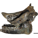
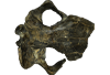
The present 3D Dataset contains the 3D models of a skull and lower jaw of the holotype of Santagnathus mariensis, described in “Old fossil findings in the Upper Triassic rocks of southern Brazil improve diversity of traversodontid cynodonts (Therapsida, Cynodontia)”
Santagnathus mariensis UFRGS-PV-1419-T View specimen
|
M3#1157Skull Type: "3D_surfaces"doi: 10.18563/m3.sf.1157 state:published |
Download 3D surface file |
|
M3#1158Lower jaw Type: "3D_surfaces"doi: 10.18563/m3.sf.1158 state:published |
Download 3D surface file |
The present 3D Dataset contains the 3D model analyzed in the following publication: occurrence of the ground sloth Nothrotheriops (Xenarthra, Folivora) in the Late Pleistocene of Uruguay: New information on its dietary and habitat preferences based on stable isotope analysis. Journal of Mammalian Evolution. https://doi.org/10.1007/s10914-023-09660-w
Nothrotheriops sp. CAV 1466 View specimen
|
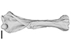
M3#1129Left humerus Type: "3D_surfaces"doi: 10.18563/m3.sf.1129 state:published |
Download 3D surface file |
The present 3D Dataset contains the 3D models analyzed in the following publication: Paulina-Carabajal, A., Ezcurra, M., Novas, F., 2019. New information on the braincase and endocranial morphology of the Late Triassic neotheropod Zupaysaurus rougieri using Computed Tomography data. Journal of Vertebrate Paleontology. https://doi.org/10.1080/02724634.2019.1630421
Zupaysaurus rougieri PULR 076 View specimen
|
M3#424The Zip contains 3 files, which correspond to: PULR_076-M1: Zupaysaurus rougieri skull, braincase and cranial endocast PULR_076-M2: Zupaysaurus rougieri braincase PULR_076-M1: Zupaysaurus rougieri brain and inner ear Type: "3D_surfaces"doi: 10.18563/m3.sf.424 state:published |
Download 3D surface file |


