3D models of Kalakocetus, the earliest Cetacea
The specimens of Speothos pacivorus
3D models related to the publication: Hidden diversity of Palaeogene metatherians: a new family of polydolopimorphian marsupials from Peruvian Amazonia
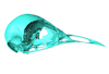
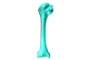
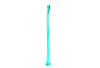
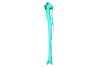
3D GM dataset of bird skeletal variation
Skeletal embryonic development in the catshark
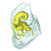
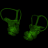
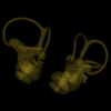
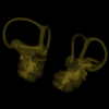
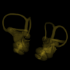
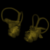
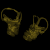
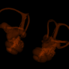
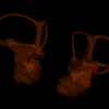
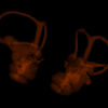
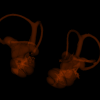
Bony connexions of the petrosal bone of extant hippos
bony labyrinth (14) , inner ear (11) , Eocene (11) , geometric morphometrics (10) , CT-scan (10) , Oligocene (9) , Micro-CT (9)
Lionel Hautier (25) , Maëva Judith Orliac (24) , Laurent Marivaux (18) , Renaud Lebrun (15) , Rodolphe Tabuce (14) , Pierre-Olivier Antoine (13) , Bastien Mennecart (13)
|
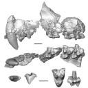
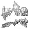
3D models related to the publication: A new large pantherine and a sabre-toothed cat (Mammalia, Carnivora, Felidae) from the late Miocene hominoid-bearing Khorat sand pits, Nakhon Ratchasima Province, northeastern Thailand.Camille Grohé
Published online: 04/09/2023 |
|
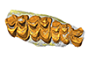
M3#1209Holotype of Pachypanthera piriyai, a left hemi-mandible with alveoli for i1-i3 and canine, roots of p3, p4 and partially broken off m1 crown. Type: "3D_surfaces"doi: 10.18563/m3.sf.1209 state:published |
Download 3D surface file |
Pachypanthera piriyai CUF-KR-2 View specimen
|
M3#1210Paratype of Pachypanthera piriyai, a right hemi-maxilla with P3-P4, alveoli of C and M1, root of P2 Type: "3D_surfaces"doi: 10.18563/m3.sf.1210 state:published |
Download 3D surface file |

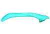
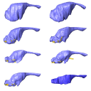
This contribution contains the 3D models analyzed in Müller et al. (2021) “Pushing the boundary? Testing the ‘functional elongation hypothesis’ of the giraffe’s neck”.
Aepyceros melampus ZFMK 2001.278 View specimen

|
M3#643Vertebrae C7, T1 Type: "3D_surfaces"doi: 10.18563/m3.sf.643 state:published |
Download 3D surface file |
Giraffa camelopardalis ZMB 66393 View specimen

|
M3#644Vertebrae Type: "3D_surfaces"doi: 10.18563/m3.sf.644 state:published |
Download 3D surface file |
Giraffa camelopardalis ZSM 1967/17 View specimen

|
M3#645Vertebrae Type: "3D_surfaces"doi: 10.18563/m3.sf.645 state:published |
Download 3D surface file |
Giraffa camelopardalis ZSM 1981/19 View specimen
|
M3#646C3, C4, C5, C6, C7, T1, T2 Type: "3D_surfaces"doi: 10.18563/m3.sf.646 state:published |
Download 3D surface file |
Giraffa camelopardalis KMDA M-10861 View specimen

|
M3#647C3, C4, C5, C6, C7, T1, T2. Acquired via laser scanner. Type: "3D_surfaces"doi: 10.18563/m3.sf.647 state:published |
Download 3D surface file |
Giraffa camelopardalis SMF 84214 View specimen

|
M3#648C7, T1. Warning : photogrammetric models (unit scale is CM, not MM). Type: "3D_surfaces"doi: 10.18563/m3.sf.648 state:published |
Download 3D surface file |
Giraffa camelopardalis SMF 78299 View specimen

|
M3#649C7, T1. Warning : unscaled photogrammetric 3D models (unknown size). Type: "3D_surfaces"doi: 10.18563/m3.sf.649 state:published |
Download 3D surface file |
Giraffa camelopardalis SMF o. N View specimen

|
M3#650C7, T1. Warning : unscaled photogrammetric 3D models (unknown size). Type: "3D_surfaces"doi: 10.18563/m3.sf.650 state:published |
Download 3D surface file |
Giraffa camelopardalis SMNS 19138 View specimen

|
M3#671C7, T1. Warning : unscaled photogrammetric 3D models (unknown size). Type: "3D_surfaces"doi: 10.18563/m3.sf.671 state:published |
Download 3D surface file |
Okapia johnstoni ZMB 62086 View specimen

|
M3#651C3, C4, C5, C6, C7, T1, T2 Type: "3D_surfaces"doi: 10.18563/m3.sf.651 state:published |
Download 3D surface file |
Okapia johnstoni ZMB 70325 View specimen

|
M3#652C3, C4, C5, C6, C7, T1, T2 Type: "3D_surfaces"doi: 10.18563/m3.sf.652 state:published |
Download 3D surface file |
Sivatherium giganteum NHMUK 15707 View specimen
|
M3#653C7. Warning : unscaled photogrammetric 3D model (unknown size). Type: "3D_surfaces"doi: 10.18563/m3.sf.653 state:published |
Download 3D surface file |
Sivatherium giganteum NHMUK 15297 View specimen

|
M3#654T1. Warning : unscaled photogrammetric 3D model (unknown size). Type: "3D_surfaces"doi: 10.18563/m3.sf.654 state:published |
Download 3D surface file |
Cervus elaphus ZMB 47502 View specimen

|
M3#655C3, C4, C5, C6, C7, T1, T2 Type: "3D_surfaces"doi: 10.18563/m3.sf.655 state:published |
Download 3D surface file |
Axis axis SMF 1450 View specimen

|
M3#656C7, T1 Type: "3D_surfaces"doi: 10.18563/m3.sf.656 state:published |
Download 3D surface file |
Cervus nippon SMF 4368 View specimen
|
M3#657C7, T1 Type: "3D_surfaces"doi: 10.18563/m3.sf.657 state:published |
Download 3D surface file |
Capreolus capreolus SMF 79852 View specimen

|
M3#658C7, T1 Type: "3D_surfaces"doi: 10.18563/m3.sf.658 state:published |
Download 3D surface file |
Capreolus capreolus ZFMK 67.237 View specimen

|
M3#659C7, T1 Type: "3D_surfaces"doi: 10.18563/m3.sf.659 state:published |
Download 3D surface file |
Muntiacus reevesi SMF 92954 View specimen

|
M3#660C7, T1 Type: "3D_surfaces"doi: 10.18563/m3.sf.660 state:published |
Download 3D surface file |
Muntiacus reevesi SMF 92332 View specimen

|
M3#661C7, T1 Type: "3D_surfaces"doi: 10.18563/m3.sf.661 state:published |
Download 3D surface file |
Alces alces SMF 35549 View specimen

|
M3#662C7, T1 Type: "3D_surfaces"doi: 10.18563/m3.sf.662 state:published |
Download 3D surface file |
Dama dama ZFMK 86.125 View specimen

|
M3#663C7, T1 Type: "3D_surfaces"doi: 10.18563/m3.sf.663 state:published |
Download 3D surface file |
Antilope cervicapra ZMB 78829 View specimen

|
M3#664C3, C4, C5, C6, C7, T1, T2 Type: "3D_surfaces"doi: 10.18563/m3.sf.664 state:published |
Download 3D surface file |
Bison bonasus SMNS 2998 View specimen

|
M3#665C7, T1. Warning : unscaled photogrammetric 3D models (unknown size). Type: "3D_surfaces"doi: 10.18563/m3.sf.665 state:published |
Download 3D surface file |
Nanger dama SMF 74435 View specimen

|
M3#666C7, T1 Type: "3D_surfaces"doi: 10.18563/m3.sf.666 state:published |
Download 3D surface file |
Litocranius walleri SMF 23747 View specimen

|
M3#667C7, T1 Type: "3D_surfaces"doi: 10.18563/m3.sf.667 state:published |
Download 3D surface file |
Litocranius walleri SMF 23749 View specimen

|
M3#669C7, T1 Type: "3D_surfaces"doi: 10.18563/m3.sf.669 state:published |
Download 3D surface file |
Tragelaphus eurycerus SMF 95875 View specimen

|
M3#670C7, T1 Type: "3D_surfaces"doi: 10.18563/m3.sf.670 state:published |
Download 3D surface file |
Bos javanicus SMF 64934 View specimen

|
M3#672C7, T1 Type: "3D_surfaces"doi: 10.18563/m3.sf.672 state:published |
Download 3D surface file |
Ovis aries ZFMK 1982.338 View specimen

|
M3#673C7, T1 Type: "3D_surfaces"doi: 10.18563/m3.sf.673 state:published |
Download 3D surface file |
Rupicapra rupicapra ZFMK 72.367 View specimen

|
M3#674C7, T1 Type: "3D_surfaces"doi: 10.18563/m3.sf.674 state:published |
Download 3D surface file |
Kobus ellipsiprymnus SMNS 4443 View specimen

|
M3#675C7, T1 Type: "3D_surfaces"doi: 10.18563/m3.sf.675 state:published |
Download 3D surface file |
Sylvicapra grimmia SMNS 15292 View specimen

|
M3#676C7, T1 Type: "3D_surfaces"doi: 10.18563/m3.sf.676 state:published |
Download 3D surface file |
Syncerus caffer SMNS 7347 View specimen

|
M3#677C7, T1. Warning : unscaled photogrammetric 3D models (unknown size). Type: "3D_surfaces"doi: 10.18563/m3.sf.677 state:published |
Download 3D surface file |
Procapra gutturosa SMNS 5796 View specimen

|
M3#678C7, T1 Type: "3D_surfaces"doi: 10.18563/m3.sf.678 state:published |
Download 3D surface file |
Damaliscus pygargus SMNS 21617 View specimen

|
M3#679C7, T1 Type: "3D_surfaces"doi: 10.18563/m3.sf.679 state:published |
Download 3D surface file |
Madoqua kirkii SMNS 4432 View specimen

|
M3#680C7, T1 Type: "3D_surfaces"doi: 10.18563/m3.sf.680 state:published |
Download 3D surface file |
Bubalus mindorensis SMNS 2054 View specimen

|
M3#681C7, T1. Warning : unscaled photogrammetric 3D models (unknown size). Type: "3D_surfaces"doi: 10.18563/m3.sf.681 state:published |
Download 3D surface file |
Capra hircus SMNS 51328 View specimen

|
M3#682C7, T1 Type: "3D_surfaces"doi: 10.18563/m3.sf.682 state:published |
Download 3D surface file |
Connochaetes taurinus SMNS 4442 View specimen

|
M3#683C7, T1. Warning : unscaled photogrammetric 3D models (unknown size). Type: "3D_surfaces"doi: 10.18563/m3.sf.683 state:published |
Download 3D surface file |
Antilocapra americana ZSM 1964/218 View specimen

|
M3#684C3, C4, C5, C6, C7, T1, T2 Type: "3D_surfaces"doi: 10.18563/m3.sf.684 state:published |
Download 3D surface file |
Antilocapra americana ZMB 77281 View specimen

|
M3#685C7, T1 Type: "3D_surfaces"doi: 10.18563/m3.sf.685 state:published |
Download 3D surface file |
Moschus moschiferus ZMB 62080 View specimen

|
M3#686C3, C4, C5, C6, C7, T1, T2 Type: "3D_surfaces"doi: 10.18563/m3.sf.686 state:published |
Download 3D surface file |
Moschus moschiferus ZMB 60367 View specimen

|
M3#687C7, T1 Type: "3D_surfaces"doi: 10.18563/m3.sf.687 state:published |
Download 3D surface file |
Moschus moschiferus ZMB 51830 View specimen

|
M3#688C7, T1 Type: "3D_surfaces"doi: 10.18563/m3.sf.688 state:published |
Download 3D surface file |
Tragulus javanicus SMF 82179 View specimen

|
M3#689C7, T1 Type: "3D_surfaces"doi: 10.18563/m3.sf.689 state:published |
Download 3D surface file |
Tragulus javanicus ZMB 86222 View specimen

|
M3#690C7, T1 Type: "3D_surfaces"doi: 10.18563/m3.sf.690 state:published |
Download 3D surface file |
Tragulus sp. ZMB o. N. View specimen

|
M3#691C7, T1 Type: "3D_surfaces"doi: 10.18563/m3.sf.691 state:published |
Download 3D surface file |
Hyemoschus aquaticus ZMB 71071 View specimen
|
M3#692C7, T1 Type: "3D_surfaces"doi: 10.18563/m3.sf.692 state:published |
Download 3D surface file |
Hyemoschus aquaticus ZMB 103235 View specimen

|
M3#693C7, T1 Type: "3D_surfaces"doi: 10.18563/m3.sf.693 state:published |
Download 3D surface file |
Vicugna vicugna SMF 94752 View specimen

|
M3#694C7, T1 Type: "3D_surfaces"doi: 10.18563/m3.sf.694 state:published |
Download 3D surface file |
Camelus dromedarius SMF 70473 View specimen

|
M3#695C7, T1. Warning : unscaled photogrammetric 3D models (unknown size). Type: "3D_surfaces"doi: 10.18563/m3.sf.695 state:published |
Download 3D surface file |
Camelus bactrianus SMF 25542 View specimen

|
M3#696C7, T1. Warning : unscaled photogrammetric 3D models (unknown size). Type: "3D_surfaces"doi: 10.18563/m3.sf.696 state:published |
Download 3D surface file |
Lama glama SMNS 31175 View specimen

|
M3#697C7, T1 Type: "3D_surfaces"doi: 10.18563/m3.sf.697 state:published |
Download 3D surface file |
Vicugna pacos SMNS 46255 View specimen

|
M3#698C7, T1 Type: "3D_surfaces"doi: 10.18563/m3.sf.698 state:published |
Download 3D surface file |
Vicugna pacos SMNS 7349 View specimen

|
M3#699C7, T1 Type: "3D_surfaces"doi: 10.18563/m3.sf.699 state:published |
Download 3D surface file |
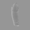
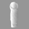
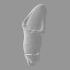
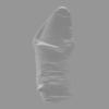
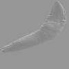
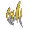
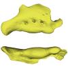
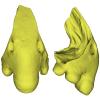
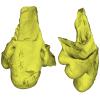
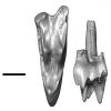
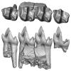
The present 3D dataset contains 3D models of new material from the middle Eocene of the Upper Subathu Formation in the Kalakot area (India), documenting the anterior dentition of the raoellid Indohyus indirae. Raoellidae are closely related to stem cetaceans and bring crucial information to understand the earliest phase of land to water transition in Cetacea.
Indohyus indirae GU/RJ/31 View specimen
|
M3#1505Right i1 Type: "3D_surfaces"doi: 10.18563/m3.sf.1505 state:published |
Download 3D surface file |
Indohyus indirae GU/RJ/32 View specimen
|
M3#1506Right i1 Type: "3D_surfaces"doi: 10.18563/m3.sf.1506 state:published |
Download 3D surface file |
Indohyus indirae GU/RJ/16 View specimen
|
M3#1507Left I3 Type: "3D_surfaces"doi: 10.18563/m3.sf.1507 state:published |
Download 3D surface file |
Indohyus indirae GU/RJ/23 View specimen
|
M3#1508Left I2 Type: "3D_surfaces"doi: 10.18563/m3.sf.1508 state:published |
Download 3D surface file |
Indohyus indirae GU/RJ/25 View specimen
|
M3#1509Left I1 Type: "3D_surfaces"doi: 10.18563/m3.sf.1509 state:published |
Download 3D surface file |
Indohyus indirae GU/RJ/26 View specimen
|
M3#1510Right I3 Type: "3D_surfaces"doi: 10.18563/m3.sf.1510 state:published |
Download 3D surface file |
Indohyus indirae GU/RJ/57 View specimen
|
M3#1511Left I1 Type: "3D_surfaces"doi: 10.18563/m3.sf.1511 state:published |
Download 3D surface file |
Indohyus indirae GU/RJ/61 View specimen
|
M3#1512Right upper canine Type: "3D_surfaces"doi: 10.18563/m3.sf.1512 state:published |
Download 3D surface file |
Indohyus indirae GU/RJ/63 View specimen

|
M3#1513Left upper canine Type: "3D_surfaces"doi: 10.18563/m3.sf.1513 state:published |
Download 3D surface file |
Indohyus indirae GU/RJ/74 View specimen
|
M3#1514Left upper canine Type: "3D_surfaces"doi: 10.18563/m3.sf.1514 state:published |
Download 3D surface file |
Indohyus indirae GU/RJ/439 View specimen
|
M3#1515Left upper canine Type: "3D_surfaces"doi: 10.18563/m3.sf.1515 state:published |
Download 3D surface file |
Indohyus indirae GU/RJ/457 View specimen
|
M3#1516Left upper canine Type: "3D_surfaces"doi: 10.18563/m3.sf.1516 state:published |
Download 3D surface file |
Indohyus indirae GU/RJ/846 View specimen
|
M3#1517Left upper canine Type: "3D_surfaces"doi: 10.18563/m3.sf.1517 state:published |
Download 3D surface file |
Indohyus indirae GU/RJ/822 View specimen
|
M3#1518Right fragmentary maxillary with decidual canine and I3 Type: "3D_surfaces"doi: 10.18563/m3.sf.1518 state:published |
Download 3D surface file |
Indohyus indirae GU/RJ/824 View specimen
|
M3#1519Right fragmentary mandible with lower canine and small part of the i3 Type: "3D_surfaces"doi: 10.18563/m3.sf.1519 state:published |
Download 3D surface file |
Indohyus indirae GU/RJ/838 View specimen
|
M3#1520Right fragmentary mandible with permanent i2, i3 and canine and small part of the root of the decidual i3 Type: "3D_surfaces"doi: 10.18563/m3.sf.1520 state:published |
Download 3D surface file |
Indohyus indirae GU/RJ/842 View specimen
|
M3#1521Left fragmentary mandible with decidual and permanent canine Type: "3D_surfaces"doi: 10.18563/m3.sf.1521 state:published |
Download 3D surface file |
Indohyus indirae GU/RJ/26,32,56,57,457,822,838,842 : Composite Anterior dentition View specimen
|
M3#15293D composite reconstruction of the anterior dentition of Indohyus indirae with GU/RJ/57 (I1), 56 (I2), 26 (I3), 457 (Upper canine), 822 (P1), 32 (i1), 838 (i2, i3 and lower canine) and 842 (p1) Type: "3D_surfaces"doi: 10.18563/m3.sf.1529 state:published |
Download 3D surface file |

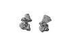
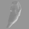
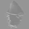
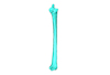
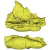
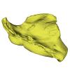
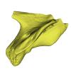
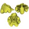
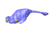
The present 3D Dataset contains the 3D models of Protocetus atavus described and figured in the following publication: Berger et al. (2025) The endocranial anatomy of Protocetids and its implications for early whale evolution.
Protocetus atavus SMNS-P-11084 View specimen
|
M3#1654Textured model of the whole skull Type: "3D_surfaces"doi: 10.18563/m3.sf.1654 state:published |
Download 3D surface file |
|
M3#1655Brain endocast Type: "3D_surfaces"doi: 10.18563/m3.sf.1655 state:published |
Download 3D surface file |
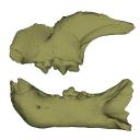
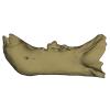
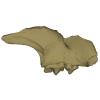
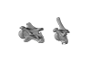
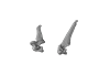
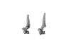
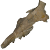
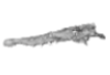
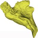
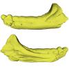
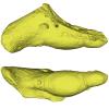
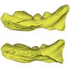
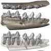
The holotype of Hamadasuchus rebouli Buffetaut 1994 from the Kem Kem beds of Morocco (Late Albian – Cenomanian) consists of a left dentary which is limited, fragmentary and reconstructed in some areas. To aid in assessing if the original diagnosis can be considered as valid, the specimen was CT scanned for the first time. This is especially important to resolve the taxonomic status of certain specimens that have been assigned to Hamadasuchus rebouli since then. The reconstructed structures in this contribution are in agreement with the original description, notably in terms of alveolar count; thus the original diagnosis of this taxon remains valid and some specimens are not referable to H. rebouli anymore.
Hamadasuchus rebouli MDE C001 View specimen

|
M3#1402Dentary and teeth Type: "3D_surfaces"doi: 10.18563/m3.sf.1402 state:published |
Download 3D surface file |

|
M3#1403Toothmarks Type: "3D_surfaces"doi: 10.18563/m3.sf.1403 state:published |
Download 3D surface file |
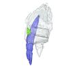
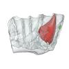
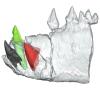
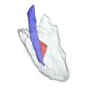
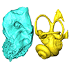
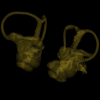
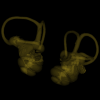
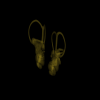
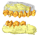
The present 3D Dataset contains the 3D models analyzed in Mennecart, B., Duranthon, F., & Costeur, L. 2024. Systematic contribution of the auditory region to the knowledge of the oldest European Bovidae (Mammalia, Ruminantia). Journal of Anatomy XXX. https://doi.org/10.1111/joa.14132
Pusillutragus montrealensis MHNT.PAL.2015.0.2261.4 View specimen
|
M3#1522Right petrosal, bony labyrinth, stapes Type: "3D_surfaces"doi: 10.18563/m3.sf.1522 state:published |
Download 3D surface file |
Pusillutragus montrealensis MHNT.PAL.2015.0.2261.9 View specimen
|
M3#1523Left petrosal and left bony labyrinth Type: "3D_surfaces"doi: 10.18563/m3.sf.1523 state:published |
Download 3D surface file |
Eotragus artenensis SMNS-P-41625 View specimen
|
M3#1524Petrosal (right), bony labyrinth (left) Type: "3D_surfaces"doi: 10.18563/m3.sf.1524 state:published |
Download 3D surface file |
Eotragus clavatus NMB San.15056 View specimen
|
M3#1528Right petrosal and right bony labyrinth Type: "3D_surfaces"doi: 10.18563/m3.sf.1528 state:published |
Download 3D surface file |
Eotragus clavatus NMB San.15055 View specimen
|
M3#1526Left Petrosal Type: "3D_surfaces"doi: 10.18563/m3.sf.1526 state:published |
Download 3D surface file |
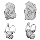
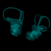
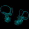
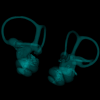
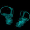
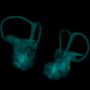
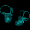
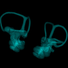
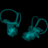
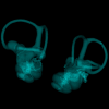
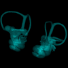
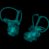
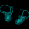
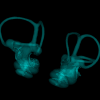
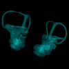
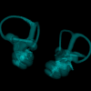
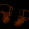
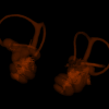
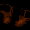
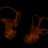
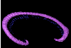
This contribution contains 3D models of left and right house mouse (Mus musculus domesticus) inner ears analyzed in Renaud et al. (2024). The studied mice belong to four groups: wild-trapped mice, wild-derived lab offspring, a typical laboratory strain (Swiss) and hybrids between wild-derived and Swiss mice. They have been analyzed to assess the impact of mobility reduction on inner ear morphology, including patterns of divergence, levels of inter-individual variance (disparity) and intra-individual variance (fluctuating asymmetry)
Mus musculus Tourch_7819 View specimen
|
M3#1366Two bony labyrinths Type: "3D_surfaces"doi: 10.18563/m3.sf.1366 state:published |
Download 3D surface file |
Mus musculus Tourch_7821 View specimen
|
M3#1367Two bony labyrinths Type: "3D_surfaces"doi: 10.18563/m3.sf.1367 state:published |
Download 3D surface file |
Mus musculus Tourch_7839 View specimen
|
M3#1368Two bony labyrinths Type: "3D_surfaces"doi: 10.18563/m3.sf.1368 state:published |
Download 3D surface file |
Mus musculus Tourch_7873 View specimen
|
M3#1369Two bony labyrinths Type: "3D_surfaces"doi: 10.18563/m3.sf.1369 state:published |
Download 3D surface file |
Mus musculus Tourch_7877 View specimen
|
M3#1370Two bony labyrinths Type: "3D_surfaces"doi: 10.18563/m3.sf.1370 state:published |
Download 3D surface file |
Mus musculus Tourch_7922 View specimen
|
M3#1371Two bony labyrinths Type: "3D_surfaces"doi: 10.18563/m3.sf.1371 state:published |
Download 3D surface file |
Mus musculus Tourch_7923 View specimen
|
M3#1372Two bony labyrinths Type: "3D_surfaces"doi: 10.18563/m3.sf.1372 state:published |
Download 3D surface file |
Mus musculus Tourch_7925 View specimen
|
M3#1373Two bony labyrinths Type: "3D_surfaces"doi: 10.18563/m3.sf.1373 state:published |
Download 3D surface file |
Mus musculus Tourch_7927 View specimen
|
M3#1374Two bony labyrinths Type: "3D_surfaces"doi: 10.18563/m3.sf.1374 state:published |
Download 3D surface file |
Mus musculus Tourch_7932 View specimen
|
M3#1375Two bony labyrinths Type: "3D_surfaces"doi: 10.18563/m3.sf.1375 state:published |
Download 3D surface file |
Mus musculus Bal02 View specimen
|
M3#1320Two bony labyrinths Type: "3D_surfaces"doi: 10.18563/m3.sf.1320 state:published |
Download 3D surface file |
Mus musculus Bal04 View specimen

|
M3#1321Two bony labyrinths Type: "3D_surfaces"doi: 10.18563/m3.sf.1321 state:published |
Download 3D surface file |
Mus musculus Bal06 View specimen
|
M3#1322Two bony labyrinths Type: "3D_surfaces"doi: 10.18563/m3.sf.1322 state:published |
Download 3D surface file |
Mus musculus Bal08 View specimen
|
M3#1323Two bony labyrinths Type: "3D_surfaces"doi: 10.18563/m3.sf.1323 state:published |
Download 3D surface file |
Mus musculus Bal11 View specimen
|
M3#1324Two bony labyrinths Type: "3D_surfaces"doi: 10.18563/m3.sf.1324 state:published |
Download 3D surface file |
Mus musculus Bal12 View specimen
|
M3#1325Two bony labyrinths Type: "3D_surfaces"doi: 10.18563/m3.sf.1325 state:published |
Download 3D surface file |
Mus musculus Bal15 View specimen
|
M3#1326Two bony labyrinths Type: "3D_surfaces"doi: 10.18563/m3.sf.1326 state:published |
Download 3D surface file |
Mus musculus Bal16 View specimen
|
M3#1327Two bony labyrinths Type: "3D_surfaces"doi: 10.18563/m3.sf.1327 state:published |
Download 3D surface file |
Mus musculus Bal17 View specimen
|
M3#1328Two bony labyrinths Type: "3D_surfaces"doi: 10.18563/m3.sf.1328 state:published |
Download 3D surface file |
Mus musculus Bal18 View specimen
|
M3#1329Two bony labyrinths Type: "3D_surfaces"doi: 10.18563/m3.sf.1329 state:published |
Download 3D surface file |
Mus musculus Bal19 View specimen
|
M3#1330Two bony labyrinths Type: "3D_surfaces"doi: 10.18563/m3.sf.1330 state:published |
Download 3D surface file |
Mus musculus Bal20 View specimen
|
M3#1331Two bony labyrinths Type: "3D_surfaces"doi: 10.18563/m3.sf.1331 state:published |
Download 3D surface file |
Mus musculus Bal21 View specimen
|
M3#1332Two bony labyrinths Type: "3D_surfaces"doi: 10.18563/m3.sf.1332 state:published |
Download 3D surface file |
Mus musculus Bal22 View specimen
|
M3#1333Two bony labyrinths Type: "3D_surfaces"doi: 10.18563/m3.sf.1333 state:published |
Download 3D surface file |
Mus musculus Bal23 View specimen
|
M3#1334Two bony labyrinths Type: "3D_surfaces"doi: 10.18563/m3.sf.1334 state:published |
Download 3D surface file |
Mus musculus Bal24 View specimen
|
M3#1335Two bony labyrinths Type: "3D_surfaces"doi: 10.18563/m3.sf.1335 state:published |
Download 3D surface file |
Mus musculus Bal25 View specimen
|
M3#1336Two bony labyrinths Type: "3D_surfaces"doi: 10.18563/m3.sf.1336 state:published |
Download 3D surface file |
Mus musculus Balan_LAB_035 View specimen
|
M3#1337Two bony labyrinths Type: "3D_surfaces"doi: 10.18563/m3.sf.1337 state:published |
Download 3D surface file |
Mus musculus Balan_LAB_046 View specimen
|
M3#1338Two bony labyrinths Type: "3D_surfaces"doi: 10.18563/m3.sf.1338 state:published |
Download 3D surface file |
Mus musculus Balan_LAB_054 View specimen
|
M3#1339Two bony labyrinths Type: "3D_surfaces"doi: 10.18563/m3.sf.1339 state:published |
Download 3D surface file |
Mus musculus Balan_LAB_056 View specimen
|
M3#1340Two bony labyrinths Type: "3D_surfaces"doi: 10.18563/m3.sf.1340 state:published |
Download 3D surface file |
Mus musculus Balan_LAB_082 View specimen
|
M3#1341Two bony labyrinths Type: "3D_surfaces"doi: 10.18563/m3.sf.1341 state:published |
Download 3D surface file |
Mus musculus Balan_LAB_086 View specimen

|
M3#1342Two bony labyrinths Type: "3D_surfaces"doi: 10.18563/m3.sf.1342 state:published |
Download 3D surface file |
Mus musculus Balan_LAB_092 View specimen
|
M3#1343Two bony labyrinths Type: "3D_surfaces"doi: 10.18563/m3.sf.1343 state:published |
Download 3D surface file |
Mus musculus Balan_LAB_319 View specimen
|
M3#1344Two bony labyrinths Type: "3D_surfaces"doi: 10.18563/m3.sf.1344 state:published |
Download 3D surface file |
Mus musculus Balan_LAB_325 View specimen
|
M3#1345Two bony labyrinths Type: "3D_surfaces"doi: 10.18563/m3.sf.1345 state:published |
Download 3D surface file |
Mus musculus Balan_LAB_329 View specimen
|
M3#1346Two bony labyrinths Type: "3D_surfaces"doi: 10.18563/m3.sf.1346 state:published |
Download 3D surface file |
Mus musculus Balan_LAB_330 View specimen
|
M3#1347Two bony labyrinths Type: "3D_surfaces"doi: 10.18563/m3.sf.1347 state:published |
Download 3D surface file |
Mus musculus Balan_LAB_F2b View specimen
|
M3#1348Two bony labyrinths Type: "3D_surfaces"doi: 10.18563/m3.sf.1348 state:published |
Download 3D surface file |
Mus musculus Balan_LAB_BB3weeks View specimen
|
M3#1349Two bony labyrinths Type: "3D_surfaces"doi: 10.18563/m3.sf.1349 state:published |
Download 3D surface file |
Mus musculus SW0ter View specimen
|
M3#1376Two bony labyrinths Type: "3D_surfaces"doi: 10.18563/m3.sf.1376 state:published |
Download 3D surface file |
Mus musculus SW343 View specimen
|
M3#1377Two bony labyrinths Type: "3D_surfaces"doi: 10.18563/m3.sf.1377 state:published |
Download 3D surface file |
Mus musculus SW1 View specimen
|
M3#1378Two bony labyrinths Type: "3D_surfaces"doi: 10.18563/m3.sf.1378 state:published |
Download 3D surface file |
Mus musculus SW2 View specimen
|
M3#1379Two bony labyrinths Type: "3D_surfaces"doi: 10.18563/m3.sf.1379 state:published |
Download 3D surface file |
Mus musculus BAL_F1_30x17_27j View specimen
|
M3#1350Two bony labyrinths Type: "3D_surfaces"doi: 10.18563/m3.sf.1350 state:published |
Download 3D surface file |
Mus musculus BAL_F1_167_48j View specimen
|
M3#1351Two bony labyrinths Type: "3D_surfaces"doi: 10.18563/m3.sf.1351 state:published |
Download 3D surface file |
Mus musculus BAL_F1_188_32j View specimen
|
M3#1352Two bony labyrinths Type: "3D_surfaces"doi: 10.18563/m3.sf.1352 state:published |
Download 3D surface file |
Mus musculus BAL_F1_192_28j View specimen
|
M3#1353Two bony labyrinths Type: "3D_surfaces"doi: 10.18563/m3.sf.1353 state:published |
Download 3D surface file |
Mus musculus BAL_F1_194_46j View specimen
|
M3#1354Two bony labyrinths Type: "3D_surfaces"doi: 10.18563/m3.sf.1354 state:published |
Download 3D surface file |
Mus musculus BAL_F1_196_44j View specimen
|
M3#1355Two bony labyrinths Type: "3D_surfaces"doi: 10.18563/m3.sf.1355 state:published |
Download 3D surface file |
Mus musculus BAL_F2_40x56_24j View specimen

|
M3#1356Two bony labyrinths Type: "3D_surfaces"doi: 10.18563/m3.sf.1356 state:published |
Download 3D surface file |
Mus musculus BAL_F2_47x61_22j View specimen
|
M3#1357Two bony labyrinths Type: "3D_surfaces"doi: 10.18563/m3.sf.1357 state:published |
Download 3D surface file |
Mus musculus Gardouch_3419 View specimen
|
M3#1358Two bony labyrinths Type: "3D_surfaces"doi: 10.18563/m3.sf.1358 state:published |
Download 3D surface file |
Mus musculus Gardouch_3432 View specimen
|
M3#1359Two bony labyrinths Type: "3D_surfaces"doi: 10.18563/m3.sf.1359 state:published |
Download 3D surface file |
Mus musculus Gardouch_3437 View specimen
|
M3#1360Two bony labyrinths Type: "3D_surfaces"doi: 10.18563/m3.sf.1360 state:published |
Download 3D surface file |
Mus musculus Gardouch_3439 View specimen
|
M3#1361Two bony labyrinths Type: "3D_surfaces"doi: 10.18563/m3.sf.1361 state:published |
Download 3D surface file |
Mus musculus Gardouch_3450 View specimen
|
M3#1362Two bony labyrinths Type: "3D_surfaces"doi: 10.18563/m3.sf.1362 state:published |
Download 3D surface file |
Mus musculus Gardouch_3453 View specimen
|
M3#1363Two bony labyrinths Type: "3D_surfaces"doi: 10.18563/m3.sf.1363 state:published |
Download 3D surface file |
Mus musculus Gardouch_3459 View specimen
|
M3#1364Two bony labyrinths Type: "3D_surfaces"doi: 10.18563/m3.sf.1364 state:published |
Download 3D surface file |
Mus musculus Gardouch_3462 View specimen
|
M3#1365Two bony labyrinths Type: "3D_surfaces"doi: 10.18563/m3.sf.1365 state:published |
Download 3D surface file |
Mus musculus SW5 View specimen
|
M3#1380Two bony labyrinths Type: "3D_surfaces"doi: 10.18563/m3.sf.1380 state:published |
Download 3D surface file |
Mus musculus SWF3 View specimen
|
M3#1381Two bony labyrinths Type: "3D_surfaces"doi: 10.18563/m3.sf.1381 state:published |
Download 3D surface file |
Mus musculus SW342 View specimen
|
M3#1382Two bony labyrinths Type: "3D_surfaces"doi: 10.18563/m3.sf.1382 state:published |
Download 3D surface file |
Mus musculus SW341 View specimen
|
M3#1383Two bony labyrinths Type: "3D_surfaces"doi: 10.18563/m3.sf.1383 state:published |
Download 3D surface file |
Mus musculus SW339 View specimen
|
M3#1384Two bony labyrinths Type: "3D_surfaces"doi: 10.18563/m3.sf.1384 state:published |
Download 3D surface file |
Mus musculus SWF4 View specimen
|
M3#1385Two bony labyrinths Type: "3D_surfaces"doi: 10.18563/m3.sf.1385 state:published |
Download 3D surface file |
Mus musculus SW0bis_350 View specimen
|
M3#1386Two bony labyrinths Type: "3D_surfaces"doi: 10.18563/m3.sf.1386 state:published |
Download 3D surface file |
Mus musculus SW0_348 View specimen
|
M3#1387Two bony labyrinths Type: "3D_surfaces"doi: 10.18563/m3.sf.1387 state:published |
Download 3D surface file |
Mus musculus SW347 View specimen
|
M3#1388Two bony labyrinths Type: "3D_surfaces"doi: 10.18563/m3.sf.1388 state:published |
Download 3D surface file |
Mus musculus SW345 View specimen
|
M3#1389Two bony labyrinths Type: "3D_surfaces"doi: 10.18563/m3.sf.1389 state:published |
Download 3D surface file |
Mus musculus hyb_125xSW_01 View specimen
|
M3#1390Two bony labyrinths Type: "3D_surfaces"doi: 10.18563/m3.sf.1390 state:published |
Download 3D surface file |
Mus musculus hyb_125xSW_02 View specimen
|
M3#1391Two bony labyrinths Type: "3D_surfaces"doi: 10.18563/m3.sf.1391 state:published |
Download 3D surface file |
Mus musculus hyb_SWx126_01 View specimen
|
M3#1392Two bony labyrinths Type: "3D_surfaces"doi: 10.18563/m3.sf.1392 state:published |
Download 3D surface file |
Mus musculus hyb_SWx126_02 View specimen
|
M3#1393Two bony labyrinths Type: "3D_surfaces"doi: 10.18563/m3.sf.1393 state:published |
Download 3D surface file |
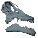
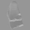
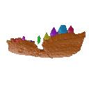
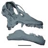
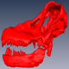
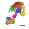
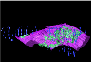
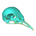
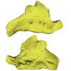
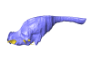
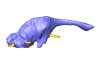
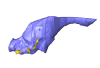
The study of titanosaur paleobiology has been severely hampered by the incomplete nature of their fossil record, particularly the scarcity of well-preserved and relatively complete cranial remains. Even the most complete titanosaur skulls are often fractured, incomplete, or deformed, which has resulted in a limited knowledge of the paleobiology related to cranial anatomy, especially functional morphology. In this context, we present the digital restoration of the skull of the Argentinean titanosaur Sarmientosaurus musacchioi, created using the open-source 3D modeling software Blender. The digitally restored model is freely accessible to other researchers, facilitating broader research and comparative studies.
Sarmientosaurus mussacchioi MDT-PV 02 View specimen
|
M3#1594Cranium and mandible of Sarmientosaurus mussacchioi Type: "3D_surfaces"doi: 10.18563/m3.sf.1594 state:published |
Download 3D surface file |
|
M3#1599Original object provided by Gabriel Casal (cranium) Type: "3D_surfaces"doi: 10.18563/m3.sf.1599 state:published |
Download 3D surface file |
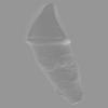
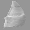
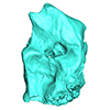
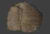
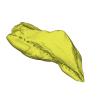
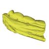
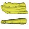
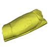
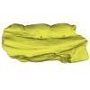
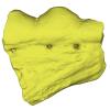
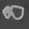
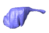
Turtles are one of the most impressive vertebrates. Much of the body is either hidden in a shell or can be drawn into it. Turtles impress with their individual longevity and their often peaceful disposition. Also, with their resilience, they have survived all extinction events since their emergence in the Late Triassic. Today's diversity of shapes is impressive and ranges from the large and high domed Galapagos turtles to the hamster-sized flat pancake turtles. The holotype of one of the oldest fossil turtles, Proganochelys quenstedtii, is housed in the paleontological collection in Tübingen/Germany. Since its discovery some years before 1873, P. quenstedtii has represented the 'prototype' of the turtle and has had an eventful scientific history. It was found in Neuenhaus (Häfner-Neuhausen in Schönbuch forest), Baden-Württemberg, Germany, and stems from Löwenstein-Formation (Weißer Keupersandstein), Late Triassic. The current catalogue number is GPIT-PV-30000. The specimen is listed in the historical inventory “Tübinger Petrefaktenverzeichnis 1841 bis 1896, [folio 326v.]“, as “[catalogue number: PV]16549, Schildkröte Weiser Keupersandstein Hafnerhausen” [turtle from White Keuper Sandstone]. Another, more recent synonym is “GPIT/RE/9396”. The same specimen was presented as uncatalogued by Gaffney (1990). Here we provide a surface scan of the steinkern for easier access of this famous specimen to the scientific community.
Proganochelys quenstedtii GPIT-PV-30000 View specimen
|
M3#967This the surface model of the steinkern of the shell of Proganochelys quenstedtii. Type: "3D_surfaces"doi: 10.18563/m3.sf.967 state:published |
Download 3D surface file |
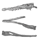
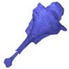
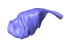
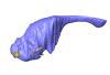
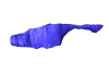
The present 3D dataset contains the 3D models of the holotype of Proterochampsa nodosa that were built and analysed in “Redescription, taxonomic revaluation, and phylogenetic affinities of Proterochampsa nodosa (Archosauriformes: Proterochampsidae), early Late Triassic of Candelaria Sequence (Santa Maria Supersequence)”.
Proterochampsa nodosa MCP 1694-PV View specimen
|
M3#9743D models of Proterochampsa nodosa. Model 1: skull. Model 2: mandible. Model 3: left mandibular ramus. Type: "3D_surfaces"doi: 10.18563/m3.sf.974 state:published |
Download 3D surface file |

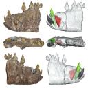
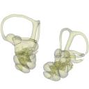
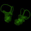
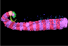
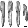
The present 3D Dataset contains the 3D models analyzed in the publication: Mummified Paleogene Spirostreptida and Julida (Arthropoda, Diplopoda) from southern France. Papers in Paleontology.
Protosilvestria sculpta NMB F1935 View specimen

|
M3#1457Paralectotype, 13 diplosegments with the proximal part of the legs Type: "3D_surfaces"doi: 10.18563/m3.sf.1457 state:published |
Download 3D surface file |

|
M3#1657CT data of NMB F1935. Images were reduced by a binning of factor 2. Type: "3D_CT"doi: 10.18563/m3.sf.1657 state:published |
Download CT data |
Protosilvestria sculpta NMB F1936 View specimen

|
M3#1458Lectotype, head with the ten following segments Type: "3D_surfaces"doi: 10.18563/m3.sf.1458 state:published |
Download 3D surface file |

|
M3#1658CT data of NMB F1936. Type: "3D_CT"doi: 10.18563/m3.sf.1658 state:published |
Download CT data |
Protosilvestria sculpta NMB F1937 View specimen

|
M3#1459Paralectotype, seven segments Type: "3D_surfaces"doi: 10.18563/m3.sf.1459 state:published |
Download 3D surface file |

|
M3#1659CT data of NMB F1937 Type: "3D_CT"doi: 10.18563/m3.sf.1659 state:published |
Download CT data |
Protosilvestria sculpta NMB F1938 View specimen

|
M3#1460Paralectotype, ten segments and the telson Type: "3D_surfaces"doi: 10.18563/m3.sf.1460 state:published |
Download 3D surface file |

|
M3#1660CT data of NMB F1938 Type: "3D_CT"doi: 10.18563/m3.sf.1660 state:published |
Download CT data |
Protosilvestria sculpta NMB F1987 View specimen

|
M3#1461Nine segments and the telson Type: "3D_surfaces"doi: 10.18563/m3.sf.1461 state:published |
Download 3D surface file |

|
M3#1661CT data of NMB F1987 Type: "3D_CT"doi: 10.18563/m3.sf.1661 state:published |
Download CT data |
Protosilvestria sculpta NMB F1988 View specimen

|
M3#1462Two parts, first part=telson and four segments, second part=five segments Type: "3D_surfaces"doi: 10.18563/m3.sf.1462 state:published |
Download 3D surface file |

|
M3#1662CT data of NMB F1988 Type: "3D_CT"doi: 10.18563/m3.sf.1662 state:published |
Download CT data |
Protosilvestria sculpta NMB F1989 View specimen

|
M3#1463Telson with 13 segments and the digestive tract Type: "3D_surfaces"doi: 10.18563/m3.sf.1463 state:published |
Download 3D surface file |

|
M3#1663CT data of NMB F1989 Type: "3D_CT"doi: 10.18563/m3.sf.1663 state:published |
Download CT data |
Protosilvestria sculpta NMB F1990 View specimen

|
M3#1464Eight segments and the telson Type: "3D_surfaces"doi: 10.18563/m3.sf.1464 state:published |
Download 3D surface file |

|
M3#1664CT data of NMB F1990 Type: "3D_CT"doi: 10.18563/m3.sf.1664 state:published |
Download CT data |
Protosilvestria sculpta NMB F3743 View specimen

|
M3#1465Seven segments and legs Type: "3D_surfaces"doi: 10.18563/m3.sf.1465 state:published |
Download 3D surface file |

|
M3#1665CT data of NMB F3743. Images were reduced by a binning of factor 2. Type: "3D_CT"doi: 10.18563/m3.sf.1665 state:published |
Download CT data |
Protosilvestria sculpta UM-SND-1704 View specimen

|
M3#1468Head and seven segments Type: "3D_surfaces"doi: 10.18563/m3.sf.1468 state:published |
Download 3D surface file |

|
M3#1666CT data of UM-SND-1704. Images were reduced by a binning of factor 2. Type: "3D_CT"doi: 10.18563/m3.sf.1666 state:published |
Download CT data |
Indet Indet UM-ROQ1-500 View specimen

|
M3#1467Head and ten segments Type: "3D_surfaces"doi: 10.18563/m3.sf.1467 state:published |
Download 3D surface file |

|
M3#1667CT data of UM-ROQ1-500. Images were reduced by a binning of factor 2. Type: "3D_CT"doi: 10.18563/m3.sf.1667 state:published |
Download CT data |

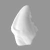
The present 3D Dataset contains the 3D model analyzed in the publication : On Roth’s “human fossil” from Baradero, Buenos Aires Province, Argentina: morphological and genetic analysis. The “human fossil” from Baradero, Buenos Aires Province, Argentina, is a collection of skeleton parts first recovered by Swiss paleontologist Santiago Roth and further studied by anthropologist Rudolf Martin. By the end of the 19th century and beginning of the 20th century it was considered as one of the oldest human skeletons from the southern cone. We studied the cranial anatomy and contextualized the ancient individual remains. We discuss the context of the finding, conducted an osteobiographical assessment and performed a 3D virtual reconstruction of the skull, using micro-CT-scans on selected skull fragments and the mandible. This was followed by the extraction of bone tissue and teeth samples for radiocarbon and genetic analyses, which brought only limited results due to poor preservation and possible contamination. We estimate that the individual from Baradero is a middle-aged adult male. We conclude that the revision of foundational collections with current methodological tools brings new insights and clarifies long held assumptions on the significance of samples that were recovered when archaeology was not yet professionalized.
Homo sapiens PIMUZ A/V 4217 View specimen
|
M3#11983D virtual reconstruction of the skull Type: "3D_surfaces"doi: 10.18563/m3.sf.1198 state:published |
Download 3D surface file |
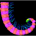
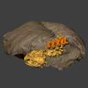
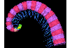
The present 3D Dataset contains the 3D models analyzed in the publication Fossils from the Montceau-les-Mines Lagerstätte (305 Ma) shed light on the anatomy, ecology and phylogeny of Carboniferous millipedes. Authors: Lheritier Mickael, Perroux Maëva, Vannier Jean, Escarguel Gilles, Wesener Thomas, Moritz Leif, Chabard Dominique, Adrien Jerome and Perrier Vincent. Journal of Systematics Palaeontology. https://doi.org/10.1080/14772019.2023.2169891
Amynilyspes fatimae MNHN.F.SOT.2134 View specimen

|
M3#1073Nearly complete specimen. Type: "3D_surfaces"doi: 10.18563/m3.sf.1073 state:published |
Download 3D surface file |
Amynilyspes fatimae MNHN.F.SOT.14983 View specimen
|
M3#1074Nearly complete specimen. Type: "3D_surfaces"doi: 10.18563/m3.sf.1074 state:published |
Download 3D surface file |
Amynilyspes fatimae MNHN.F.SOT.2129 View specimen
|
M3#1075Nearly complete specimen. Type: "3D_surfaces"doi: 10.18563/m3.sf.1075 state:published |
Download 3D surface file |
Blanzilius parriati MNHN.F.SOT.2114A View specimen
|
M3#1076Front part. Type: "3D_surfaces"doi: 10.18563/m3.sf.1076 state:published |
Download 3D surface file |
Blanzilius parriati MNHN.F.SOT.5148 View specimen
|
M3#1077Front part. Type: "3D_surfaces"doi: 10.18563/m3.sf.1077 state:published |
Download 3D surface file |
Blanzilius parriati MNHN.F.SOT.2113 View specimen
|
M3#1078Fragment with legs, sternites and possible tracheal openings. Type: "3D_surfaces"doi: 10.18563/m3.sf.1078 state:published |
Download 3D surface file |
Blanzilius parriati MNHN.F.SOT.81522 View specimen
|
M3#1079Nealry complete specimen. Type: "3D_surfaces"doi: 10.18563/m3.sf.1079 state:published |
Download 3D surface file |
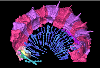
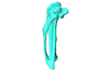
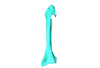
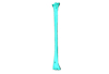
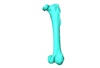
Macroevolution is integral to understanding the patterns of the diversification of life. As the life sciences increasingly use big data approaches, large multivariate datasets are required to test fundamental macroevolutionary hypotheses. In vertebrate evolution, large datasets have been created to quantify morphological variation, largely focusing on particular areas of the skeleton. We provide a landmarking protocol to quantify morphological variation in skeletal elements across the head, trunk, hindlimb and forelimb using 3-dimensional landmarks and semilandmarks, and present a large pan-skeletal database of bird morphology for 149 taxa across avian phylogeny using CT scan data. This large collection of 3D models and geometric morphometric data is open access and can be used in the future for new research, teaching and outreach. The 3D models and CT scans of the 149 specimens related to this project can be downloaded at MorphoSource (https://www.morphosource.org/projects/00000C420)
Menura novaehollandiae FMNH 336751 View specimen
|
M3#5613D model of the left carpometacarpus of the superb lyrebird, Menura novaehollandia (displayed as a mirror image in the 3DHOP viewer). Type: "3D_surfaces"doi: 10.18563/m3.sf.561 state:published |
Download 3D surface file |

|
M3#5623D model of the mandible of the superb lyrebird, Menura novaehollandiae. Type: "3D_surfaces"doi: 10.18563/m3.sf.562 state:published |
Download 3D surface file |
|
M3#5633D model of the right coracoid of the superb lyrebird, Menura novaehollandiae. Type: "3D_surfaces"doi: 10.18563/m3.sf.563 state:published |
Download 3D surface file |
|
M3#5643D model of the right scapula of the superb lyrebird, Menura novaehollandiae. Type: "3D_surfaces"doi: 10.18563/m3.sf.564 state:published |
Download 3D surface file |
|
M3#5653D model of the right tarsometatarsus of the superb lyrebird, Menura novaehollandiae. Type: "3D_surfaces"doi: 10.18563/m3.sf.565 state:published |
Download 3D surface file |
|
M3#5663D model of the sternum of the superb lyrebird, Menura novaehollandiae. Type: "3D_surfaces"doi: 10.18563/m3.sf.566 state:published |
Download 3D surface file |
|
M3#5673D model of the left femur of the superb lyrebird, Menura novaehollandiae (displayed as a mirror image in the 3DHOP viewer). Type: "3D_surfaces"doi: 10.18563/m3.sf.567 state:published |
Download 3D surface file |
|
M3#5683D model of the skull of the superb lyrebird, Menura novaehollandiae. Type: "3D_surfaces"doi: 10.18563/m3.sf.568 state:published |
Download 3D surface file |
|
M3#5693D model of the left humerus of the superb lyrebird, Menura novaehollandiae (displayed as a mirror image in the 3DHOP viewer). Type: "3D_surfaces"doi: 10.18563/m3.sf.569 state:published |
Download 3D surface file |

|
M3#5703D model of the synsacrum of the superb lyrebird, Menura novaehollandiae. Type: "3D_surfaces"doi: 10.18563/m3.sf.570 state:published |
Download 3D surface file |
|
M3#5713D model of the left radius of the superb lyrebird, Menura novaehollandiae (displayed as a mirror image in the 3DHOP viewer). Type: "3D_surfaces"doi: 10.18563/m3.sf.571 state:published |
Download 3D surface file |
|
M3#5723D model of the left tibiotarsus of the superb lyrebird, Menura novaehollandiae (displayed as a mirror image in the 3DHOP viewer). Type: "3D_surfaces"doi: 10.18563/m3.sf.572 state:published |
Download 3D surface file |
|
M3#5733D model of the left ulna of the superb lyrebird, Menura novaehollandiae (displayed as a mirror image in the 3DHOP viewer). Type: "3D_surfaces"doi: 10.18563/m3.sf.573 state:published |
Download 3D surface file |
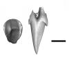
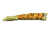
This contribution contains the 3D models described and figured in the following publication: Georgalis, G.L., K.T. Smith, L. Marivaux, A. Herrel, E.M. Essid, H.K. Ammar, W. Marzougui, R. Temani and R. Tabuce. 2024. The world’s largest worm lizard: a new giant trogonophid (Squamata: Amphisbaenia) with extreme dental adaptations from the Eocene of Chambi, Tunisia. Zoological Journal of the Linnean Society. https://doi.org/10.1093/zoolinnean/zlae133
Terastiodontosaurus marcelosanchezi ONM CBI-1-645 View specimen
|
M3#1561Holotype maxilla ONM CBI-1-645 of Terastiodontosaurus marcelosanchezi from the Eocene of Chambi Type: "3D_surfaces"doi: 10.18563/m3.sf.1561 state:published |
Download 3D surface file |
Terastiodontosaurus marcelosanchezi ONM CBI-1-646 View specimen
|
M3#1560Paratype dentary ONM CBI-1-646 of Terastiodontosaurus marcelosanchezi from the Eocene of Chambi Type: "3D_surfaces"doi: 10.18563/m3.sf.1560 state:published |
Download 3D surface file |
Terastiodontosaurus marcelosanchezi ONM CBI-1-648 View specimen
|
M3#1562Maxilla ONM CBI-1-648 of Terastiodontosaurus marcelosanchezi from the Eocene of Chambi Type: "3D_surfaces"doi: 10.18563/m3.sf.1562 state:published |
Download 3D surface file |
Terastiodontosaurus marcelosanchezi ONM CBI-1-649 View specimen
|
M3#1559Maxilla ONM CBI-1-649 of Terastiodontosaurus marcelosanchezi from the Eocene of Chambi Type: "3D_surfaces"doi: 10.18563/m3.sf.1559 state:published |
Download 3D surface file |
Terastiodontosaurus marcelosanchezi ONM CBI-1-650 View specimen
|
M3#1563Maxilla ONM CBI-1-650 of Terastiodontosaurus marcelosanchezi from the Eocene of Chambi Type: "3D_surfaces"doi: 10.18563/m3.sf.1563 state:published |
Download 3D surface file |
Terastiodontosaurus marcelosanchezi ONM CBI-1-651 View specimen
|
M3#1564Maxilla ONM CBI-1-651 of Terastiodontosaurus marcelosanchezi from the Eocene of Chambi Type: "3D_surfaces"doi: 10.18563/m3.sf.1564 state:published |
Download 3D surface file |
Terastiodontosaurus marcelosanchezi ONM CBI-1-653 View specimen
|
M3#1565Maxilla ONM CBI-1-653 of Terastiodontosaurus marcelosanchezi from the Eocene of Chambi Type: "3D_surfaces"doi: 10.18563/m3.sf.1565 state:published |
Download 3D surface file |
Terastiodontosaurus marcelosanchezi ONM CBI-1-654 View specimen
|
M3#1576Maxilla ONM CBI-1-654 of Terastiodontosaurus marcelosanchezi from the Eocene of Chambi Type: "3D_surfaces"doi: 10.18563/m3.sf.1576 state:published |
Download 3D surface file |
Terastiodontosaurus marcelosanchezi ONM CBI-1-657 View specimen

|
M3#1566Dentary ONM CBI-1-657 of Terastiodontosaurus marcelosanchezi from the Eocene of Chambi Type: "3D_surfaces"doi: 10.18563/m3.sf.1566 state:published |
Download 3D surface file |
Terastiodontosaurus marcelosanchezi ONM CBI-1-658 View specimen
|
M3#1567Premaxilla ONM CBI-1-658 of Terastiodontosaurus marcelosanchezi from the Eocene of Chambi Type: "3D_surfaces"doi: 10.18563/m3.sf.1567 state:published |
Download 3D surface file |
Terastiodontosaurus marcelosanchezi ONM CBI-1-659 View specimen
|
M3#1568Dentary ONM CBI-1-659 of Terastiodontosaurus marcelosanchezi from the Eocene of Chambi Type: "3D_surfaces"doi: 10.18563/m3.sf.1568 state:published |
Download 3D surface file |
Terastiodontosaurus marcelosanchezi ONM CBI-1-660 View specimen
|
M3#1569Dentary ONM CBI-1-660 of Terastiodontosaurus marcelosanchezi from the Eocene of Chambi Type: "3D_surfaces"doi: 10.18563/m3.sf.1569 state:published |
Download 3D surface file |
Terastiodontosaurus marcelosanchezi ONM CBI-1-661 View specimen
|
M3#1570Dentary ONM CBI-1-661 of Terastiodontosaurus marcelosanchezi from the Eocene of Chambi Type: "3D_surfaces"doi: 10.18563/m3.sf.1570 state:published |
Download 3D surface file |
Terastiodontosaurus marcelosanchezi ONM CBI-1-668 View specimen
|
M3#1571Dentary ONM CBI-1-668 of Terastiodontosaurus marcelosanchezi from the Eocene of Chambi Type: "3D_surfaces"doi: 10.18563/m3.sf.1571 state:published |
Download 3D surface file |
Terastiodontosaurus marcelosanchezi ONM CBI-1-670 View specimen
|
M3#1572Dentary ONM CBI-1-670 of Terastiodontosaurus marcelosanchezi from the Eocene of Chambi Type: "3D_surfaces"doi: 10.18563/m3.sf.1572 state:published |
Download 3D surface file |
Terastiodontosaurus marcelosanchezi ONM CBI-1-672 View specimen
|
M3#1573Premaxilla ONM CBI-1-672 of Terastiodontosaurus marcelosanchezi from the Eocene of Chambi Type: "3D_surfaces"doi: 10.18563/m3.sf.1573 state:published |
Download 3D surface file |
Terastiodontosaurus marcelosanchezi ONM CBI-1-711 View specimen
|
M3#1574Premaxilla ONM CBI-1-711 of Terastiodontosaurus marcelosanchezi from the Eocene of Chambi Type: "3D_surfaces"doi: 10.18563/m3.sf.1574 state:published |
Download 3D surface file |
Todrasaurus gheerbranti UM THR 407 View specimen
|
M3#1575Holotype dentary UM THR 407 of Todrasaurus gheerbranti Type: "3D_surfaces"doi: 10.18563/m3.sf.1575 state:published |
Download 3D surface file |
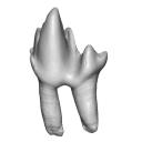
This contribution contains the 3D model described and figured in the following publication: Martin, T., Averianov, A. O., Schultz, J. A., & Schwermann, A. H. (2023). A stem therian mammal from the Lower Cretaceous of Germany. Journal of Vertebrate Paleontology, e2224848.
Spelaeomolitor speratus WMNM P99101 View specimen

|
M3#12573D_model_Spelaeomolitor_lower_molar Type: "3D_surfaces"doi: 10.18563/m3.sf.1257 state:published |
Download 3D surface file |
|
M3#1258CT imagestack (jpgs) and info data sheet (pca file) in one zip folder Type: "3D_CT"doi: 10.18563/m3.sf.1258 state:published |
Download CT data |
The present 3D Dataset contains the 3D models analyzed in the following manuscript: L. Roese-Miron, M.E.H. Jones, J.D. Ferreira and A.S. Hsiou., 2023. Virtual endocasts of Clevosaurus brasiliensis and the tuatara: Rhynchocephalian neuroanatomy and the oldest endocranial record for Lepidosauria.
Sphenodon punctatus CM 30660 View specimen
|
M3#10993D surface model of the cranial endocast of specimen CM 30660 (Sphenodon punctatus). Type: "3D_surfaces"doi: 10.18563/m3.sf.1099 state:published |
Download 3D surface file |
Sphenodon punctatus KCLZJ 001 View specimen
|
M3#11003D surface models of the cranial endocast and the initial trunks of the cranial nerves of specimen KCLZJ 001 (Sphenodon punctatus). Type: "3D_surfaces"doi: 10.18563/m3.sf.1100 state:published |
Download 3D surface file |
Sphenodon punctatus LDUCZ x0036 View specimen
|
M3#11013D surface models of the cranial endocast and the initial trunks of the cranial nerves of specimen LDUCZ x0036 (Sphenodon punctatus). Type: "3D_surfaces"doi: 10.18563/m3.sf.1101 state:published |
Download 3D surface file |
Sphenodon punctatus LDUCZ x1126 View specimen
|
M3#11023D surface model of the cranial endocast of specimen LDUCZ x1126 (Sphenodon punctatus). Type: "3D_surfaces"doi: 10.18563/m3.sf.1102 state:published |
Download 3D surface file |
Clevosaurus brasiliensis MCN PV 2852 View specimen
|
M3#11033D surface model of the cranial endocast of specimen MCN PV 2852 (Clevosaurus brasiliensis). Type: "3D_surfaces"doi: 10.18563/m3.sf.1103 state:published |
Download 3D surface file |
Sphenodon punctatus SAMA 70524 View specimen
|
M3#11043D surface models of the cranial endocast, brain, endosseous labyrinth and initial trunks of the cranial nerves of specimen SAMA 70524 (Sphenodon punctatus). Type: "3D_surfaces"doi: 10.18563/m3.sf.1104 state:published |
Download 3D surface file |
Sphenodon punctatus SU1 View specimen
|
M3#11053D surface models of the cranial endocast and the initial trunks of the cranial nerves of specimen SU1 (Sphenodon punctatus). Type: "3D_surfaces"doi: 10.18563/m3.sf.1105 state:published |
Download 3D surface file |
Sphenodon punctatus YPM HERR 009194 View specimen
|
M3#11063D surface models of the cranial endocast and the initial trunks of the cranial nerves of specimen YPM HERR 009194 (Sphenodon punctatus). Type: "3D_surfaces"doi: 10.18563/m3.sf.1106 state:published |
Download 3D surface file |
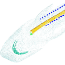
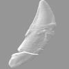
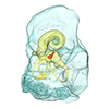
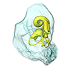
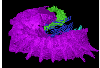
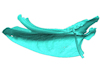
Current knowledge on the skeletogenesis of Chondrichthyes is scarce compared with their extant sister group, the bony fishes. Most of the previously described developmental tables in Chondrichthyes have focused on embryonic external morphology only. Due to its small body size and relative simplicity to raise eggs in laboratory conditions, the small-spotted catshark Scyliorhinus canicula has emerged as a reference species to describe developmental mechanisms in the Chondrichthyes lineage. Here we investigate the dynamic of mineralization in a set of six embryonic specimens using X-ray microtomography and describe the developing units of both the dermal skeleton (teeth and dermal scales) and endoskeleton (vertebral axis). This preliminary data on skeletogenesis in the catshark sets the first bases to a more complete investigation of the skeletal developmental in Chondrichthyes. It should provide comparison points with data known in osteichthyans and could thus be used in the broader context of gnathostome skeletal evolution.
Scyliorhinus canicula SC6_2_2015_03_20 View specimen

|
M3#50Mineralized skeleton of a 6,2 cm long embryo of Scyliorhinus canicula Type: "3D_surfaces"doi: 10.18563/m3.sf.50 state:published |
Download 3D surface file |
Scyliorhinus canicula SC6_7_2015_03_20 View specimen

|
M3#51Mineralized skeleton of a 6,7 cm long embryo of Scyliorhinus canicula Type: "3D_surfaces"doi: 10.18563/m3.sf.51 state:published |
Download 3D surface file |
Scyliorhinus canicula SC7_1_2015_04_03 View specimen

|
M3#52Mineralized skeleton of a 7,1 cm long embryo of Scyliorhinus canicula Type: "3D_surfaces"doi: 10.18563/m3.sf.52 state:published |
Download 3D surface file |
Scyliorhinus canicula SC7_5_2015_03_13 View specimen

|
M3#53Mineralized skeleton of a 7,5 cm long embryo of Scyliorhinus canicula Type: "3D_surfaces"doi: 10.18563/m3.sf.53 state:published |
Download 3D surface file |
Scyliorhinus canicula SC8_2015_03_20 View specimen

|
M3#54Mineralized skeleton of a 8 cm long embryo of Scyliorhinus canicula Type: "3D_surfaces"doi: 10.18563/m3.sf.54 state:published |
Download 3D surface file |
Scyliorhinus canicula SC10_2015_02_27 View specimen

|
M3#55Mineralized skeleton of a 10 cm long embryo of Scyliorhinus canicula Type: "3D_surfaces"doi: 10.18563/m3.sf.55 state:published |
Download 3D surface file |
This contribution contains the three-dimensional models of the most complete and/or informative fossil materials attributed to Peradectes crocheti Gernelle, 2024, the earliest peradectid metatherian species of Europe, from its type locality (Palette, Provence, ~55 Ma). These specimens were analyzed and discussed in: Gernelle et al. (2024), Taxonomy and evolutionary history of peradectids (Metatheria): new data from the early Eocene of France. https://doi.org/10.1007/s10914-024-09724-5
Peradectes crocheti MHN.AIX.PV.2018.26.14 View specimen
|
M3#14993D surface model of MHN.AIX.PV.2018.26.14, fragmentary left maxilla with C-P1, anterior root of P2, and M1-M3 Type: "3D_surfaces"doi: 10.18563/m3.sf.1499 state:published |
Download 3D surface file |
Peradectes crocheti MHN.AIX.PV.2017.6.6 View specimen
|
M3#15003D surface model of MHN.AIX.PV.2017.6.6, left P2 Type: "3D_surfaces"doi: 10.18563/m3.sf.1500 state:published |
Download 3D surface file |
Peradectes crocheti MHN.AIX.PV.2017.6.7 View specimen
|
M3#15013D surface model of MHN.AIX.PV.2017.6.7, left M3 Type: "3D_surfaces"doi: 10.18563/m3.sf.1501 state:published |
Download 3D surface file |
Peradectes crocheti MHN.AIX.PV.2017.6.8 View specimen
|
M3#15023D surface model of MHN.AIX.PV.2017.6.8, right hemi-mandible fragment with canine alveolus, posterior root of p1, partial p2, p3, partial m1, and m2-m3 Type: "3D_surfaces"doi: 10.18563/m3.sf.1502 state:published |
Download 3D surface file |
Peradectes crocheti MHN.AIX.PV.2017.6.9 View specimen
|
M3#15033D surface model of MHN.AIX.PV.2017.6.9, leftm1-m4 row with fragments of dentary Type: "3D_surfaces"doi: 10.18563/m3.sf.1503 state:published |
Download 3D surface file |
Peradectes crocheti MHN.AIX.PV.2017.6.14 View specimen
|
M3#15043D surface model of MHN.AIX.PV.2017.6.14, right astragalus Type: "3D_surfaces"doi: 10.18563/m3.sf.1504 state:published |
Download 3D surface file |
The present 3D Dataset contains 3D models of the holotypes described in Aiglstorfer et al. (2023a). Miocene Moschidae (Mammalia, Ruminantia) from the Linxia Basin (China) connect Europe and Asia and show early evolutionary diversity of a today monogeneric family. Palaeogeography, Palaeoclimatology, Palaeoecology.
Micromeryx? caoi CUGB GV 87045 View specimen
|
M3#11123D models of the holotype of “Micromeryx” caoi (CUGB GV87045) including the models of the teeth, the mandibule, and the sediment. Type: "3D_surfaces"doi: 10.18563/m3.sf.1112 state:published |
Download 3D surface file |
Hispanomeryx linxiaensis IVPP V28596 View specimen
|
M3#11133D models of the holotype of Hispanomeryx linxiaensis (IVPP V28596) including the models of the teeth, the mandibule, and the sediment. Type: "3D_surfaces"doi: 10.18563/m3.sf.1113 state:published |
Download 3D surface file |


