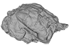
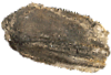
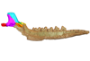
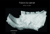
Holotype of Hamadasuchus rebouli
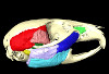
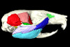
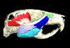
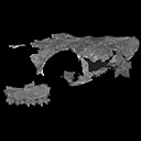
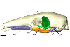
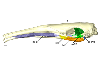
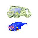
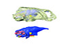
3D models of the endocranial anatomy of Voay robustus and comparative specimens
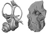
Inner ear morphology in wild vs laboratory mice
3D GM dataset of bird skeletal variation
Skeletal embryonic development in the catshark
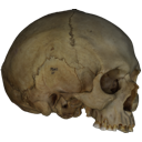
Bony connexions of the petrosal bone of extant hippos
bony labyrinth (11) , inner ear (10) , South America (8) , Eocene (8) , skull (7) , Paleobiogeography (6) , brain (6)
Maëva Judith Orliac (17) , Lionel Hautier (17) , Bastien Mennecart (12) , Pierre-Olivier Antoine (11) , Laurent Marivaux (11) , Leonardo Kerber (10) , Renaud Lebrun (9)
|
3D model and accompanying dataset related to the publication: A new, exceptionally preserved juvenile specimen of Eusaurosphargis dalsassoi (Diapsida) and implications for Mesozoic marine diapsid phylogenyTorsten M. Scheyer
Published online: 30/06/2017 |
|
M3#17994 extracted surfaces of skeletal elements of PIMUZ A/III 4380 Type: "3D_surfaces"doi: 10.18563/m3.sf.179 state:published |
Download 3D surface file |
|
M3#180Accompanying CT scan dataset Type: "3D_CT"doi: 10.18563/m3.sf.180 state:published |
Download CT data |
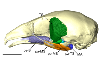
The present 3D Dataset contains the 3D models analyzed in: Amson et al., Under review. Evolutionary Adaptation to Aquatic Lifestyle in Extinct Sloths Can Lead to Systemic Alteration of Bone Structure doi:10.1098/rspb.2018.0270.
Bradypus tridactylus MNHN ZM-MO-1999-1065 View specimen
|
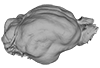
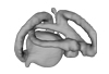
M3#337Brain endocast Type: "3D_surfaces"doi: 10.18563/m3.sf.337 state:published |
Download 3D surface file |
Choloepus didactylus MNHN-ZM-MO-1996-594 View specimen
|
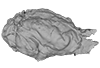
M3#338Brain endocast Type: "3D_surfaces"doi: 10.18563/m3.sf.338 state:published |
Download 3D surface file |
Thalassocnus natans MNHN-F-SAS-734 View specimen
|
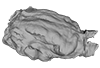
M3#339Brain endocast Type: "3D_surfaces"doi: 10.18563/m3.sf.339 state:published |
Download 3D surface file |
Thalassocnus littoralis MNHN-F-SAS-1610 View specimen
|
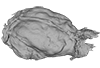
M3#340Brain endocast Type: "3D_surfaces"doi: 10.18563/m3.sf.340 state:published |
Download 3D surface file |
Thalassocnus littoralis MNHN-F-SAS-1615 View specimen
|
M3#341Brain endocast Type: "3D_surfaces"doi: 10.18563/m3.sf.341 state:published |
Download 3D surface file |
Thalassocnus carolomartini SMNK-3814 View specimen
|
M3#342Brain endocast lacking right olfactory bulb Type: "3D_surfaces"doi: 10.18563/m3.sf.342 state:published |
Download 3D surface file |
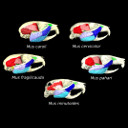
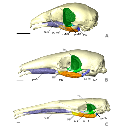
The present 3D Dataset contains the 3D models analyzed in the article entitled "One skull to rule them all? Descriptive and comparative anatomy of the masticatory apparatus in five mice species based on traditional and digital dissections" (Ginot et al. 2018, Journal of Morphology, https://doi.org/10.1002/jmor.20845).
Mus cervicolor R7314 View specimen
|
M3#343.ply surfaces of the skull and masticatory muscles of Mus cervicolor. Created with MorphoDig, .pos and .ntw files also included. Scans were obtained thanks to the Institut des Sciences de l'Evolution de Montpellier MRI platform. Type: "3D_surfaces"doi: 10.18563/m3.sf.343 state:published |
Download 3D surface file |
Mus caroli R7264 View specimen
|
M3#344.ply surfaces of the skull and masticatory muscles of Mus caroli. Created with MorphoDig, .pos and .ntw files also included. Scans were obtained thanks to the Institut des Sciences de l'Evolution de Montpellier MRI platform. Type: "3D_surfaces"doi: 10.18563/m3.sf.344 state:published |
Download 3D surface file |
Mus fragilicauda R7260 View specimen
|
M3#345.ply surfaces of the skull and masticatory muscles of Mus fragilicauda. Created with MorphoDig, .pos and .ntw files also included. Scans were obtained thanks to the Institut des Sciences de l'Evolution de Montpellier MRI platform. Type: "3D_surfaces"doi: 10.18563/m3.sf.345 state:published |
Download 3D surface file |
Mus pahari R7226 View specimen
|
M3#346.ply surfaces of the skull and masticatory muscles of Mus pahari. Created with MorphoDig, .pos and .ntw files also included. Scans were obtained thanks to the Institut des Sciences de l'Evolution de Montpellier MRI platform. Type: "3D_surfaces"doi: 10.18563/m3.sf.346 state:published |
Download 3D surface file |
Mus minutoides minutoides-1 View specimen
|
M3#347.ply surfaces of the skull and masticatory muscles of Mus minutoides. Created with MorphoDig, .pos and .ntw files also included. Scans were obtained thanks to the Institut des Sciences de l'Evolution de Montpellier MRI platform. Type: "3D_surfaces"doi: 10.18563/m3.sf.347 state:published |
Download 3D surface file |
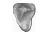
This contribution contains the 3D models of the isolated teeth of Canaanimico amazonensis, a new stem platyrrhine primate, described and figured in the following publication: Marivaux et al. (2016), Neotropics provide insights into the emergence of New World monkeys: new dental evidence from the late Oligocene of Peruvian Amazonia. Journal of Human Evolution. http://dx.doi.org/10.1016/j.jhevol.2016.05.011
Canaanimico amazonensis MUSM-2499 View specimen
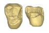
|
M3#2893D model of left upper M2 Type: "3D_surfaces"doi: 10.18563/m3.sf.289 state:published |
Download 3D surface file |
Canaanimico amazonensis MUSM-2500 View specimen
|
M3#2903D model of left upper M1 (lingual part) Type: "3D_surfaces"doi: 10.18563/m3.sf.290 state:published |
Download 3D surface file |
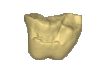
This contribution contains the 3D models described and figured in: The Neogene record of northern South American native ungulates. Smithsonian Contributions to Paleobiology. Doi: 10.5479/si.1943-6688.101
Hilarcotherium miyou IGMp 881327 View specimen
|
M3#318Right upper M2 Type: "3D_surfaces"doi: 10.18563/m3.sf.318 state:published |
Download 3D surface file |
Hilarcotherium miyou MUN-STRI 34216 View specimen
|
M3#319Right upper P4 Type: "3D_surfaces"doi: 10.18563/m3.sf.319 state:published |
Download 3D surface file |
|
M3#320Right upper M2 Type: "3D_surfaces"doi: 10.18563/m3.sf.320 state:published |
Download 3D surface file |
Falcontoxodon aguilerai AMU-CURS 585 View specimen
|
M3#321Maxilla with left M3-P2 and right I2 Type: "3D_surfaces"doi: 10.18563/m3.sf.321 state:published |
Download 3D surface file |
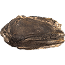
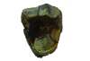
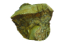
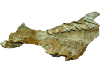
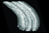
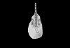
The presented dataset contains the 3D surface scan of the holotype of Birgeria americana, a partial skull described and depicted in: Romano, C., Jenks, J.F., Jattiot, R., Scheyer, T.M., Bylund, K.G. & Bucher, H. 2017. Marine Early Triassic Actinopterygii from Elko County (Nevada, USA): implications for the Smithian equatorial vertebrate eclipse. Journal of Paleontology. https://doi.org/10.1017/jpa.2017.36 .
Birgeria americana NMMNH P-66225 View specimen
|
M3#175NMMNH P-66225 is from upper lower Smithian to lower upper Smithian beds (Thaynes Group). The collecting site is located about 2.75 km south-southeast of the Winecup Ranch, east-central Elko County, Nevada, USA. P-66225 is a partial skull preserved within a large limestone nodule, with its right side exposed. It preserves the portion between the cleithrum posteriorly, and the level of the hind margin of the orbital opening anteriorly. The fossil has a length of 26 cm. Type: "3D_surfaces"doi: 10.18563/m3.sf.175 state:published |
Download 3D surface file |
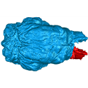
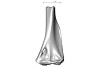
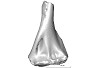
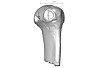
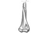
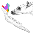
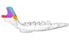
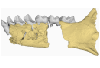
This contribution contains the 3D models described and figured in the following publication: Mennecart B., de Perthuis Ad., Rössner G.E., Guzmán J.A., de Perthuis Au., Costeur L. The first French tragulid skull (Mammalia, Ruminantia, Tragulidae) and associated tragulid remains from the Middle Miocene of Contres (Loir-et-Cher, France). Comptes Rendus Palévol. https://doi.org/10.1016/j.crpv.2017.08.004
Dorcatherium crassum NMB Fa.213.abg View specimen
|
M3#181The 3D surface files of the specimen NMB Fa.213 are the reconstructions of the main skull fragments, the right petrosal bone, and the left bony labyrinth. Type: "3D_surfaces"doi: 10.18563/m3.sf.181 state:published |
Download 3D surface file |
This contribution contains the 3D models described and figured in the following publications:
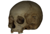
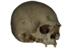
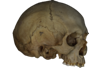
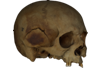
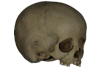
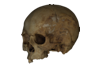
- Marini E., Lussu P., 2020. A virtual physical anthropology lab. Teaching in the time of coronavirus, in prep.;
- Lussu P., Bratzu D., Marini E., 2020. Cloud-based ultra close-range digital photogrammetry: validation of an approach for the effective virtual reconstruction of skeletal remains, in prep.
Homo sapiens MSAE 59 View specimen

|
M3#509MSAE 59 Type: "3D_surfaces"doi: 10.18563/m3.sf.509 state:published |
Download 3D surface file |
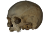
Homo sapiens MSAE 62 View specimen
|
M3#510MSAE 62 Type: "3D_surfaces"doi: 10.18563/m3.sf.510 state:published |
Download 3D surface file |
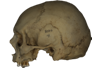
Homo sapiens MSAE 63 View specimen
|
M3#512MSAE 63 Type: "3D_surfaces"doi: 10.18563/m3.sf.512 state:published |
Download 3D surface file |
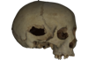
Homo sapiens MSAE 78 View specimen
|
M3#514MSAE 78 Type: "3D_surfaces"doi: 10.18563/m3.sf.514 state:published |
Download 3D surface file |
Homo sapiens MSAE 95 View specimen
|
M3#515MSAE 95 Type: "3D_surfaces"doi: 10.18563/m3.sf.515 state:published |
Download 3D surface file |
Homo sapiens MSAE 1852 View specimen
|
M3#516MSAE 1852 Type: "3D_surfaces"doi: 10.18563/m3.sf.516 state:published |
Download 3D surface file |
Homo sapiens MSAE 6426 View specimen
|
M3#517MSAE 6426 Type: "3D_surfaces"doi: 10.18563/m3.sf.517 state:published |
Download 3D surface file |
Homo sapiens MSAE 6428 View specimen
|
M3#518MSAE 6428 Type: "3D_surfaces"doi: 10.18563/m3.sf.518 state:published |
Download 3D surface file |
Homo sapiens MSAE 6992 View specimen
|
M3#519MSAE 6992 Type: "3D_surfaces"doi: 10.18563/m3.sf.519 state:published |
Download 3D surface file |
Homo sapiens MSAE 7688 View specimen
|
M3#520MSAE 7688 Type: "3D_surfaces"doi: 10.18563/m3.sf.520 state:published |
Download 3D surface file |
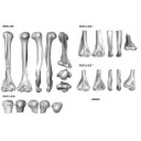
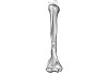
The present 3D Dataset contains the 3D models analyzed in the publication “Systematic and locomotor diversification of the Adapis group (Primates, Adapiformes) in the late Eocene of the Quercy (Southwest France), revealed by humeral remains”. In this paper, twenty humeral specimens from the old and new Quercy collections attributed to the fossil primates Adapis and Palaeolemur are described and analysed together. In this dataset only the scans of the fossils belonging to the collections of Université de Montpellier are provided.
In our paper (Marigó et al., 2019) we provide a qualitative and quantitative analysis of the different humeri, revealing that high variability is present within the “Adapis group” sample. Six different morphotypes are identified, confirming that what has often been called “Adapis parisiensis” is a mix of different species that present different locomotor adaptations.
Adapis sp. UM ROS 2-95 View specimen
|
M3#356Complete right humerus ROS 2-95 attributed to the Adapis group Type: "3D_surfaces"doi: 10.18563/m3.sf.356 state:published |
Download 3D surface file |
Adapis sp. UM ROS 2-536 View specimen
|
M3#357Proximal end of the right humerus ROS 2-536 attributed to the Adapis group Type: "3D_surfaces"doi: 10.18563/m3.sf.357 state:published |
Download 3D surface file |
Adapis sp. UM ROS 2-534 View specimen
|
M3#358Distal end of the left humerus ROS 2-534 attributed to the Adapis group Type: "3D_surfaces"doi: 10.18563/m3.sf.358 state:published |
Download 3D surface file |
Adapis sp. UM ROS 2-535 View specimen
|
M3#359Distal end of the left humerus ROS 2-535 attributed to the Adapis group Type: "3D_surfaces"doi: 10.18563/m3.sf.359 state:published |
Download 3D surface file |
Adapis sp. UM ROS 2-80 View specimen
|
M3#360Proximal end of the right humerus ROS 2-80 attributed to the Adapis group Type: "3D_surfaces"doi: 10.18563/m3.sf.360 state:published |
Download 3D surface file |
Adapis sp. UM ROS 2-79 View specimen
|
M3#361Distal end of the right humerus ROS 2-79 attributed to the Adapis group Type: "3D_surfaces"doi: 10.18563/m3.sf.361 state:published |
Download 3D surface file |
Adapis sp. UM ECA 1364 View specimen
|
M3#362Distal end of the left humerus ECA 1364 attributed to the Adapis group Type: "3D_surfaces"doi: 10.18563/m3.sf.362 state:published |
Download 3D surface file |
Adapis sp. UM ACQ-262 View specimen

|
M3#3733D model of ACQ 262. Humerus Type: "3D_surfaces"doi: 10.18563/m3.sf373 state:published |
Download 3D surface file |
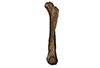
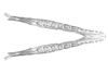
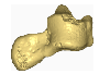
This note presents the 3D model of the hemi-mandible UM-PAT 159 of the MP7 Diacodexis species D. cf. gigasei and 3D models corresponding to the restoration of the ascending ramus, broken on the original specimen, and to a restoration of a complete mandible based on the preserved left hemi-mandible.
Diacodexis cf. gigasei UMPAT159 View specimen
|
M3#3153D models of UM PAT 159 after the restoration of the ascending ramus Type: "3D_surfaces"doi: 10.18563/m3.sf.315 state:published |
Download 3D surface file |
|
M3#316restoration of a complete mandible based on the preserved left hemi-mandible UM PAT 159 Type: "3D_surfaces"doi: 10.18563/m3.sf.316 state:published |
Download 3D surface file |
|
M3#3173D model of the hemi-mandible UM PAT 159 Type: "3D_surfaces"doi: 10.18563/m3.sf.317 state:published |
Download 3D surface file |
This contribution contains the 3D models of the fossil remains (maxilla, dentary, and talus) attributed to Djebelemur martinezi, a ca. 50 Ma primate from Tunisia (Djebel Chambi), described and figured in the following publication: Marivaux et al. (2013), Djebelemur, a tiny pre-tooth-combed primate from the Eocene of Tunisia: a glimpse into the origin of crown strepsirhines. PLoS ONE. https://doi.org/10.1371/journal.pone.0080778
Djebelemur martinezi CBI-1-544 View specimen
|
M3#365CBI-1-544, left maxilla preserving P3-M3 and alveoli for P2 and C1 Type: "3D_surfaces"doi: 10.18563/m3.sf.365 state:published |
Download 3D surface file |
Djebelemur martinezi CBI-1-567 View specimen
|
M3#363Isolated left upper P4 Type: "3D_surfaces"doi: 10.18563/m3.sf.363 state:published |
Download 3D surface file |
Djebelemur martinezi CBI-1-565-577-587-580 View specimen
|
M3#366- CBI-1-565, a damaged right mandible, which consists of three isolated pieces found together and reassembled here: the anterior part of the dentary bears the p3 and m1, and alveoli for p4, p2 and c, while the posterior part preserves m3 and a portion of the ascending ramus; the m2 was found isolated but in the same small calcareous block treated by acid processing. - CBI-1-577, isolated right lower p4. - CBI-1-587, isolated left lower p2 (reversed). - CBI-1-580, isolated left lower canine (reversed). Type: "3D_surfaces"doi: 10.18563/m3.sf.366 state:published |
Download 3D surface file |
Djebelemur martinezi CBI-1-545 View specimen
|
M3#364Right Talus Type: "3D_surfaces"doi: 10.18563/m3.sf.364 state:published |
Download 3D surface file |
The present 3D Dataset contains the 3D models analyzed in Velazco P. M., Grohé C. 2017. Comparative anatomy of the bony labyrinth of the bats Platalina genovensium (Phyllostomidae, Lonchophyllinae) and Tomopeas ravus (Molossidae, Tomopeatinae). Biotempo 14(2).
Platalina genovensium 278520 View specimen

|
M3#276Right bony labyrinth surface positioned (.PLY) Labels associated (.FLG) Type: "3D_surfaces"doi: 10.18563/m3.sf.276 state:published |
Download 3D surface file |
Tomopeas ravus 278525 View specimen

|
M3#277Right bony labyrinth surface (.PLY) Labels associated (.FLG) Type: "3D_surfaces"doi: 10.18563/m3.sf.277 state:published |
Download 3D surface file |
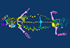
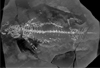
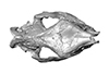
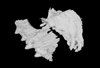
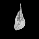
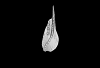
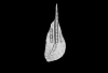
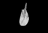
The present 3D Dataset contains the 3D models analyzed in: "a giant dapediid from the Late Triassic of Switzerland and insights into neopterygian phylogeny", Royal Society Open Science, https://doi.org/10.1098/rsos.180497
Scopulipiscis saxciput PIMUZ A/I 3026 View specimen
|
M3#1773D surfaces of the skull and endocranial spaces inside neurocranium, including the aortic canal, braincase, fossa bridgei, lateral cranial canal, nerves and other passageways, notochord, posterior myodome, and right semicircular canals. Type: "3D_surfaces"doi: 10.18563/m3.sf.177 state:published |
Download 3D surface file |

|
M3#178Scan of the neurocranium of PIMUZ A/I 3026 Type: "3D_CT"doi: 10.18563/m3.sf.178 state:published |
Download CT data |
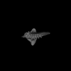
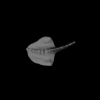
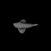
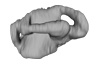
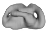
The present 3D Dataset contains the 3D models analyzed in the publication ‘Ontogenetic development of the otic region in the new model organism, Leucoraja erinacea (Chondrichthyes; Rajidae)’, https://doi.org/10.1017/S1755691018000993
Leucoraja erinacea 2018.9.26.1 View specimen
|
M3#3673D model of the right skeletal labyrinth of the adult specimen of Leucoraja erincea. T Type: "3D_surfaces"doi: 10.18563/m3.sf.367 state:published |
Download 3D surface file |
Leucoraja erinacea 2018.9.25.2 View specimen
|
M3#3683D model of the right skeletal labyrinth of the stage 34 specimen of Leucoraja erincea. Type: "3D_surfaces"doi: 10.18563/m3.sf.368 state:published |
Download 3D surface file |
Leucoraja erinacea 2018.9.25.3 View specimen

|
M3#3693D model of the right skeletal labyrinth of the stage 32 specimen of Leucoraja erinacea. Type: "3D_surfaces"doi: 10.18563/m3.sf.369 state:published |
Download 3D surface file |
|
M3#3723D model of the right membranous system of stage 32 of Leucoraja erincea. Type: "3D_surfaces"doi: 10.18563/m3.sf.372 state:published |
Download 3D surface file |
Leucoraja erinacea 2018.9.25.4 View specimen
|
M3#3703D model of the right skeletal labyrinth of the stage 31 specimen of Leucoraja erinacea. Type: "3D_surfaces"doi: 10.18563/m3.sf.370 state:published |
Download 3D surface file |
Leucoraja erinacea 2018.9.26.5 View specimen

|
M3#3763D model of the right skeletal labyrinth of the stage 29 specimen of Leucoraja erinacea. Type: "3D_surfaces"doi: 10.18563/m3.sf.376 state:published |
Download 3D surface file |
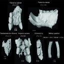
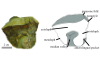
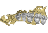
This contribution contains 3D models of extinct rodents Dinomyidae from Miocene and Quaternary of Brazil. The Miocene specimens that were digitalized include the holotypes of Potamarchus adamiae, Pseudopotamarchus villanuevai, and Ferigolomys pacarana collected in the Solimões Formation (Upper Miocene), northern Brazil. The Quaternary specimens are the holotype and paratype of Niedemys piauiensis, found in Upper Pleistocene deposits from northeast Brazil.
Potamarchus adamiae UFAC-CS 011 View specimen
|
M3#410UFAC-CS 011 – holotype, palatal region of the skull with cheek teeth Type: "3D_surfaces"doi: 10.18563/m3.sf.410 state:published |
Download 3D surface file |
Potamarchus adamiae UFAC-CS 043 View specimen
|
M3#411UFAC-CS 043, left dentary with cheek teeth Type: "3D_surfaces"doi: 10.18563/m3.sf.411 state:published |
Download 3D surface file |
Pseudopotamarchus villanuevai UFAC 4762 View specimen
|
M3#412UFAC 4762 – holotype, incomplete right maxilla with cheek teeth Type: "3D_surfaces"doi: 10.18563/m3.sf.412 state:published |
Download 3D surface file |
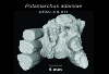
Ferigolomys pacarana UFAC 6460 View specimen
|
M3#413UFAC 6460 – holotype, palatal region of the skull with cheek teeth Type: "3D_surfaces"doi: 10.18563/m3.sf.413 state:published |
Download 3D surface file |
Drytomomys sp. UFAC 2742 View specimen

|
M3#414UFAC 2742, right dentary with cheek teeth Type: "3D_surfaces"doi: 10.18563/m3.sf.414 state:published |
Download 3D surface file |
Niedemys piauiensis FUMDHAM 113-146365-2 View specimen
|
M3#418FUMDHAM 113-146365-2 - holotype, upper right tooth Type: "3D_surfaces"doi: 10.18563/m3.sf.418 state:published |
Download 3D surface file |
Niedemys piauiensis FUMDHAM 113-145304-2 View specimen
|
M3#419FUMDHAM 113-145304-2 - paratype, left lower molar Type: "3D_surfaces"doi: 10.18563/m3.sf.419 state:published |
Download 3D surface file |
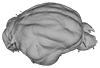
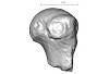
This contribution provides for the first time the 3D model of the type specimen of Molassitherium delemontense (Mammalia, Rhinocerotidae) described in the following publication: Becker et al. (2013), Journal of Systematic Palaeontology, Vol. 11, Issue 8, 947–972, https://doi.org/10.1080/14772019.2012.699007. Conservation issues of the specimen and solutions using 3D model and 3D prints are detailed.
Molassitherium delemontense MJSN POI007–245 View specimen
|
M3#384Skull of Molassitherium delemontense Becker and Antoine, 2013 (in Becker et al. 2013): holotype Type: "3D_surfaces"doi: 10.18563/m3.sf.384 state:published |
Download 3D surface file |
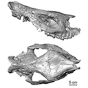
The present 3D Dataset contains the 3D model analyzed in the following publication: Solé et al. (2018), Niche partitioning of the European carnivorous mammals during the paleogene. Palaios. https://doi.org/10.2110/palo.2018.022
Hyaenodon leptorhynchus FSL848325 View specimen
|
M3#336The specimen FSL848325 is separated in two fragments: the anterior part bears the incisors, the deciduous and permanent canines, while the posterior part bears the right P3, P4, M1 and M2. The P2 is isolated. When combined, the cranium length is approximatively 10.5 cm long. The anterior part is 6.9 cm long and 2.15 cm wide (taken at the level of the P1). The posterior part is 4.8 cm long. The anterior part of the cranium is very narrow. Type: "3D_surfaces"doi: 10.18563/m3.sf.336 state:published |
Download 3D surface file |
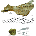
The present 3D Dataset contains the 3D models of the holotype and the paratypes of the new species Siphonodella leiosa described and analyzed in the following publication: L. Souquet, C. Corradini, C. Girard: Siphonodella leiosa (Conodonta), a new unornamented species from the Tournaisian (lower Carboniferous) of Puech de la Suque (Montagne Noire, France). Geobios, https://doi.org/10.1016/j.geobios.2020.06.004.
Siphonodella leiosa UM PSQ 1 View specimen
|
M3#525Siphonodella leiosa, paratype, dextral P1 element Type: "3D_surfaces"doi: 10.18563/m3.sf.525 state:published |
Download 3D surface file |
Siphonodella leiosa UM PSQ 2 View specimen
|
M3#526Siphonodella leiosa, holotype, dextral P1 element Type: "3D_surfaces"doi: 10.18563/m3.sf.526 state:published |
Download 3D surface file |
Siphonodella leiosa UM PSQ 3 View specimen

|
M3#527Siphonodella leiosa, paratype, dextral P1 element Type: "3D_surfaces"doi: 10.18563/m3.sf.527 state:published |
Download 3D surface file |
Siphonodella leiosa UM PSQ 4 View specimen
|
M3#528Siphonodella leiosa, paratype, dextral P1 element Type: "3D_surfaces"doi: 10.18563/m3.sf.528 state:published |
Download 3D surface file |
Siphonodella leiosa UM PSQ 5 View specimen
|
M3#529Siphonodella leiosa, paratype, sinistral P1 element Type: "3D_surfaces"doi: 10.18563/m3.sf.529 state:published |
Download 3D surface file |
Siphonodella leiosa UM PSQ 6 View specimen
|
M3#530Siphonodella leiosa, paratype, dextral P1 element Type: "3D_surfaces"doi: 10.18563/m3.sf.530 state:published |
Download 3D surface file |
Siphonodella leiosa UM PSQ 7 View specimen
|
M3#531Siphonodella leiosa, paratype, dextral P1 element Type: "3D_surfaces"doi: 10.18563/m3.sf.531 state:published |
Download 3D surface file |
Siphonodella leiosa UM PSQ 8 View specimen
|
M3#532Siphonodella leiosa, paratype, sinistral P1 element Type: "3D_surfaces"doi: 10.18563/m3.sf.532 state:published |
Download 3D surface file |
Siphonodella leiosa UM PSQ 9 View specimen
|
M3#533Siphonodella leiosa, paratype, dextral P1 element Type: "3D_surfaces"doi: 10.18563/m3.sf.533 state:published |
Download 3D surface file |
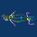
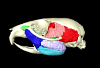
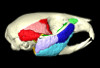
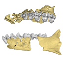
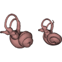
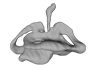
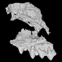
The present 3D Dataset contains the 3D models described in “Comparative masticatory myology in anteaters and its implications for interpreting morphological convergence in myrmecophagous placentals”.
Cyclopes didactylus M1571_JAG View specimen
|
M3#522Skull, mandible, and muscles of Cyclopes didactylus Type: "3D_surfaces"doi: 10.18563/m3.sf.522 state:published |
Download 3D surface file |
Tamandua tetradactyla M3075_JAG View specimen
|
M3#524Skull, left mandibles, and muscles of Tamandua tetradactyla. Type: "3D_surfaces"doi: 10.18563/m3.sf.524 state:published |
Download 3D surface file |
Myrmecophaga tridactyla M3023_JAG View specimen
|
M3#523Skull, left mandible and muscles of Myrmecophaga tridactyla. Type: "3D_surfaces"doi: 10.18563/m3.sf.523 state:published |
Download 3D surface file |
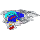
This contribution contains the 3D model described and figured in the following publication: New turtles from the Late Cretaceous of Monte Alto-SP, Brazil, including cranial osteology, neuroanatomy and phylogenetic position of a new taxon. PalZ. https://doi.org/10.1007/s12542-017-0397-x
Yuraramirim montealtensis 04-0008/89 View specimen
|
M3#2783D surfaces related to specimen MPMA 04-0008/89. Type: "3D_surfaces"doi: 10.18563/m3.sf.278 state:published |
Download 3D surface file |