Holotype of Hamadasuchus rebouli
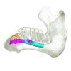
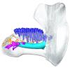
3D models of the endocranial anatomy of Voay robustus and comparative specimens
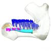
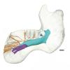
Inner ear morphology in wild vs laboratory mice
3D GM dataset of bird skeletal variation
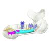
Skeletal embryonic development in the catshark
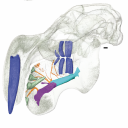
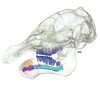
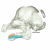
Bony connexions of the petrosal bone of extant hippos
bony labyrinth (11) , inner ear (10) , Eocene (8) , South America (8) , skull (7) , Oligocene (6) , phylogeny (6)
Maëva Judith Orliac (17) , Lionel Hautier (17) , Bastien Mennecart (12) , Laurent Marivaux (11) , Pierre-Olivier Antoine (11) , Leonardo Kerber (10) , Rodolphe Tabuce (9)
|
A surface scan of the "Tübingen Steinkern", Holotype of Proganochelys quenstedtii (Testudinata), with some historical remarks.Ingmar Werneburg
Published online: 08/08/2022 |
|
M3#967This the surface model of the steinkern of the shell of Proganochelys quenstedtii. Type: "3D_surfaces"doi: 10.18563/m3.sf.967 state:published |
Download 3D surface file |
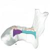
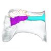
This contribution contains the 3D models described and figured in the following publication: Hautier L, Gomes Rodrigues H, Ferreira-Cardoso S, Emerling CA, Porcher M-L, Asher R, Portela Miguez R, Delsuc F. 2023. From teeth to pad: tooth loss and development of keratinous structures in sirenians. Proceedings of the Royal Society B. https://doi.org/10.1098/rspb.2023.1932
Dugong dugon 2005.51 View specimen

|
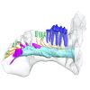
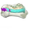
M3#1275Internal mandibular morphology. Orange = dorsal canaliculi; purple = mental branches; cyan = mandibular canal; dark blue = teeth; green = tooth alveoli. Type: "3D_surfaces"doi: 10.18563/m3.sf.1275 state:published |
Download 3D surface file |
Dugong dugon 2023.66 View specimen
|
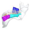
M3#1274Internal mandibular morphology. Orange = dorsal canaliculi; purple = mental branches; cyan = mandibular canal; dark blue = teeth; green = tooth alveoli. Type: "3D_surfaces"doi: 10.18563/m3.sf.1274 state:published |
Download 3D surface file |
Dugong dugon 5386 View specimen
|
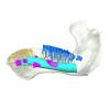
M3#1276Internal mandibular morphology. Orange = dorsal canaliculi; purple = mental branches; cyan = mandibular canal; dark blue = teeth; green = tooth alveoli. Type: "3D_surfaces"doi: 10.18563/m3.sf.1276 state:published |
Download 3D surface file |
Dugong dugon 1848.8.29.7/GERM 1027g View specimen
|
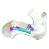
M3#1277Internal mandibular morphology. Orange = dorsal canaliculi; purple = mental branches; cyan = mandibular canal; dark blue = teeth. Type: "3D_surfaces"doi: 10.18563/m3.sf.1277 state:published |
Download 3D surface file |
Dugong dugon 1991.413 View specimen
|
M3#1278Internal mandibular morphology. Orange = dorsal canaliculi; purple = mental branches; cyan = mandibular canal; dark blue = teeth. Type: "3D_surfaces"doi: 10.18563/m3.sf.1278 state:published |
Download 3D surface file |
Dugong dugon 1991.427 View specimen

|
M3#1279Internal mandibular morphology. Orange = dorsal canaliculi; purple = mental branches; cyan = mandibular canal; dark blue = teeth. Type: "3D_surfaces"doi: 10.18563/m3.sf.1279 state:published |
Download 3D surface file |
Dugong dugon 2017-3-9 View specimen
|
M3#1280Internal mandibular morphology. Orange = dorsal canaliculi; purple = mental branches; cyan = mandibular canal; dark blue = teeth. Type: "3D_surfaces"doi: 10.18563/m3.sf.1280 state:published |
Download 3D surface file |
Eosiren lybica 1913-22 View specimen
|
M3#1281Internal mandibular morphology. Orange = dorsal canaliculi; purple = mental branches; cyan = mandibular canal; dark blue = teeth; green = tooth alveoli. Type: "3D_surfaces"doi: 10.18563/m3.sf.1281 state:published |
Download 3D surface file |
Halitherium taulannense RGHP C001 View specimen
|
M3#1282Internal mandibular morphology. Cyan = mandibular canal; dark blue = teeth. Type: "3D_surfaces"doi: 10.18563/m3.sf.1282 state:published |
Download 3D surface file |
Halitherium taulannense RGHP C009 View specimen
|
M3#1283Internal mandibular morphology. Orange = dorsal canaliculi; purple = mental branches; cyan = mandibular canal; dark blue = teeth; green = tooth alveoli. Type: "3D_surfaces"doi: 10.18563/m3.sf.1283 state:published |
Download 3D surface file |
Hydrodamalis gigas 1947.10.21.1 View specimen
|
M3#1284Internal mandibular morphology. Orange = dorsal canaliculi; purple = mental branches; cyan = mandibular canal Type: "3D_surfaces"doi: 10.18563/m3.sf.1284 state:published |
Download 3D surface file |
Hydrodamalis gigas C1021 View specimen
|
M3#1285Anterior part of the mandible Type: "3D_surfaces"doi: 10.18563/m3.sf.1285 state:published |
Download 3D surface file |

|
M3#1286Posterior part of the mandible Type: "3D_surfaces"doi: 10.18563/m3.sf.1286 state:published |
Download 3D surface file |
Hydrodamalis gigas 2023.67 View specimen
|
M3#1287Internal mandibular morphology. Orange = dorsal canaliculi; purple = mental branches; cyan = mandibular canal Type: "3D_surfaces"doi: 10.18563/m3.sf.1287 state:published |
Download 3D surface file |
Libysiren sickenbergi M.82429 View specimen
|
M3#1288Internal mandibular morphology. Orange = dorsal canaliculi; purple = mental branches; cyan = mandibular canal; dark blue = teeth; green = tooth alveoli. Type: "3D_surfaces"doi: 10.18563/m3.sf.1288 state:published |
Download 3D surface file |
Libysiren sickenbergi M.45675 View specimen
|
M3#1289Internal mandibular morphology. Orange = dorsal canaliculi; purple = mental branches; cyan = mandibular canal; dark blue = teeth; green = tooth alveoli. Type: "3D_surfaces"doi: 10.18563/m3.sf.1289 state:published |
Download 3D surface file |
Prorastomus sirenoides OR.448976 View specimen
|
M3#1290Internal morphology of the left mandible. Orange = dorsal canaliculi; purple = mental branches; cyan = mandibular canal; dark blue = teeth; green = tooth alveoli. Type: "3D_surfaces"doi: 10.18563/m3.sf.1290 state:published |
Download 3D surface file |
|
M3#1304Internal morphology of the right mandible. Orange = dorsal canaliculi; purple = mental branches; cyan = mandibular canal; dark blue = teeth. Type: "3D_surfaces"doi: 10.18563/m3.sf.1304 state:published |
Download 3D surface file |
Ribodon limbatus M.7073 View specimen
|
M3#1292Internal mandibular morphology. Orange = dorsal canaliculi; purple = mental branches; cyan = mandibular canal; dark blue = teeth; green = tooth alveoli. Type: "3D_surfaces"doi: 10.18563/m3.sf.1292 state:published |
Download 3D surface file |
Rytiodus capgrandi PAL2017-8-1 View specimen
|
M3#1293Internal mandibular morphology. Orange = dorsal canaliculi; purple = mental branches; cyan = mandibular canal; dark blue = teeth; green = tooth alveoli. Type: "3D_surfaces"doi: 10.18563/m3.sf.1293 state:published |
Download 3D surface file |
Trichechus inunguis 1868.12.19.2 View specimen
|
M3#1294Internal mandibular morphology. Orange = dorsal canaliculi; purple = mental branches; cyan = mandibular canal; dark blue = teeth; green = tooth alveoli. Type: "3D_surfaces"doi: 10.18563/m3.sf.1294 state:published |
Download 3D surface file |
Trichechus manatus 1843.3.10.12 View specimen
|
M3#1295Internal mandibular morphology. Orange = dorsal canaliculi; purple = mental branches; cyan = mandibular canal; dark blue = teeth. Type: "3D_surfaces"doi: 10.18563/m3.sf.1295 state:published |
Download 3D surface file |
Trichechus manatus 1864.6.5.1 View specimen
|
M3#1296Internal mandibular morphology. Orange = dorsal canaliculi; purple = mental branches; cyan = mandibular canal; dark blue = teeth; green = tooth alveoli. Type: "3D_surfaces"doi: 10.18563/m3.sf.1296 state:published |
Download 3D surface file |
Trichechus manatus 1950.1.23.1 View specimen
|
M3#1297Internal mandibular morphology. Orange = dorsal canaliculi; purple = mental branches; cyan = mandibular canal; dark blue = teeth; green = tooth alveoli. Type: "3D_surfaces"doi: 10.18563/m3.sf.1297 state:published |
Download 3D surface file |
Trichechus senegalensis 1885.6.30.2 View specimen
|
M3#1298Internal mandibular morphology. Orange = dorsal canaliculi; purple = mental branches; cyan = mandibular canal; dark blue = teeth; green = tooth alveoli. Type: "3D_surfaces"doi: 10.18563/m3.sf.1298 state:published |
Download 3D surface file |
Trichechus senegalensis 1894.7.25.8 View specimen
|
M3#1299Internal mandibular morphology. Orange = dorsal canaliculi; purple = mental branches; cyan = mandibular canal; dark blue = teeth. Type: "3D_surfaces"doi: 10.18563/m3.sf.1299 state:published |
Download 3D surface file |
Trichechus senegalensis V97 View specimen
|
M3#1302Mandibular internal morphology. Orange = dorsal canaliculi; purple = mental branches; cyan = mandibular canal; dark blue = teeth. Type: "3D_surfaces"doi: 10.18563/m3.sf.1302 state:published |
Download 3D surface file |
Trichechus sp. 65.4.28.9 View specimen
|
M3#1300Internal mandibular morphology. Orange = dorsal canaliculi; purple = mental branches; cyan = mandibular canal; dark blue = teeth; green = tooth alveoli. Type: "3D_surfaces"doi: 10.18563/m3.sf.1300 state:published |
Download 3D surface file |
Dugong dugon 1946.8.6.2 View specimen
|
M3#1301Mandibular internal morphology. Orange = dorsal canaliculi; purple = mental branches; cyan = mandibular canal; dark blue = teeth. Type: "3D_surfaces"doi: 10.18563/m3.sf.1301 state:published |
Download 3D surface file |
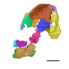
The present 3D dataset contains the 3D models of the holotype of Proterochampsa nodosa that were built and analysed in “Redescription, taxonomic revaluation, and phylogenetic affinities of Proterochampsa nodosa (Archosauriformes: Proterochampsidae), early Late Triassic of Candelaria Sequence (Santa Maria Supersequence)”.
Proterochampsa nodosa MCP 1694-PV View specimen
|
M3#9743D models of Proterochampsa nodosa. Model 1: skull. Model 2: mandible. Model 3: left mandibular ramus. Type: "3D_surfaces"doi: 10.18563/m3.sf.974 state:published |
Download 3D surface file |
The present 3D Dataset contains the 3D models analyzed in the following manuscript: L. Roese-Miron, M.E.H. Jones, J.D. Ferreira and A.S. Hsiou., 2023. Virtual endocasts of Clevosaurus brasiliensis and the tuatara: Rhynchocephalian neuroanatomy and the oldest endocranial record for Lepidosauria.
Sphenodon punctatus CM 30660 View specimen
|
M3#10993D surface model of the cranial endocast of specimen CM 30660 (Sphenodon punctatus). Type: "3D_surfaces"doi: 10.18563/m3.sf.1099 state:published |
Download 3D surface file |
Sphenodon punctatus KCLZJ 001 View specimen
|
M3#11003D surface models of the cranial endocast and the initial trunks of the cranial nerves of specimen KCLZJ 001 (Sphenodon punctatus). Type: "3D_surfaces"doi: 10.18563/m3.sf.1100 state:published |
Download 3D surface file |
Sphenodon punctatus LDUCZ x0036 View specimen
|
M3#11013D surface models of the cranial endocast and the initial trunks of the cranial nerves of specimen LDUCZ x0036 (Sphenodon punctatus). Type: "3D_surfaces"doi: 10.18563/m3.sf.1101 state:published |
Download 3D surface file |
Sphenodon punctatus LDUCZ x1126 View specimen
|
M3#11023D surface model of the cranial endocast of specimen LDUCZ x1126 (Sphenodon punctatus). Type: "3D_surfaces"doi: 10.18563/m3.sf.1102 state:published |
Download 3D surface file |
Clevosaurus brasiliensis MCN PV 2852 View specimen
|
M3#11033D surface model of the cranial endocast of specimen MCN PV 2852 (Clevosaurus brasiliensis). Type: "3D_surfaces"doi: 10.18563/m3.sf.1103 state:published |
Download 3D surface file |
Sphenodon punctatus SAMA 70524 View specimen
|
M3#11043D surface models of the cranial endocast, brain, endosseous labyrinth and initial trunks of the cranial nerves of specimen SAMA 70524 (Sphenodon punctatus). Type: "3D_surfaces"doi: 10.18563/m3.sf.1104 state:published |
Download 3D surface file |
Sphenodon punctatus SU1 View specimen
|
M3#11053D surface models of the cranial endocast and the initial trunks of the cranial nerves of specimen SU1 (Sphenodon punctatus). Type: "3D_surfaces"doi: 10.18563/m3.sf.1105 state:published |
Download 3D surface file |
Sphenodon punctatus YPM HERR 009194 View specimen
|
M3#11063D surface models of the cranial endocast and the initial trunks of the cranial nerves of specimen YPM HERR 009194 (Sphenodon punctatus). Type: "3D_surfaces"doi: 10.18563/m3.sf.1106 state:published |
Download 3D surface file |
The present 3D Dataset contains the 3D model of the skin of Allosaurus described in Hendrickx, C. et al. in press. Morphology and distribution of scales, dermal ossifications, and other non-feather integumentary structures in non-avialan theropod dinosaurs. Biological Reviews.
Allosaurus jimmadseni UMNH VP C481 View specimen
|
M3#902The material consists of a 3D reconstruction of the counterpart of a 30 cm2 patch of skin impression associated with the anterior dorsal ribs/pectoral region of the specimen of Allosaurus jimmadseni UMNH VP C481. The skin shows a semi-uniform basement of 1-2 mm diameter pebbles with a smaller number of slightly larger (up to 3 mm) ovoid scales. The irregular shape, distribution, and overall small size of these larger scales suggest that they are not classifiable as feature scales but rather as variations in the basement scales. Type: "3D_surfaces"doi: 10.18563/m3.sf.902 state:published |
Download 3D surface file |
The present 3D Dataset contains the 3D model analyzed in Solé F., Lesport J.-F., Heitz A., and Mennecart B. minor revision. A new gigantic carnivore (Carnivora, Amphicyonidae) from the late middle Miocene of France. PeerJ.
Tartarocyon cazanavei MHNBx 2020.20.1 View specimen
|
M3#903Surface scan (ply) and texture (png) of the holotype of Tartarocyon cazanavei (MHNBx 2020.20.1) Type: "3D_surfaces"doi: 10.18563/m3.sf.903 state:published |
Download 3D surface file |
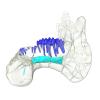
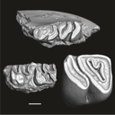
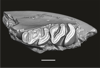
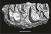
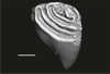
This contribution contains 3D models of upper molar rows of house mice (Mus musculus domesticus) belonging to Western European commensal and Sub-Antarctic feral populations. These two groups are characterized by different patterns of wear and alignment of the three molars along the row, related to contrasted masticatory demand in relation with their diet. These models are analyzed in the following publication: Renaud et al 2023, “Molar wear in house mice, insight into diet preferences at an ecological time scale?”, https://doi.org/10.1093/biolinnean/blad091
Mus musculus G09_06 View specimen
|
M3#1166right upper molar row Type: "3D_surfaces"doi: 10.18563/m3.sf.1166 state:published |
Download 3D surface file |
Mus musculus G09_10 View specimen
|
M3#1168right upper molar row Type: "3D_surfaces"doi: 10.18563/m3.sf.1168 state:published |
Download 3D surface file |
Mus musculus G09_15 View specimen
|
M3#1169right upper molar row Type: "3D_surfaces"doi: 10.18563/m3.sf.1169 state:published |
Download 3D surface file |
Mus musculus G09_16 View specimen
|
M3#1170right upper molar row Type: "3D_surfaces"doi: 10.18563/m3.sf.1170 state:published |
Download 3D surface file |
Mus musculus G09_17 View specimen
|
M3#1171right upper molar row Type: "3D_surfaces"doi: 10.18563/m3.sf.1171 state:published |
Download 3D surface file |
Mus musculus G09_21 View specimen
|
M3#1172right upper molar row Type: "3D_surfaces"doi: 10.18563/m3.sf.1172 state:published |
Download 3D surface file |
Mus musculus G09_26 View specimen
|
M3#1173right upper molar row Type: "3D_surfaces"doi: 10.18563/m3.sf.1173 state:published |
Download 3D surface file |
Mus musculus G09_27 View specimen
|
M3#1174right upper molar row Type: "3D_surfaces"doi: 10.18563/m3.sf.1174 state:published |
Download 3D surface file |
Mus musculus G09_29 View specimen
|
M3#1175right upper molar row Type: "3D_surfaces"doi: 10.18563/m3.sf.1175 state:published |
Download 3D surface file |
Mus musculus G09_65 View specimen
|
M3#1176right upper molar row Type: "3D_surfaces"doi: 10.18563/m3.sf.1176 state:published |
Download 3D surface file |
Mus musculus G09_66 View specimen
|
M3#1177right upper molar row Type: "3D_surfaces"doi: 10.18563/m3.sf.1177 state:published |
Download 3D surface file |
Mus musculus G93_03 View specimen
|
M3#1178right upper molar row Type: "3D_surfaces"doi: 10.18563/m3.sf.1178 state:published |
Download 3D surface file |
Mus musculus G93_04 View specimen
|
M3#1179right upper molar row Type: "3D_surfaces"doi: 10.18563/m3.sf.1179 state:published |
Download 3D surface file |
Mus musculus G93_10 View specimen
|
M3#1180right upper molar row Type: "3D_surfaces"doi: 10.18563/m3.sf.1180 state:published |
Download 3D surface file |
Mus musculus G93_11 View specimen
|
M3#1181right upper molar row Type: "3D_surfaces"doi: 10.18563/m3.sf.1181 state:published |
Download 3D surface file |
Mus musculus G93_13 View specimen
|
M3#1182right upper molar row Type: "3D_surfaces"doi: 10.18563/m3.sf.1182 state:published |
Download 3D surface file |
Mus musculus G93_14 View specimen
|
M3#1183right upper molar row Type: "3D_surfaces"doi: 10.18563/m3.sf.1183 state:published |
Download 3D surface file |
Mus musculus G93_15 View specimen
|
M3#1184right upper molar row Type: "3D_surfaces"doi: 10.18563/m3.sf.1184 state:published |
Download 3D surface file |
Mus musculus G93_24 View specimen
|
M3#1185left molar row Type: "3D_surfaces"doi: 10.18563/m3.sf.1185 state:published |
Download 3D surface file |
Mus musculus Tourch_7819 View specimen
|
M3#1186right upper molar row Type: "3D_surfaces"doi: 10.18563/m3.sf.1186 state:published |
Download 3D surface file |
Mus musculus G93_25 View specimen
|
M3#1187right upper molar row Type: "3D_surfaces"doi: 10.18563/m3.sf.1187 state:published |
Download 3D surface file |
Mus musculus Tourch_7821 View specimen
|
M3#1188right upper molar row Type: "3D_surfaces"doi: 10.18563/m3.sf.1188 state:published |
Download 3D surface file |
Mus musculus Tourch_7839 View specimen
|
M3#1189right upper molar row Type: "3D_surfaces"doi: 10.18563/m3.sf.1189 state:published |
Download 3D surface file |
Mus musculus Tourch_7873 View specimen
|
M3#1190right upper molar row Type: "3D_surfaces"doi: 10.18563/m3.sf.1190 state:published |
Download 3D surface file |
Mus musculus Tourch_7877 View specimen
|
M3#1196right upper molar row Type: "3D_surfaces"doi: 10.18563/m3.sf.1196 state:published |
Download 3D surface file |
Mus musculus Tourch_7922 View specimen
|
M3#1191right upper molar row Type: "3D_surfaces"doi: 10.18563/m3.sf.1191 state:published |
Download 3D surface file |
Mus musculus Tourch_7923 View specimen
|
M3#1192right upper molar row Type: "3D_surfaces"doi: 10.18563/m3.sf.1192 state:published |
Download 3D surface file |
Mus musculus Tourch_7925 View specimen
|
M3#1193right upper molar row Type: "3D_surfaces"doi: 10.18563/m3.sf.1193 state:published |
Download 3D surface file |
Mus musculus Tourch_7927 View specimen
|
M3#1194right upper molar row Type: "3D_surfaces"doi: 10.18563/m3.sf.1194 state:published |
Download 3D surface file |
Mus musculus Tourch_7932 View specimen
|
M3#1195right upper molar row Type: "3D_surfaces"doi: 10.18563/m3.sf.1195 state:published |
Download 3D surface file |
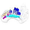
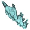
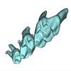
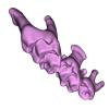
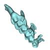
The present 3D Dataset contains the 3D models of extant Chiropteran endocranial casts, documenting 16 of the 19 extant bat families. They are used by Maugoust & Orliac (2023) to assess the correspondences between the brain and brain-surrounding tissues (i.e., neural tissues, blood vessels, meninges) and their imprint on the braincase, allowing for eventually proposing a Chiroptera-scale nomenclature of the endocast.
Balantiopteryx plicata UMMZ 102659 View specimen
|
M3#1132Endocranial cast of the corresponding cranium of Balantiopteryx plicata Type: "3D_surfaces"doi: 10.18563/m3.sf.1132 state:published |
Download 3D surface file |
Nycteris macrotis AMNH M-187705 View specimen
|
M3#1133Endocranial cast of the corresponding cranium of Nycteris macrotis Type: "3D_surfaces"doi: 10.18563/m3.sf.1133 state:published |
Download 3D surface file |
Thyroptera tricolor UMMZ 53240 View specimen
|
M3#1134Endocranial cast of the corresponding cranium of Thyroptera tricolor Type: "3D_surfaces"doi: 10.18563/m3.sf.1134 state:published |
Download 3D surface file |
Noctilio albiventris UMMZ 105827 View specimen
|
M3#1135Endocranial cast of the corresponding cranium of Noctilio albiventris Type: "3D_surfaces"doi: 10.18563/m3.sf.1135 state:published |
Download 3D surface file |
Mormoops blainvillii AMNH M-271513 View specimen
|
M3#1136Endocranial cast of the corresponding cranium of Mormoops blainvillii Type: "3D_surfaces"doi: 10.18563/m3.sf.1136 state:published |
Download 3D surface file |
Macrotus waterhousii UMMZ 95718 View specimen
|
M3#1137Endocranial cast of the corresponding cranium of Macrotus waterhousii Type: "3D_surfaces"doi: 10.18563/m3.sf.1137 state:published |
Download 3D surface file |
Nyctiellus lepidus UMMZ 105767 View specimen
|
M3#1138Endocranial cast of the corresponding cranium of Nyctiellus lepidus Type: "3D_surfaces"doi: 10.18563/m3.sf.1138 state:published |
Download 3D surface file |
Cheiromeles torquatus AMNH M-247585 View specimen
|
M3#1139Endocranial cast of the corresponding cranium of Cheiromeles torquatus Type: "3D_surfaces"doi: 10.18563/m3.sf.1139 state:published |
Download 3D surface file |
Miniopterus schreibersii UMMZ 156998 View specimen
|
M3#1140Endocranial cast of the corresponding cranium of Miniopterus schreibersii Type: "3D_surfaces"doi: 10.18563/m3.sf.1140 state:published |
Download 3D surface file |
Kerivoula pellucida UMMZ 161396 View specimen
|
M3#1141Endocranial cast of the corresponding cranium of Kerivoula pellucida Type: "3D_surfaces"doi: 10.18563/m3.sf.1141 state:published |
Download 3D surface file |
Scotophilus kuhlii UMMZ 157013 View specimen
|
M3#1142Endocranial cast of the corresponding cranium of Scotophilus kuhlii Type: "3D_surfaces"doi: 10.18563/m3.sf.1142 state:published |
Download 3D surface file |
Rhinolophus luctus MNHN CG-2006-87 View specimen
|
M3#1143Endocranial cast of the corresponding cranium of Rhinolophus luctus Type: "3D_surfaces"doi: 10.18563/m3.sf.1143 state:published |
Download 3D surface file |
Triaenops persicus AM RG-38552 View specimen
|
M3#1144Endocranial cast of the corresponding cranium of Triaenops persicus Type: "3D_surfaces"doi: 10.18563/m3.sf.1144 state:published |
Download 3D surface file |
Hipposideros armiger UM ZOOL-762-V View specimen
|
M3#1145Endocranial cast of the corresponding cranium of Hipposideros armiger Type: "3D_surfaces"doi: 10.18563/m3.sf.1145 state:published |
Download 3D surface file |
Lavia frons AM RG-12268 View specimen
|
M3#1146Endocranial cast of the corresponding cranium of Lavia frons Type: "3D_surfaces"doi: 10.18563/m3.sf.1146 state:published |
Download 3D surface file |
Rhinopoma hardwickii AM RG-M31166 View specimen
|
M3#1147Endocranial cast of the corresponding cranium of Rhinopoma hardwickii Type: "3D_surfaces"doi: 10.18563/m3.sf.1147 state:published |
Download 3D surface file |
Sphaerias blanfordi AMNH M-274330 View specimen
|
M3#1148Endocranial cast of the corresponding cranium of Sphaerias blanfordi Type: "3D_surfaces"doi: 10.18563/m3.sf.1148 state:published |
Download 3D surface file |
Rousettus aegyptiacus UMMZ 161026 View specimen
|
M3#1149Endocranial cast of the corresponding cranium of Rousettus aegyptiacus Type: "3D_surfaces"doi: 10.18563/m3.sf.1149 state:published |
Download 3D surface file |
Pteropus pumilus UMMZ 162253 View specimen
|
M3#1150Endocranial cast of the corresponding cranium of Pteropus pumilus Type: "3D_surfaces"doi: 10.18563/m3.sf.1150 state:published |
Download 3D surface file |
The present 3D Dataset contains the 3D models analyzed in: Abel P., Pommery Y., Ford D. P., Koyabu D., Werneburg I. 2022. Skull sutures and cranial mechanics in the Permian reptile Captorhinus aguti and the evolution of the temporal region in early amniotes. Frontiers in Ecology and Evolution. https://doi.org/10.3389/fevo.2022.841784
Captorhinus aguti OMNH 44816 View specimen
|
M3#965Segmented cranial bone surfaces of OMNH 44816 Type: "3D_surfaces"doi: 10.18563/m3.sf.965 state:published |
Download 3D surface file |
This contribution contains the 3D model of an endocranial cast analyzed in “A 10 ka intentionally deformed human skull from Northeast Asia”. There are many studies on the morphological characteristics of intentional cranial deformation (ICD), but few related 3D models were published. Here, we present the surface model of an intentionally deformed 10 ka human cranium for further research on ICD practice. The 3D model of the endocranial cast of this ICD cranium was discovered near Harbin City, Province Heilongjiang, Northeast China. The fossil preserved only the frontal, parietal, and occipital bones. To complete the endocast model of the specimen, we printed a 3D model and used modeling clay to reconstruct the missing part based on the general form of the modern human endocast morphology.
Homo sapiens IVPP-PA1616 View specimen
|
M3#972The frontal region of the endocast is flattened, probably formed by the constant pressure on the frontal bone during growth. There is a well-developed frontal crest on the endocranial surface. The endocast widens posteriorly from the frontal lobe. The widest point of the endocast is at the lateral border of the parietal lobe. The lower parietal areas display a marked lateral expansion. The overall shape of the endocast is asymmetrical, with the left side of the parietal lobe being more laterally expanded than the right side. Like the frontal lobe, the occipital lobe is also anteroposteriorly flattened. Type: "3D_surfaces"doi: 10.18563/m3.sf.972 state:published |
Download 3D surface file |
|
M3#976The original endocranial cast model (with texture) of IVPP-PA1616. It shows the original structures of the specimen, and was not altered in any way. Type: "3D_surfaces"doi: 10.18563/m3.sf.976 state:published |
Download 3D surface file |
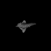
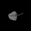
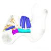
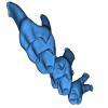
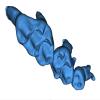
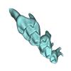
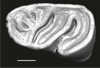
This contribution contains the three-dimensional models of the inner ear of the hetaxodontid rodents Amblyrhiza, Clidomys and Elasmodontomys from the West Indies. These specimens were analyzed and discussed in : The inner ear of caviomorph rodents: phylogenetic implications and application to extinct West Indian taxa.
Amblyrhiza inundata 11842 View specimen
|
M3#11543D surface of the left-oriented inner ear of Amblyrhiza. Type: "3D_surfaces"doi: 10.18563/m3.sf.1154 state:published |
Download 3D surface file |
Clidomys sp NA View specimen
|
M3#11553D surface of the left-oriented inner ear of Clidomys sp. Type: "3D_surfaces"doi: 10.18563/m3.sf.1155 state:published |
Download 3D surface file |
Elasmodontomys obliquus 17127 View specimen
|
M3#11563D surface of the left-oriented inner ear of Elasmodontomys obliquus. Type: "3D_surfaces"doi: 10.18563/m3.sf.1156 state:published |
Download 3D surface file |
The present 3D Dataset contains the 3D model analyzed in Gaetano, L. C., Abdala, F., Seoane, F. D., Tartaglione, A., Schulz, M., Otero, A., Leardi, J. M., Apaldetti, C., Krapovickas, V., and Steinbach, E. 2021. A new cynodont from the Upper Triassic Los Colorados Formation (Argentina, South America) reveals a novel paleobiogeographic context for mammalian ancestors. Scientific Reports.
Tessellatia bonapartei PULR-V121 View specimen
|
M3#9603D surface model of PULR-V121 Type: "3D_surfaces"doi: 10.18563/m3.sf.960 state:published |
Download 3D surface file |
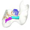
This contribution contains the 3D models described and figured in the following publication: Bonis, L. de, Grohé, C., Surault, J., Gardin, A. 2022. Description of the first cranium and endocranial structures of Stenoplesictis minor (Mammalia, Carnivora), an early aeluroid from the Oligocene of the Quercy Phosphorites (southwestern France). Historical Biology. https://doi.org/10.1080/08912963.2022.2045980
Stenoplesictis minor UM-ACQ 6705 View specimen
|
M3#961Endocranium Type: "3D_surfaces"doi: 10.18563/m3.sf.961 state:published |
Download 3D surface file |
|
M3#962Right bony labyrinth Type: "3D_surfaces"doi: 10.18563/m3.sf.962 state:published |
Download 3D surface file |
|
M3#963Left bony labyrinth Type: "3D_surfaces"doi: 10.18563/m3.sf.963 state:published |
Download 3D surface file |
|
M3#964Cranium in transparency with endocranial structures Type: "3D_surfaces"doi: 10.18563/m3.sf.964 state:published |
Download 3D surface file |
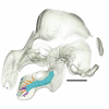
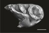
The present 3D Dataset contains 3D models of the cranium surface and of the bony labyrinth endocast of the stem bat Vielasia sigei. They are used by (Hand et al., 2023) to explore the phylogenetic position of this species, to infer its laryngeal echolocating capabilities, and to eventually discuss chiropteran evolution before the crown clade diversification.
Vielasia sigei UM VIE-250 View specimen
|
M3#1269External surface of the cranium Type: "3D_surfaces"doi: 10.18563/m3.sf.1269 state:published |
Download 3D surface file |
|
M3#1270Virtual endocast of the right bony labyrinth Type: "3D_surfaces"doi: 10.18563/m3.sf.1270 state:published |
Download 3D surface file |

The present 3D Dataset contains the 3D model analyzed in the publication : On Roth’s “human fossil” from Baradero, Buenos Aires Province, Argentina: morphological and genetic analysis. The “human fossil” from Baradero, Buenos Aires Province, Argentina, is a collection of skeleton parts first recovered by Swiss paleontologist Santiago Roth and further studied by anthropologist Rudolf Martin. By the end of the 19th century and beginning of the 20th century it was considered as one of the oldest human skeletons from the southern cone. We studied the cranial anatomy and contextualized the ancient individual remains. We discuss the context of the finding, conducted an osteobiographical assessment and performed a 3D virtual reconstruction of the skull, using micro-CT-scans on selected skull fragments and the mandible. This was followed by the extraction of bone tissue and teeth samples for radiocarbon and genetic analyses, which brought only limited results due to poor preservation and possible contamination. We estimate that the individual from Baradero is a middle-aged adult male. We conclude that the revision of foundational collections with current methodological tools brings new insights and clarifies long held assumptions on the significance of samples that were recovered when archaeology was not yet professionalized.
Homo sapiens PIMUZ A/V 4217 View specimen
|
M3#11983D virtual reconstruction of the skull Type: "3D_surfaces"doi: 10.18563/m3.sf.1198 state:published |
Download 3D surface file |
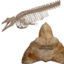
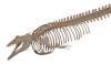
The present 3D Dataset contains the 3D models analyzed in Bianucci et al. 2023, A heavyweight early whale pushes the boundaries of vertebrate morphology, Nature. These include bones of the holotype of new species Perucetus colossus (MUSM 3248), as well as the articulated skeleton of Cynthiacetus peruvianus (holotype, MNHN.F.PRU10). The latter was used to estimate the total skeleton volume of P. colossus.
Perucetus colossus MUSM 3248 View specimen
|
M3#1131Thirteen vertebrae, rib, and innominate of Perucetus colossus (holotype, MUSM NNNN). Type: "3D_surfaces"doi: 10.18563/m3.sf.1131 state:published |
Download 3D surface file |
Cynthiacetus peruvianus MNHN.F.PRU10 View specimen
|
M3#1130Articulated skeleton of the holotype of Cynthiacetus peruvianus MNHN.F.PRU10 Type: "3D_surfaces"doi: 10.18563/m3.sf.1130 state:published |
Download 3D surface file |
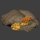
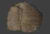
This contribution contains the three-dimensional digital models of a part of the dental fossil material (the large specimens) of caviomorph rodents, discovered in late middle Miocene detrital deposits of the TAR-31 locality in Peruvian Amazonia (San Martín, Peru). These fossils were described, figured and discussed in the following publication: Boivin, Marivaux et al. (2021), Late middle Miocene caviomorph rodents from Tarapoto, Peruvian Amazonia. PLoS ONE 16(11): e0258455. https://doi.org/10.1371/journal.pone.0258455
Microscleromys paradoxalis MUSM 4643 View specimen
|
M3#1115Fragment of left mandibule preserving dp4, m1 and a portion of incisor Type: "3D_surfaces"doi: 10.18563/m3.sf.1115 state:published |
Download 3D surface file |
Ricardomys longidens MUSM 4375 View specimen
|
M3#1116Fragment of left maxillary preserving DP4 and M1 (or M1 and M2) Type: "3D_surfaces"doi: 10.18563/m3.sf.1116 state:published |
Download 3D surface file |
"Scleromys" sp. MUSM 4272 View specimen
|
M3#1117Isolated left upper molar Type: "3D_surfaces"doi: 10.18563/m3.sf.1117 state:published |
Download 3D surface file |
"Scleromys" sp. MUSM 4275 View specimen
|
M3#1118Isolated right upper molar Type: "3D_surfaces"doi: 10.18563/m3.sf.1118 state:published |
Download 3D surface file |
"Scleromys" sp. MUSM 4273 View specimen
|
M3#1119Isolated left upper molar Type: "3D_surfaces"doi: 10.18563/m3.sf.1119 state:published |
Download 3D surface file |
"Scleromys" sp. MUSM 4276 View specimen
|
M3#1120Isolated right upper molar Type: "3D_surfaces"doi: 10.18563/m3.sf.1120 state:published |
Download 3D surface file |
"Scleromys" sp. MUSM 4282 View specimen
|
M3#1121Isolated right lower molar Type: "3D_surfaces"doi: 10.18563/m3.sf.1121 state:published |
Download 3D surface file |
"Scleromys" sp. MUSM 4281 View specimen
|
M3#1122Isolated right lower molar Type: "3D_surfaces"doi: 10.18563/m3.sf.1122 state:published |
Download 3D surface file |
"Scleromys" sp. MUSM 4280 View specimen
|
M3#1123Isolated left p4 Type: "3D_surfaces"doi: 10.18563/m3.sf.1123 state:published |
Download 3D surface file |
"Scleromys" sp. MUSM 4277 View specimen
|
M3#1124Isolated left lower dp4 Type: "3D_surfaces"doi: 10.18563/m3.sf.1124 state:published |
Download 3D surface file |
"Scleromys" sp. MUSM 4279 View specimen
|
M3#1125Isolated right lower dp4 (mesial fragment) Type: "3D_surfaces"doi: 10.18563/m3.sf.1125 state:published |
Download 3D surface file |
gen.indet sp. indet MUSM 4283 View specimen
|
M3#1126Isolated right lower p4 Type: "3D_surfaces"doi: 10.18563/m3.sf.1126 state:published |
Download 3D surface file |
Microscleromys sp. MUSM 4658 View specimen
|
M3#1127Isolated left tarsal bone (astragalus) Type: "3D_surfaces"doi: 10.18563/m3.sf.1127 state:published |
Download 3D surface file |
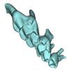
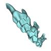
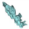
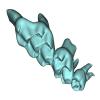
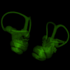
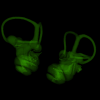
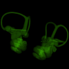
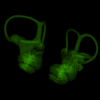
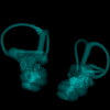
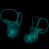
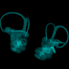
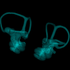
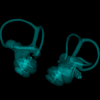
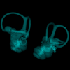
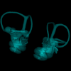
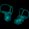
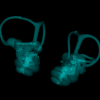
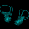
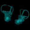
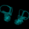
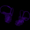
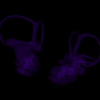
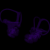
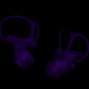
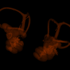
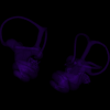
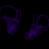
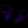
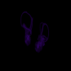
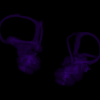
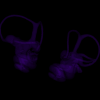
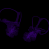
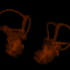
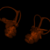
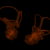
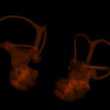
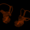
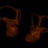
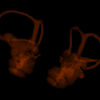
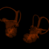
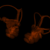
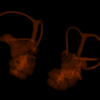
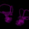
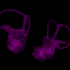
This contribution contains 3D models of left and right house mouse (Mus musculus domesticus) inner ears analyzed in Renaud et al. (2024). The studied mice belong to four groups: wild-trapped mice, wild-derived lab offspring, a typical laboratory strain (Swiss) and hybrids between wild-derived and Swiss mice. They have been analyzed to assess the impact of mobility reduction on inner ear morphology, including patterns of divergence, levels of inter-individual variance (disparity) and intra-individual variance (fluctuating asymmetry)
Mus musculus Tourch_7819 View specimen
|
M3#1366Two bony labyrinths Type: "3D_surfaces"doi: 10.18563/m3.sf.1366 state:in_press |
Download 3D surface file |
Mus musculus Tourch_7821 View specimen
|
M3#1367Two bony labyrinths Type: "3D_surfaces"doi: 10.18563/m3.sf.1367 state:in_press |
Download 3D surface file |
Mus musculus Tourch_7839 View specimen
|
M3#1368Two bony labyrinths Type: "3D_surfaces"doi: 10.18563/m3.sf.1368 state:in_press |
Download 3D surface file |
Mus musculus Tourch_7873 View specimen
|
M3#1369Two bony labyrinths Type: "3D_surfaces"doi: 10.18563/m3.sf.1369 state:in_press |
Download 3D surface file |
Mus musculus Tourch_7877 View specimen
|
M3#1370Two bony labyrinths Type: "3D_surfaces"doi: 10.18563/m3.sf.1370 state:in_press |
Download 3D surface file |
Mus musculus Tourch_7922 View specimen
|
M3#1371Two bony labyrinths Type: "3D_surfaces"doi: 10.18563/m3.sf.1371 state:in_press |
Download 3D surface file |
Mus musculus Tourch_7923 View specimen
|
M3#1372Two bony labyrinths Type: "3D_surfaces"doi: 10.18563/m3.sf.1372 state:in_press |
Download 3D surface file |
Mus musculus Tourch_7925 View specimen
|
M3#1373Two bony labyrinths Type: "3D_surfaces"doi: 10.18563/m3.sf.1373 state:in_press |
Download 3D surface file |
Mus musculus Tourch_7927 View specimen
|
M3#1374Two bony labyrinths Type: "3D_surfaces"doi: 10.18563/m3.sf.1374 state:in_press |
Download 3D surface file |
Mus musculus Tourch_7932 View specimen
|
M3#1375Two bony labyrinths Type: "3D_surfaces"doi: 10.18563/m3.sf.1375 state:in_press |
Download 3D surface file |
Mus musculus Bal02 View specimen
|
M3#1320Two bony labyrinths Type: "3D_surfaces"doi: 10.18563/m3.sf.1320 state:in_press |
Download 3D surface file |
Mus musculus Bal04 View specimen

|
M3#1321Two bony labyrinths Type: "3D_surfaces"doi: 10.18563/m3.sf.1321 state:in_press |
Download 3D surface file |
Mus musculus Bal06 View specimen
|
M3#1322Two bony labyrinths Type: "3D_surfaces"doi: 10.18563/m3.sf.1322 state:in_press |
Download 3D surface file |
Mus musculus Bal08 View specimen
|
M3#1323Two bony labyrinths Type: "3D_surfaces"doi: 10.18563/m3.sf.1323 state:in_press |
Download 3D surface file |
Mus musculus Bal11 View specimen
|
M3#1324Two bony labyrinths Type: "3D_surfaces"doi: 10.18563/m3.sf.1324 state:in_press |
Download 3D surface file |
Mus musculus Bal12 View specimen
|
M3#1325Two bony labyrinths Type: "3D_surfaces"doi: 10.18563/m3.sf.1325 state:in_press |
Download 3D surface file |
Mus musculus Bal15 View specimen
|
M3#1326Two bony labyrinths Type: "3D_surfaces"doi: 10.18563/m3.sf.1326 state:in_press |
Download 3D surface file |
Mus musculus Bal16 View specimen
|
M3#1327Two bony labyrinths Type: "3D_surfaces"doi: 10.18563/m3.sf.1327 state:in_press |
Download 3D surface file |
Mus musculus Bal17 View specimen
|
M3#1328Two bony labyrinths Type: "3D_surfaces"doi: 10.18563/m3.sf.1328 state:in_press |
Download 3D surface file |
Mus musculus Bal18 View specimen
|
M3#1329Two bony labyrinths Type: "3D_surfaces"doi: 10.18563/m3.sf.1329 state:in_press |
Download 3D surface file |
Mus musculus Bal19 View specimen
|
M3#1330Two bony labyrinths Type: "3D_surfaces"doi: 10.18563/m3.sf.1330 state:in_press |
Download 3D surface file |
Mus musculus Bal20 View specimen
|
M3#1331Two bony labyrinths Type: "3D_surfaces"doi: 10.18563/m3.sf.1331 state:in_press |
Download 3D surface file |
Mus musculus Bal21 View specimen
|
M3#1332Two bony labyrinths Type: "3D_surfaces"doi: 10.18563/m3.sf.1332 state:in_press |
Download 3D surface file |
Mus musculus Bal22 View specimen
|
M3#1333Two bony labyrinths Type: "3D_surfaces"doi: 10.18563/m3.sf.1333 state:in_press |
Download 3D surface file |
Mus musculus Bal23 View specimen
|
M3#1334Two bony labyrinths Type: "3D_surfaces"doi: 10.18563/m3.sf.1334 state:in_press |
Download 3D surface file |
Mus musculus Bal24 View specimen
|
M3#1335Two bony labyrinths Type: "3D_surfaces"doi: 10.18563/m3.sf.1335 state:in_press |
Download 3D surface file |
Mus musculus Bal25 View specimen
|
M3#1336Two bony labyrinths Type: "3D_surfaces"doi: 10.18563/m3.sf.1336 state:in_press |
Download 3D surface file |
Mus musculus Balan_LAB_035 View specimen
|
M3#1337Two bony labyrinths Type: "3D_surfaces"doi: 10.18563/m3.sf.1337 state:in_press |
Download 3D surface file |
Mus musculus Balan_LAB_046 View specimen
|
M3#1338Two bony labyrinths Type: "3D_surfaces"doi: 10.18563/m3.sf.1338 state:in_press |
Download 3D surface file |
Mus musculus Balan_LAB_054 View specimen
|
M3#1339Two bony labyrinths Type: "3D_surfaces"doi: 10.18563/m3.sf.1339 state:in_press |
Download 3D surface file |
Mus musculus Balan_LAB_056 View specimen
|
M3#1340Two bony labyrinths Type: "3D_surfaces"doi: 10.18563/m3.sf.1340 state:in_press |
Download 3D surface file |
Mus musculus Balan_LAB_082 View specimen
|
M3#1341Two bony labyrinths Type: "3D_surfaces"doi: 10.18563/m3.sf.1341 state:in_press |
Download 3D surface file |
Mus musculus Balan_LAB_086 View specimen

|
M3#1342Two bony labyrinths Type: "3D_surfaces"doi: 10.18563/m3.sf.1342 state:in_press |
Download 3D surface file |
Mus musculus Balan_LAB_092 View specimen
|
M3#1343Two bony labyrinths Type: "3D_surfaces"doi: 10.18563/m3.sf.1343 state:in_press |
Download 3D surface file |
Mus musculus Balan_LAB_319 View specimen
|
M3#1344Two bony labyrinths Type: "3D_surfaces"doi: 10.18563/m3.sf.1344 state:in_press |
Download 3D surface file |
Mus musculus Balan_LAB_325 View specimen
|
M3#1345Two bony labyrinths Type: "3D_surfaces"doi: 10.18563/m3.sf.1345 state:in_press |
Download 3D surface file |
Mus musculus Balan_LAB_329 View specimen
|
M3#1346Two bony labyrinths Type: "3D_surfaces"doi: 10.18563/m3.sf.1346 state:in_press |
Download 3D surface file |
Mus musculus Balan_LAB_330 View specimen
|
M3#1347Two bony labyrinths Type: "3D_surfaces"doi: 10.18563/m3.sf.1347 state:in_press |
Download 3D surface file |
Mus musculus Balan_LAB_F2b View specimen
|
M3#1348Two bony labyrinths Type: "3D_surfaces"doi: 10.18563/m3.sf.1348 state:in_press |
Download 3D surface file |
Mus musculus Balan_LAB_BB3weeks View specimen
|
M3#1349Two bony labyrinths Type: "3D_surfaces"doi: 10.18563/m3.sf.1349 state:in_press |
Download 3D surface file |
Mus musculus SW0ter View specimen
|
M3#1376Two bony labyrinths Type: "3D_surfaces"doi: 10.18563/m3.sf.1376 state:in_press |
Download 3D surface file |
Mus musculus SW343 View specimen
|
M3#1377Two bony labyrinths Type: "3D_surfaces"doi: 10.18563/m3.sf.1377 state:in_press |
Download 3D surface file |
Mus musculus SW1 View specimen
|
M3#1378Two bony labyrinths Type: "3D_surfaces"doi: 10.18563/m3.sf.1378 state:in_press |
Download 3D surface file |
Mus musculus SW2 View specimen
|
M3#1379Two bony labyrinths Type: "3D_surfaces"doi: 10.18563/m3.sf.1379 state:in_press |
Download 3D surface file |
Mus musculus BAL_F1_30x17_27j View specimen
|
M3#1350Two bony labyrinths Type: "3D_surfaces"doi: 10.18563/m3.sf.1350 state:in_press |
Download 3D surface file |
Mus musculus BAL_F1_167_48j View specimen
|
M3#1351Two bony labyrinths Type: "3D_surfaces"doi: 10.18563/m3.sf.1351 state:in_press |
Download 3D surface file |
Mus musculus BAL_F1_188_32j View specimen
|
M3#1352Two bony labyrinths Type: "3D_surfaces"doi: 10.18563/m3.sf.1352 state:in_press |
Download 3D surface file |
Mus musculus BAL_F1_192_28j View specimen
|
M3#1353Two bony labyrinths Type: "3D_surfaces"doi: 10.18563/m3.sf.1353 state:in_press |
Download 3D surface file |
Mus musculus BAL_F1_194_46j View specimen
|
M3#1354Two bony labyrinths Type: "3D_surfaces"doi: 10.18563/m3.sf.1354 state:in_press |
Download 3D surface file |
Mus musculus BAL_F1_196_44j View specimen
|
M3#1355Two bony labyrinths Type: "3D_surfaces"doi: 10.18563/m3.sf.1355 state:in_press |
Download 3D surface file |
Mus musculus BAL_F2_40x56_24j View specimen

|
M3#1356Two bony labyrinths Type: "3D_surfaces"doi: 10.18563/m3.sf.1356 state:in_press |
Download 3D surface file |
Mus musculus BAL_F2_47x61_22j View specimen
|
M3#1357Two bony labyrinths Type: "3D_surfaces"doi: 10.18563/m3.sf.1357 state:in_press |
Download 3D surface file |
Mus musculus Gardouch_3419 View specimen
|
M3#1358Two bony labyrinths Type: "3D_surfaces"doi: 10.18563/m3.sf.1358 state:in_press |
Download 3D surface file |
Mus musculus Gardouch_3432 View specimen
|
M3#1359Two bony labyrinths Type: "3D_surfaces"doi: 10.18563/m3.sf.1359 state:in_press |
Download 3D surface file |
Mus musculus Gardouch_3437 View specimen
|
M3#1360Two bony labyrinths Type: "3D_surfaces"doi: 10.18563/m3.sf.1360 state:in_press |
Download 3D surface file |
Mus musculus Gardouch_3439 View specimen
|
M3#1361Two bony labyrinths Type: "3D_surfaces"doi: 10.18563/m3.sf.1361 state:in_press |
Download 3D surface file |
Mus musculus Gardouch_3450 View specimen
|
M3#1362Two bony labyrinths Type: "3D_surfaces"doi: 10.18563/m3.sf.1362 state:in_press |
Download 3D surface file |
Mus musculus Gardouch_3453 View specimen
|
M3#1363Two bony labyrinths Type: "3D_surfaces"doi: 10.18563/m3.sf.1363 state:in_press |
Download 3D surface file |
Mus musculus Gardouch_3459 View specimen
|
M3#1364Two bony labyrinths Type: "3D_surfaces"doi: 10.18563/m3.sf.1364 state:in_press |
Download 3D surface file |
Mus musculus Gardouch_3462 View specimen
|
M3#1365Two bony labyrinths Type: "3D_surfaces"doi: 10.18563/m3.sf.1365 state:in_press |
Download 3D surface file |
Mus musculus SW5 View specimen
|
M3#1380Two bony labyrinths Type: "3D_surfaces"doi: 10.18563/m3.sf.1380 state:in_press |
Download 3D surface file |
Mus musculus SWF3 View specimen
|
M3#1381Two bony labyrinths Type: "3D_surfaces"doi: 10.18563/m3.sf.1381 state:in_press |
Download 3D surface file |
Mus musculus SW342 View specimen
|
M3#1382Two bony labyrinths Type: "3D_surfaces"doi: 10.18563/m3.sf.1382 state:in_press |
Download 3D surface file |
Mus musculus SW341 View specimen
|
M3#1383Two bony labyrinths Type: "3D_surfaces"doi: 10.18563/m3.sf.1383 state:in_press |
Download 3D surface file |
Mus musculus SW339 View specimen
|
M3#1384Two bony labyrinths Type: "3D_surfaces"doi: 10.18563/m3.sf.1384 state:in_press |
Download 3D surface file |
Mus musculus SWF4 View specimen
|
M3#1385Two bony labyrinths Type: "3D_surfaces"doi: 10.18563/m3.sf.1385 state:in_press |
Download 3D surface file |
Mus musculus SW0bis_350 View specimen
|
M3#1386Two bony labyrinths Type: "3D_surfaces"doi: 10.18563/m3.sf.1386 state:in_press |
Download 3D surface file |
Mus musculus SW0_348 View specimen
|
M3#1387Two bony labyrinths Type: "3D_surfaces"doi: 10.18563/m3.sf.1387 state:in_press |
Download 3D surface file |
Mus musculus SW347 View specimen
|
M3#1388Two bony labyrinths Type: "3D_surfaces"doi: 10.18563/m3.sf.1388 state:in_press |
Download 3D surface file |
Mus musculus SW345 View specimen
|
M3#1389Two bony labyrinths Type: "3D_surfaces"doi: 10.18563/m3.sf.1389 state:in_press |
Download 3D surface file |
Mus musculus hyb_125xSW_01 View specimen
|
M3#1390Two bony labyrinths Type: "3D_surfaces"doi: 10.18563/m3.sf.1390 state:in_press |
Download 3D surface file |
Mus musculus hyb_125xSW_02 View specimen
|
M3#1391Two bony labyrinths Type: "3D_surfaces"doi: 10.18563/m3.sf.1391 state:in_press |
Download 3D surface file |
Mus musculus hyb_SWx126_01 View specimen
|
M3#1392Two bony labyrinths Type: "3D_surfaces"doi: 10.18563/m3.sf.1392 state:in_press |
Download 3D surface file |
Mus musculus hyb_SWx126_02 View specimen
|
M3#1393Two bony labyrinths Type: "3D_surfaces"doi: 10.18563/m3.sf.1393 state:in_press |
Download 3D surface file |
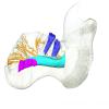
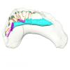
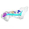
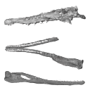
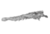
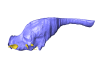
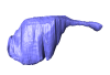
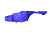
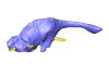
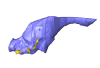
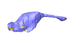
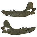
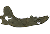
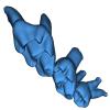
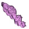
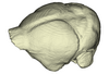
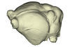
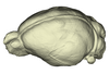
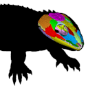
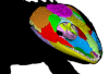
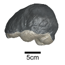
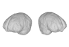
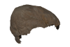
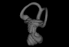
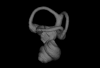
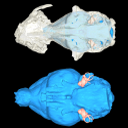
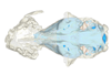
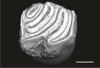
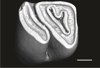
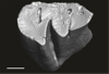
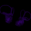
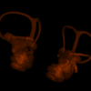
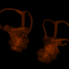
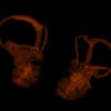
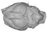
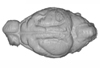
The holotype of Hamadasuchus rebouli Buffetaut 1994 from the Kem Kem beds of Morocco (Late Albian – Cenomanian) consists of a left dentary which is limited, fragmentary and reconstructed in some areas. To aid in assessing if the original diagnosis can be considered as valid, the specimen was CT scanned for the first time. This is especially important to resolve the taxonomic status of certain specimens that have been assigned to Hamadasuchus rebouli since then. The reconstructed structures in this contribution are in agreement with the original description, notably in terms of alveolar count; thus the original diagnosis of this taxon remains valid and some specimens are not referable to H. rebouli anymore.
Hamadasuchus rebouli MDE C001 View specimen

|
M3#1402Dentary and teeth Type: "3D_surfaces"doi: 10.18563/m3.sf.1402 state:in_press |
Download 3D surface file |

|
M3#1403Toothmarks Type: "3D_surfaces"doi: 10.18563/m3.sf.1403 state:in_press |
Download 3D surface file |
The present 3D Dataset contains the 3D models illustrated and described in the chapter “Paleoneurology of Artiodactyla, an overview of the evolution of the artiodactyl brain” (Orliac et al. 2022) published in "Paleoneurology of amniotes: new directions in the study of fossil endocasts", edited by Dozo, Paulina-Carabajal, Macrini and Walsh.
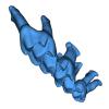
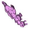
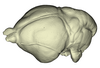
Homacodon vagans AMNH 12695 View specimen
|
M3#1063Endocranial cast Type: "3D_surfaces"doi: 10.18563/m3.sf.1063 state:published |
Download 3D surface file |
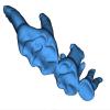
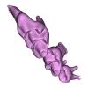
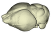
Helohyus sp. AMNH 13079 View specimen
|
M3#1064Endocranial cast Type: "3D_surfaces"doi: 10.18563/m3.sf.1064 state:published |
Download 3D surface file |
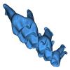
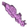
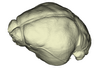
Leptauchenia sp. AMNH 45508 View specimen
|
M3#1065endocranial cast Type: "3D_surfaces"doi: 10.18563/m3.sf.1065 state:published |
Download 3D surface file |
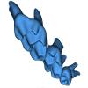
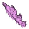
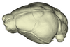
Agriochoerus sp. AMNH 95330 View specimen
|
M3#1067endocranial cast Type: "3D_surfaces"doi: 10.18563/m3.sf.1067 state:published |
Download 3D surface file |
Mouillacitherium elegans UM ACQ 6625 View specimen
|
M3#1068endocranial cast Type: "3D_surfaces"doi: 10.18563/m3.sf.1068 state:published |
Download 3D surface file |
Caenomeryx filholi UM PDS 2570 View specimen
|
M3#1069endocranial cast Type: "3D_surfaces"doi: 10.18563/m3.sf.1069 state:published |
Download 3D surface file |
Dichobune leporina MNHN.F.QU16586 View specimen
|
M3#1070endocranial cast Type: "3D_surfaces"doi: 10.18563/m3.sf.1070 state:published |
Download 3D surface file |
Anoplotherium sp. not numbered View specimen
|
M3#1071endocranial cast Type: "3D_surfaces"doi: 10.18563/m3.sf.1071 state:published |
Download 3D surface file |