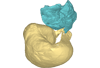
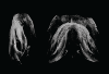
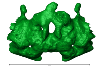
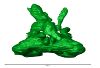
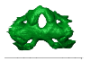
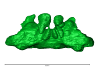
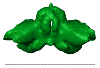
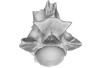
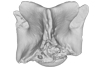
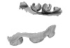
3D models of Cainotheriids Ossicular chain
Explodable 3D Dog Skull for Veterinary Education
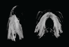
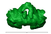
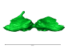
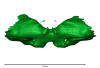
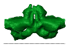
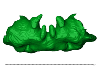
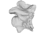
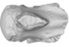
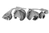
3D models of Kalakocetus, the earliest Cetacea
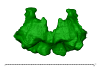
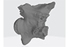
3D GM dataset of bird skeletal variation
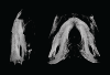
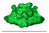
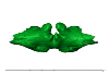
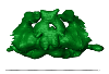
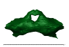
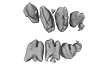
Skeletal embryonic development in the catshark
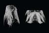
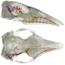
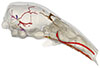
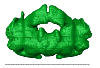
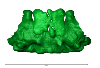
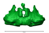
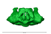
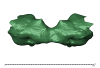
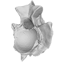
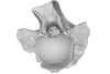
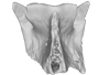
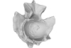
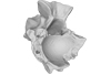
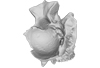
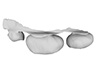
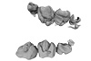
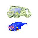
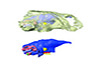
Bony connexions of the petrosal bone of extant hippos
bony labyrinth (14) , inner ear (11) , Eocene (11) , geometric morphometrics (10) , CT-scan (10) , Oligocene (9) , Micro-CT (9)
Lionel Hautier (25) , Maëva Judith Orliac (24) , Laurent Marivaux (18) , Renaud Lebrun (15) , Rodolphe Tabuce (15) , Pierre-Olivier Antoine (13) , Bastien Mennecart (13)
|
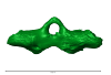
3D models related to the publication: The early evolution of cranial appendages in Bovoidea revealed by new species of Amphimoschus (Mammalia: Ruminantia) from ChinaYi-Kun Li, Bastien Mennecart
Published online: 07/09/2021 |
|
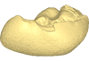
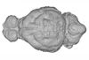
M3#803the holotype, a right hemimandible with tooth row p2 to m3 Type: "3D_surfaces"doi: 10.18563/m3.sf.803 state:published |
Download 3D surface file |
Amphimoschus xishuiensis IVPP V 25521.2 View specimen
|
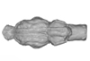
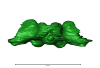
M3#804referred material, anterior part of a skull with right P4-M3 and left P3-M2 Type: "3D_surfaces"doi: 10.18563/m3.sf.804 state:published |
Download 3D surface file |
The present 3D Dataset contains the 3D models analyzed in Mennecart B., Métais G., Costeur L., Ginsburg L, and Rössner G. 2021, Reassessment of the enigmatic ruminant Miocene genus Amphimoschus Bourgeois, 1873 (Mammalia, Artiodactyla, Pecora). PlosOne. https://doi.org/10.1371/journal.pone.0244661
Amphimoschus ponteleviensis MNHN.F.AR3266 View specimen
|
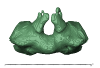
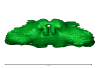
M3#701Surface scan of the cast of the skull of Amphimoschus ponteleviensis MNHN.F.AR3266 from Artenay (France) Type: "3D_surfaces"doi: 10.18563/m3.sf.701 state:published |
Download 3D surface file |
|
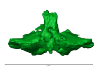
M3#702Right petrosal bone and bony labyrinth of the skull MNHN.F.AR3266 from Artenay (France) Type: "3D_surfaces"doi: 10.18563/m3.sf.702 state:published |
Download 3D surface file |
Amphimoschus ponteleviensis SMNS40693 View specimen
|
M3#704Left petrosal bone and bony labyrinth of the skull SMNS40693 from Langenau 1 (Germany) Type: "3D_surfaces"doi: 10.18563/m3.sf.704 state:published |
Download 3D surface file |
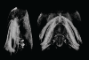
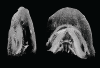
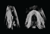
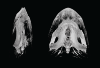
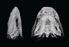
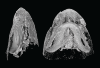
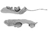
This contribution contains the 3D models described and figured in the following publication: Mourlam, M., Orliac, M. J. (2017), Protocetid (Cetacea, Artiodactyla) bullae and petrosals from the Middle Eocene locality of Kpogamé, Togo: new insights into the early history of cetacean hearing. Journal of Systematic Palaeontology https://doi.org/10.1080/14772019.2017.1328378
?Carolinacetus indet. UM KPG-M 164 View specimen
|
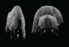
M3#132left petrosal of ?Carolinacetus sp. from the locality of Kpogamé, Togo Type: "3D_surfaces"doi: 10.18563/m3.sf.132 state:published |
Download 3D surface file |
indet. indet. UM KPG-M 73 View specimen

|
M3#133labelled surface of the left petrosal Type: "3D_surfaces"doi: 10.18563/m3.sf.133 state:published |
Download 3D surface file |
|
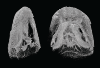
M3#134left bullaof Protocetidae indeterminate from Kpogamé, Togo Type: "3D_surfaces"doi: 10.18563/m3.sf.134 state:published |
Download 3D surface file |
|
M3#135petrotympanic complex of Protocetidae indeterminate from Kpogamé, Togo Type: "3D_surfaces"doi: 10.18563/m3.sf.135 state:published |
Download 3D surface file |
?Carolinacetus indet. UM KPG-M 33 View specimen
|
M3#136left auditory bulla of a juvenile specimen of ?Carolinacetus sp. from Kpogamé, Togo Type: "3D_surfaces"doi: 10.18563/m3.sf.136 state:published |
Download 3D surface file |
Togocetus traversei UM KPG-M 80 View specimen

|
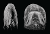
M3#137fragmentary right auditory bulla of Togocetus traversei from Kpogamé, Togo Type: "3D_surfaces"doi: 10.18563/m3.sf.137 state:published |
Download 3D surface file |
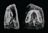
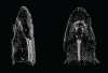
This contribution contains the 3D model described and figured in the following publication: Albino, A., Carrillo-Briceño, J. D. & Neenan, J. M. 2016. An enigmatic aquatic snake from the Cenomanian of northern South America. PeerJ 4:e2027 http://dx.doi.org/10.7717/peerj.2027
Lunaophis aquaticus MCNC-1827-F View specimen
|
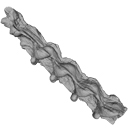
M3#116Articulated precloacal vertebrae of Lunaophis aquaticus Type: "3D_surfaces"doi: 10.18563/m3.sf.116 state:published |
Download 3D surface file |
The present dataset contains the 3D models analyzed in Berio, F., Bayle, Y., Baum, D., Goudemand, N., and Debiais-Thibaud, M. 2022. Hide and seek shark teeth in Random Forests: Machine learning applied to Scyliorhinus canicula. It contains the head surfaces of 56 North Atlantic and Mediterranean small-spotted catsharks Scyliorhinus canicula, from which tooth surfaces were further extracted to perform geometric morphometrics and machine learning.
Scyliorhinus canicula 081118A View specimen
|
M3#941Head of a 10.6 cm long Scyliorhinus canicula female from a North Atlantic population. Type: "3D_surfaces"doi: 10.18563/m3.sf.941 state:published |
Download 3D surface file |
Scyliorhinus canicula 081118B View specimen
|
M3#942Head of a 11.0 cm long Scyliorhinus canicula female from a North Atlantic population. Type: "3D_surfaces"doi: 10.18563/m3.sf.942 state:published |
Download 3D surface file |
Scyliorhinus canicula 200118I View specimen
|
M3#959Head of a 45.0 cm long Scyliorhinus canicula female from a Mediterranean population. Type: "3D_surfaces"doi: 10.18563/m3.sf.959 state:published |
Download 3D surface file |
Scyliorhinus canicula 200118H View specimen
|
M3#958Head of a 47.0 cm long Scyliorhinus canicula female from a Mediterranean population. Type: "3D_surfaces"doi: 10.18563/m3.sf.958 state:published |
Download 3D surface file |
Scyliorhinus canicula 200118G View specimen
|
M3#957Head of a 40.0 cm long Scyliorhinus canicula female from a Mediterranean population. Type: "3D_surfaces"doi: 10.18563/m3.sf.957 state:published |
Download 3D surface file |
Scyliorhinus canicula 081118C View specimen
|
M3#940Head of a 11.2 cm long Scyliorhinus canicula female from a North Atlantic population. Type: "3D_surfaces"doi: 10.18563/m3.sf.940 state:published |
Download 3D surface file |
Scyliorhinus canicula 081118D View specimen
|
M3#939Head of a 10.2 cm long Scyliorhinus canicula female from a North Atlantic population. Type: "3D_surfaces"doi: 10.18563/m3.sf.939 state:published |
Download 3D surface file |
Scyliorhinus canicula 081118E View specimen
|
M3#938Head of a 12.0 cm long Scyliorhinus canicula male from a North Atlantic population. Type: "3D_surfaces"doi: 10.18563/m3.sf.938 state:published |
Download 3D surface file |
Scyliorhinus canicula 081118F View specimen
|
M3#937Head of a 10.7 cm long Scyliorhinus canicula male from a North Atlantic population. Type: "3D_surfaces"doi: 10.18563/m3.sf.937 state:published |
Download 3D surface file |
Scyliorhinus canicula 081118G View specimen
|
M3#936Head of a 10.8 cm long Scyliorhinus canicula male from a North Atlantic population. Type: "3D_surfaces"doi: 10.18563/m3.sf.936 state:published |
Download 3D surface file |
Scyliorhinus canicula 200118F View specimen
|
M3#935Head of a 41.5 cm long Scyliorhinus canicula female from a Mediterranean population. Type: "3D_surfaces"doi: 10.18563/m3.sf.935 state:published |
Download 3D surface file |
Scyliorhinus canicula 200118E View specimen
|
M3#934Head of a 40.0 cm long Scyliorhinus canicula female from a Mediterranean population. Type: "3D_surfaces"doi: 10.18563/m3.sf.934 state:published |
Download 3D surface file |
Scyliorhinus canicula 200118D View specimen
|
M3#933Head of a 42.0 cm long Scyliorhinus canicula male from a Mediterranean population. Type: "3D_surfaces"doi: 10.18563/m3.sf.933 state:published |
Download 3D surface file |
Scyliorhinus canicula 200118C View specimen
|
M3#943Head of a 41.0 cm long Scyliorhinus canicula male from a Mediterranean population. Type: "3D_surfaces"doi: 10.18563/m3.sf.943 state:published |
Download 3D surface file |
Scyliorhinus canicula 200118B View specimen
|
M3#945Head of a 44.0 cm long Scyliorhinus canicula male from a Mediterranean population. Type: "3D_surfaces"doi: 10.18563/m3.sf.945 state:published |
Download 3D surface file |
Scyliorhinus canicula 200118A View specimen
|
M3#944Head of a 46.0 cm long Scyliorhinus canicula male from a Mediterranean population. Type: "3D_surfaces"doi: 10.18563/m3.sf.944 state:published |
Download 3D surface file |
Scyliorhinus canicula 030418A View specimen
|
M3#956Head of a 13.9 cm long Scyliorhinus canicula female from a North Atlantic population. Type: "3D_surfaces"doi: 10.18563/m3.sf.956 state:published |
Download 3D surface file |
Scyliorhinus canicula 030418B View specimen
|
M3#955Head of a 13.6 cm long Scyliorhinus canicula female from a North Atlantic population. Type: "3D_surfaces"doi: 10.18563/m3.sf.955 state:published |
Download 3D surface file |
Scyliorhinus canicula 030418C View specimen
|
M3#954Head of a 13.4 cm long Scyliorhinus canicula male from a North Atlantic population. Type: "3D_surfaces"doi: 10.18563/m3.sf.954 state:published |
Download 3D surface file |
Scyliorhinus canicula 030418D View specimen
|
M3#953Head of a 13.2 cm long Scyliorhinus canicula male from a North Atlantic population. Type: "3D_surfaces"doi: 10.18563/m3.sf.953 state:published |
Download 3D surface file |
Scyliorhinus canicula 071118A View specimen
|
M3#952Head of a 36.0 cm long Scyliorhinus canicula female from a North Atlantic population. Type: "3D_surfaces"doi: 10.18563/m3.sf.952 state:published |
Download 3D surface file |
Scyliorhinus canicula 071118B View specimen
|
M3#951Head of a 33.0 cm long Scyliorhinus canicula female from a North Atlantic population. Type: "3D_surfaces"doi: 10.18563/m3.sf.951 state:published |
Download 3D surface file |
Scyliorhinus canicula 071118C View specimen
|
M3#950Head of a 32.0 cm long Scyliorhinus canicula female from a North Atlantic population. Type: "3D_surfaces"doi: 10.18563/m3.sf.950 state:published |
Download 3D surface file |
Scyliorhinus canicula 071118D View specimen
|
M3#949Head of a 35.0 cm long Scyliorhinus canicula male from a North Atlantic population. Type: "3D_surfaces"doi: 10.18563/m3.sf.949 state:published |
Download 3D surface file |
Scyliorhinus canicula 071118E View specimen
|
M3#948Head of a 35.0 cm long Scyliorhinus canicula male from a North Atlantic population. Type: "3D_surfaces"doi: 10.18563/m3.sf.948 state:published |
Download 3D surface file |
Scyliorhinus canicula 071118F View specimen
|
M3#947Head of a 33.0 cm long Scyliorhinus canicula male from a North Atlantic population. Type: "3D_surfaces"doi: 10.18563/m3.sf.947 state:published |
Download 3D surface file |
Scyliorhinus canicula 121118G View specimen
|
M3#946Head of a 36.0 cm long Scyliorhinus canicula female from a North Atlantic population. Type: "3D_surfaces"doi: 10.18563/m3.sf.946 state:published |
Download 3D surface file |
Scyliorhinus canicula 121118H View specimen
|
M3#932Head of a 35.0 cm long Scyliorhinus canicula female from a North Atlantic population. Type: "3D_surfaces"doi: 10.18563/m3.sf.932 state:published |
Download 3D surface file |
Scyliorhinus canicula 121118I View specimen
|
M3#931Head of a 33.0 cm long Scyliorhinus canicula male from a North Atlantic population. Type: "3D_surfaces"doi: 10.18563/m3.sf.931 state:published |
Download 3D surface file |
Scyliorhinus canicula 121118J View specimen
|
M3#917Head of a 36.0 cm long Scyliorhinus canicula male from a North Atlantic population. Type: "3D_surfaces"doi: 10.18563/m3.sf.917 state:published |
Download 3D surface file |
Scyliorhinus canicula 180118A View specimen
|
M3#916Head of a 57.0 cm long Scyliorhinus canicula female from a North Atlantic population. Type: "3D_surfaces"doi: 10.18563/m3.sf.916 state:published |
Download 3D surface file |
Scyliorhinus canicula 180118B View specimen
|
M3#915Head of a 58.0 cm long Scyliorhinus canicula female from a North Atlantic population. Type: "3D_surfaces"doi: 10.18563/m3.sf.915 state:published |
Download 3D surface file |
Scyliorhinus canicula 180118C View specimen
|
M3#911Head of a 58.5 cm long Scyliorhinus canicula female from a North Atlantic population. Type: "3D_surfaces"doi: 10.18563/m3.sf.911 state:published |
Download 3D surface file |
Scyliorhinus canicula 180118D View specimen
|
M3#914Head of a 56.0 cm long Scyliorhinus canicula male from a North Atlantic population. Type: "3D_surfaces"doi: 10.18563/m3.sf.914 state:published |
Download 3D surface file |
Scyliorhinus canicula 180118E View specimen
|
M3#913Head of a 58.0 cm long Scyliorhinus canicula male from a North Atlantic population. Type: "3D_surfaces"doi: 10.18563/m3.sf.913 state:published |
Download 3D surface file |
Scyliorhinus canicula 180118F View specimen
|
M3#912Head of a 59.0 cm long Scyliorhinus canicula male from a North Atlantic population. Type: "3D_surfaces"doi: 10.18563/m3.sf.912 state:published |
Download 3D surface file |
Scyliorhinus canicula 270918A View specimen
|
M3#910Head of a 56.0 cm long Scyliorhinus canicula male from a North Atlantic population. Type: "3D_surfaces"doi: 10.18563/m3.sf.910 state:published |
Download 3D surface file |
Scyliorhinus canicula 270918B View specimen
|
M3#908Head of a 59.5 cm long Scyliorhinus canicula male from a North Atlantic population. Type: "3D_surfaces"doi: 10.18563/m3.sf.908 state:published |
Download 3D surface file |
Scyliorhinus canicula 270918C View specimen
|
M3#909Head of a 63.0 cm long Scyliorhinus canicula female from a North Atlantic population. Type: "3D_surfaces"doi: 10.18563/m3.sf.909 state:published |
Download 3D surface file |
Scyliorhinus canicula 270918D View specimen
|
M3#907Head of a 64.0 cm long Scyliorhinus canicula female from a North Atlantic population. Type: "3D_surfaces"doi: 10.18563/m3.sf.907 state:published |
Download 3D surface file |
Scyliorhinus canicula 12111931 View specimen
|
M3#905Head of a 9.5 cm long Scyliorhinus canicula male from a Mediterranean population. Type: "3D_surfaces"doi: 10.18563/m3.sf.905 state:published |
Download 3D surface file |
Scyliorhinus canicula 12111933 View specimen
|
M3#906Head of a 9.5 cm long Scyliorhinus canicula female from a Mediterranean population. Type: "3D_surfaces"doi: 10.18563/m3.sf.906 state:published |
Download 3D surface file |
Scyliorhinus canicula 190118A View specimen
|
M3#918Head of a 8.8 cm long Scyliorhinus canicula female from a Mediterranean population. Type: "3D_surfaces"doi: 10.18563/m3.sf.918 state:published |
Download 3D surface file |
Scyliorhinus canicula 190118C View specimen
|
M3#930Head of a 9.0 cm long Scyliorhinus canicula female from a Mediterranean population. Type: "3D_surfaces"doi: 10.18563/m3.sf.930 state:published |
Download 3D surface file |
Scyliorhinus canicula 190118D View specimen
|
M3#929Head of a 8.9 cm long Scyliorhinus canicula male from a Mediterranean population. Type: "3D_surfaces"doi: 10.18563/m3.sf.929 state:published |
Download 3D surface file |
Scyliorhinus canicula 190118F View specimen
|
M3#928Head of a 9.1 cm long Scyliorhinus canicula male from a Mediterranean population. Type: "3D_surfaces"doi: 10.18563/m3.sf.928 state:published |
Download 3D surface file |
Scyliorhinus canicula 060718A View specimen
|
M3#927Head of a 25.5 cm long Scyliorhinus canicula male from a Mediterranean population. Type: "3D_surfaces"doi: 10.18563/m3.sf.927 state:published |
Download 3D surface file |
Scyliorhinus canicula 060718B View specimen
|
M3#926Head of a 23.0 cm long Scyliorhinus canicula female from a Mediterranean population. Type: "3D_surfaces"doi: 10.18563/m3.sf.926 state:published |
Download 3D surface file |
Scyliorhinus canicula 060718C View specimen
|
M3#925Head of a 28.0 cm long Scyliorhinus canicula male from a Mediterranean population. Type: "3D_surfaces"doi: 10.18563/m3.sf.925 state:published |
Download 3D surface file |
Scyliorhinus canicula 060718D View specimen
|
M3#924Head of a 21.0 cm long Scyliorhinus canicula male from a Mediterranean population. Type: "3D_surfaces"doi: 10.18563/m3.sf.924 state:published |
Download 3D surface file |
Scyliorhinus canicula 060718E View specimen

|
M3#923Head of a 23.5 cm long Scyliorhinus canicula male from a Mediterranean population. Type: "3D_surfaces"doi: 10.18563/m3.sf.923 state:published |
Download 3D surface file |
Scyliorhinus canicula 060718F View specimen
|
M3#922Head of a 22.5 cm long Scyliorhinus canicula female from a Mediterranean population. Type: "3D_surfaces"doi: 10.18563/m3.sf.922 state:published |
Download 3D surface file |
Scyliorhinus canicula 121218A View specimen
|
M3#921Head of a 31.0 cm long Scyliorhinus canicula female from a Mediterranean population. Type: "3D_surfaces"doi: 10.18563/m3.sf.921 state:published |
Download 3D surface file |
Scyliorhinus canicula 121218B View specimen
|
M3#920Head of a 31.0 cm long Scyliorhinus canicula female from a Mediterranean population. Type: "3D_surfaces"doi: 10.18563/m3.sf.920 state:published |
Download 3D surface file |
Scyliorhinus canicula 121218C View specimen
|
M3#919Head of a 31.0 cm long Scyliorhinus canicula female from a Mediterranean population. Type: "3D_surfaces"doi: 10.18563/m3.sf.919 state:published |
Download 3D surface file |
Scyliorhinus canicula 121218D View specimen
|
M3#904Head of a 31.0 cm long Scyliorhinus canicula male from a Mediterranean population. Type: "3D_surfaces"doi: 10.18563/m3.sf.904 state:published |
Download 3D surface file |
The present 3D Dataset contains the 3D models analyzed in the following publication: Le Verger K., González Ruiz L.R., Billet G. 2021. Comparative anatomy and phylogenetic contribution of intracranial osseous canals and cavities in armadillos and glyptodonts (Xenarthra, Cingulata). Journal of Anatomy 00: 1-30 p. https://doi.org/10.1111/joa.13512
Bradypus tridactylus MNHN ZM-MO-1999-1065 View specimen
|
M3#844Bradypus tridactylus MNHN ZM-MO-1999-1065: cranium, cranial canals & alveolar cavities. Type: "3D_surfaces"doi: 10.18563/m3.sf.844 state:published |
Download 3D surface file |
Tamandua tetradactyla NHMUK ZD-1903.7.7.135 View specimen
|
M3#845Tamandua tetradactyla NHMUK ZD-1903.7.7.135: cranium, cranial canals & alveolar cavities. Type: "3D_surfaces"doi: 10.18563/m3.sf.845 state:published |
Download 3D surface file |
Dasypus novemcinctus AMNH 33150 View specimen
|
M3#846Dasypus novemcinctus AMNH 33150: cranium, cranial canals & alveolar cavities. Type: "3D_surfaces"doi: 10.18563/m3.sf.846 state:published |
Download 3D surface file |
Dasypus novemcinctus AMNH 133261 View specimen
|
M3#847Dasypus novemcinctus AMNH 133261: cranium, cranial canals & alveolar cavities. Type: "3D_surfaces"doi: 10.18563/m3.sf.847 state:published |
Download 3D surface file |
Dasypus novemcinctus AMNH 133328 View specimen
|
M3#848Dasypus novemcinctus AMNH 133328: cranium, cranial canals & alveolar cavities. Type: "3D_surfaces"doi: 10.18563/m3.sf.848 state:published |
Download 3D surface file |
Zaedyus pichiy ZMB-MAM-49039 View specimen
|
M3#849Zaedyus pichiy ZMB-MAM-49039: cranium, cranial canals & alveolar cavities. Type: "3D_surfaces"doi: 10.18563/m3.sf.849 state:published |
Download 3D surface file |
Zaedyus pichiy MHNG 1627.053 View specimen

|
M3#850Zaedyus pichiy MHNG 1627.053: cranium, cranial canals & alveolar cavities. Type: "3D_surfaces"doi: 10.18563/m3.sf.850 state:published |
Download 3D surface file |
Zaedyus pichiy MHNG 1276.076 View specimen
|
M3#851Zaedyus pichiy MHNG 1276.076: cranium, cranial canals & alveolar cavities. Type: "3D_surfaces"doi: 10.18563/m3.sf.851 state:published |
Download 3D surface file |
Cabassous unicinctus NBC_ZMA.MAM.26326.a View specimen
|
M3#852Cabassous unicinctus NBC ZMA.MAM.26326.a: cranium, cranial canals & alveolar cavities. Type: "3D_surfaces"doi: 10.18563/m3.sf.852 state:published |
Download 3D surface file |
Cabassous unicinctus MNHN-CG-1999-1044 View specimen
|
M3#853Cabassous unicinctus MNHN-CG-1999-1044: cranium, cranial canals & alveolar cavities. Type: "3D_surfaces"doi: 10.18563/m3.sf.853 state:published |
Download 3D surface file |
Vassallia maxima FMNH P14424 View specimen
|
M3#854Vassallia maxima FMNH P14424: cranium, cranial canals & alveolar cavities. Type: "3D_surfaces"doi: 10.18563/m3.sf.854 state:published |
Download 3D surface file |
Glyptodon sp. MNHN-F-PAM-759 View specimen
|
M3#855Glyptodon sp. MNHN-F-PAM-759: cranium, cranial canals & alveolar cavities. Type: "3D_surfaces"doi: 10.18563/m3.sf.855 state:published |
Download 3D surface file |
Glyptodon sp. MNHN-F-PAM-760 View specimen

|
M3#856Glyptodon sp. MNHN-F-PAM-760: cranium, cranial canals & alveolar cavities. Type: "3D_surfaces"doi: 10.18563/m3.sf.856 state:published |
Download 3D surface file |
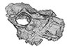
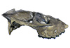
The present 3D Dataset contains the 3D models described and figured in the following publication: Grohé C., Bonis L. de, Chaimanee Y., Chavasseau O., Rugbumrung M., Yamee C., Suraprasit K., Gibert C., Surault J., Blondel C., Jaeger J.-J. 2020. The late middle Miocene Mae Moh Basin of northern Thailand: the richest Neogene assemblage of Carnivora from Southeast Asia and a paleobiogeographic analysis of Miocene Asian carnivorans. American Museum Novitates. http://digitallibrary.amnh.org/handle/2246/7223
Siamogale bounosa MM-54 View specimen
|
M3#5053D model of the skull of Siamogale bounosa The zip file contains: - the 3D surface in PLY - the orientation files in .pos and .ori - the project in .ntw Type: "3D_surfaces"doi: 10.18563/m3.sf.505 state:published |
Download 3D surface file |
Vishnuonyx maemohensis MM-78 View specimen
|
M3#5063D model of the skull of Vishnuonyx maemohensis The zip file contains: - the 3D surface in PLY - the orientation files in .pos and .ori - the project in .ntw Type: "3D_surfaces"doi: 10.18563/m3.sf.506 state:published |
Download 3D surface file |
|
M3#5073D model of the reconstructed upper teeth of Vishnuonyx maemohensis The zip file contains: - the 3D surface in PLY - the orientation files in .pos and .ori - the project in .ntw Type: "3D_surfaces"doi: 10.18563/m3.sf.507 state:published |
Download 3D surface file |
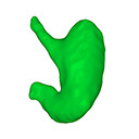
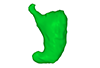
The present 3D Dataset contains the 3D models analyzed in: Kaigai N et al. Morphogenesis and three-dimensional movement of the stomach during the human embryonic period, Anat Rec (Hoboken). 2014 May;297(5):791-797. doi: 10.1002/ar.22833.
Homo sapiens KC-CS16STM27159 View specimen
|
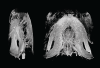
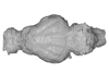
M3#56computationally reconstructed stomach of the human embryo (M3#56_KC-CS16STM27159) at Carnegie Stage 16 (Crown Rump Length= 9.9mm). Type: "3D_surfaces"doi: 10.18563/m3.sf56 state:published |
Download 3D surface file |
Homo sapiens KC-CS17STM20383 View specimen
|
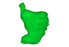
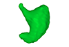
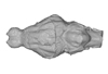
M3#57computationally reconstructed stomach of the human embryo (M3#57_KC-CS17STM20383) at Carnegie Stage 17 (Crown Rump Length= 12.3mm). Type: "3D_surfaces"doi: 10.18563/m3.sf57 state:published |
Download 3D surface file |
Homo sapiens KC-CS18STM21807 View specimen
|
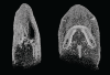
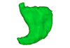
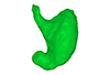
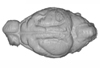
M3#58computationally reconstructed stomach of the human embryo (M3#58_KC-CS18STM21807) at Carnegie Stage 18 (Crown Rump Length= 14.7mm). Type: "3D_surfaces"doi: 10.18563/m3.sf58 state:published |
Download 3D surface file |
Homo sapiens KC-CS19STM17998 View specimen
|
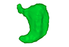
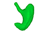
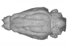
M3#59computationally reconstructed stomach of the human embryo (M3#59_KC-CS19STM17998) at Carnegie Stage 19 (Crown Rump Length was unmeasured ). Type: "3D_surfaces"doi: 10.18563/m3.sf59 state:published |
Download 3D surface file |
Homo sapiens KC-CS20STM20785 View specimen
|
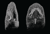
M3#60computationally reconstructed stomach of the human embryo (M3#60_KC-CS20STM20785) at Carnegie Stage 20 (Crown Rump Length= 18.7 mm). Type: "3D_surfaces"doi: 10.18563/m3.sf60 state:published |
Download 3D surface file |
Homo sapiens KC-CS21STM24728 View specimen
|
M3#61computationally reconstructed stomach of the human embryo (M3#61_KC-CS21STM24728) at Carnegie Stage 21 (Crown Rump Length= 20.9 mm). Type: "3D_surfaces"doi: 10.18563/m3.sf61 state:published |
Download 3D surface file |
Homo sapiens KC-CS22STM26438 View specimen
|
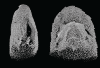
M3#62computationally reconstructed stomach of the human embryo (M3#62_KC-CS22STM26438) at Carnegie Stage 22 (Crown Rump Length= 21.5 mm). Type: "3D_surfaces"doi: 10.18563/m3.sf62 state:published |
Download 3D surface file |
Homo sapiens KC-CS23STM20018 View specimen
|
M3#63computationally reconstructed stomach of the human embryo (M3#63_KC-CS23STM20018) at Carnegie Stage 23 (Crown Rump Length= 23.1 mm). Type: "3D_surfaces"doi: 10.18563/m3.sf63 state:published |
Download 3D surface file |
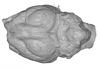
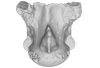
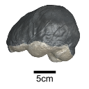
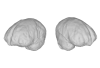
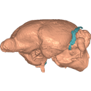
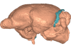
The present 3D Dataset contains the 3D models illustrated and described in the chapter “Paleoneurology of Artiodactyla, an overview of the evolution of the artiodactyl brain” (Orliac et al. 2022) published in "Paleoneurology of amniotes: new directions in the study of fossil endocasts", edited by Dozo, Paulina-Carabajal, Macrini and Walsh.
Homacodon vagans AMNH 12695 View specimen
|
M3#1063Endocranial cast Type: "3D_surfaces"doi: 10.18563/m3.sf.1063 state:published |
Download 3D surface file |
Helohyus sp. AMNH 13079 View specimen
|
M3#1064Endocranial cast Type: "3D_surfaces"doi: 10.18563/m3.sf.1064 state:published |
Download 3D surface file |
Leptauchenia sp. AMNH 45508 View specimen
|
M3#1065endocranial cast Type: "3D_surfaces"doi: 10.18563/m3.sf.1065 state:published |
Download 3D surface file |
Agriochoerus sp. AMNH 95330 View specimen
|
M3#1067endocranial cast Type: "3D_surfaces"doi: 10.18563/m3.sf.1067 state:published |
Download 3D surface file |
Mouillacitherium elegans UM ACQ 6625 View specimen
|
M3#1068endocranial cast Type: "3D_surfaces"doi: 10.18563/m3.sf.1068 state:published |
Download 3D surface file |
Caenomeryx filholi UM PDS 2570 View specimen
|
M3#1069endocranial cast Type: "3D_surfaces"doi: 10.18563/m3.sf.1069 state:published |
Download 3D surface file |
Dichobune leporina MNHN.F.QU16586 View specimen
|
M3#1070endocranial cast Type: "3D_surfaces"doi: 10.18563/m3.sf.1070 state:published |
Download 3D surface file |
Anoplotherium sp. not numbered View specimen
|
M3#1071endocranial cast Type: "3D_surfaces"doi: 10.18563/m3.sf.1071 state:published |
Download 3D surface file |
The present 3D Dataset contains the 3D models analyzed in: Perrichon et al. 2023. Ontogenetic variability of the intertympanic sinus distinguishes lineages within Crocodylia.
Mecistops sp. ag SVSTUA 022001 View specimen
|
M3#980Intertympanic sinus system (expressed in meters) Type: "3D_surfaces"doi: 10.18563/m3.sf.980 state:published |
Download 3D surface file |
Crocodylus niloticus ag SVSTUA 022002 View specimen
|
M3#981Intertympanic sinus system Type: "3D_surfaces"doi: 10.18563/m3.sf.981 state:published |
Download 3D surface file |
Mecistops sp. AMU Zoo 04721 View specimen
|
M3#982Intertympanic sinus system Type: "3D_surfaces"doi: 10.18563/m3.sf.982 state:published |
Download 3D surface file |
Crocodylus sp. MHNL QV14 View specimen
|
M3#983Intertympanic sinus system Type: "3D_surfaces"doi: 10.18563/m3.sf.983 state:published |
Download 3D surface file |
Crocodylus rhombifer MHNL 42006506 View specimen

|
M3#1008Intertympanic sinus system (incomplete) Type: "3D_surfaces"doi: 10.18563/m3.sf.1008 state:published |
Download 3D surface file |
Crocodylus rhombifer MHNL 42006507 View specimen
|
M3#1007Intertympanic sinus system (incomplete) Type: "3D_surfaces"doi: 10.18563/m3.sf.1007 state:published |
Download 3D surface file |
Crocodylus niloticus MHNL 50001387 View specimen
|
M3#1006Intertympanic sinus system Type: "3D_surfaces"doi: 10.18563/m3.sf.1006 state:published |
Download 3D surface file |
Crocodylus palustris MHNL 50001388 View specimen
|
M3#1004Intertympanic sinus system Type: "3D_surfaces"doi: 10.18563/m3.sf.1004 state:published |
Download 3D surface file |
Crocodylus porosus MHNL 50001389 View specimen
|
M3#1009Intertympanic sinus system Type: "3D_surfaces"doi: 10.18563/m3.sf.1009 state:published |
Download 3D surface file |
Mecistops sp. MHNL 50001393 View specimen
|
M3#1010Intertympanic sinus system Type: "3D_surfaces"doi: 10.18563/m3.sf.1010 state:published |
Download 3D surface file |
Crocodylus niloticus MHNL 50001397 View specimen
|
M3#1018Intertympanic sinus system Type: "3D_surfaces"doi: 10.18563/m3.sf.1018 state:published |
Download 3D surface file |
Crocodylus porosus MHNL 50001398 View specimen
|
M3#1017Intertympanic sinus system Type: "3D_surfaces"doi: 10.18563/m3.sf.1017 state:published |
Download 3D surface file |
Crocodylus niloticus MHNL 50001405 View specimen
|
M3#1016Intertympanic sinus system Type: "3D_surfaces"doi: 10.18563/m3.sf.1016 state:published |
Download 3D surface file |
Gavialis gangeticus MHNL 50001407 View specimen
|
M3#1015Intertympanic sinus system Type: "3D_surfaces"doi: 10.18563/m3.sf.1015 state:published |
Download 3D surface file |
Crocodylus niloticus MHNL 90001850 View specimen
|
M3#1014Intertympanic sinus system Type: "3D_surfaces"doi: 10.18563/m3.sf.1014 state:published |
Download 3D surface file |
Crocodylus niloticus MHNL 90001851 View specimen
|
M3#1013Intertympanic sinus system Type: "3D_surfaces"doi: 10.18563/m3.sf.1013 state:published |
Download 3D surface file |
Crocodylus niloticus MHNL 90001855 View specimen
|
M3#1012Intertympanic sinus system Type: "3D_surfaces"doi: 10.18563/m3.sf.1012 state:published |
Download 3D surface file |
Osteolaemus tetraspis MHNM.9095.0 View specimen
|
M3#1011Intertympanic sinus system Type: "3D_surfaces"doi: 10.18563/m3.sf.1011 state:published |
Download 3D surface file |
Voay robustus MNHN F.1908-5 View specimen
|
M3#1003Intertympanic sinus system Type: "3D_surfaces"doi: 10.18563/m3.sf.1003 state:published |
Download 3D surface file |
Crocodylus sp. MNHN-F.1908-5-2 View specimen
|
M3#1005Intertympanic sinus system Type: "3D_surfaces"doi: 10.18563/m3.sf.1005 state:published |
Download 3D surface file |
Osteolaemus tetraspis MZS Cro 040 View specimen
|
M3#1002Intertympanic sinus system Type: "3D_surfaces"doi: 10.18563/m3.sf.1002 state:published |
Download 3D surface file |
Crocodylus acutus MZS Cro 055 View specimen
|
M3#991Intertympanic sinus system Type: "3D_surfaces"doi: 10.18563/m3.sf.991 state:published |
Download 3D surface file |
Melanosuchus niger MZS Cro 073 View specimen
|
M3#989Intertympanic sinus system Type: "3D_surfaces"doi: 10.18563/m3.sf.989 state:published |
Download 3D surface file |
Mecistops sp. MZS Cro 083 View specimen

|
M3#990Intertympanic sinus system Type: "3D_surfaces"doi: 10.18563/m3.sf.990 state:published |
Download 3D surface file |
Tomistoma schlegelii MZS Cro 094 View specimen
|
M3#988Intertympanic sinus system Type: "3D_surfaces"doi: 10.18563/m3.sf.988 state:published |
Download 3D surface file |
Gavialis gangeticus NHMUK 1846.1.7.3 View specimen
|
M3#987Intertympanic sinus system Type: "3D_surfaces"doi: 10.18563/m3.sf.987 state:published |
Download 3D surface file |
Osteolaemus tetraspis NHMUK 1862.6.30.5 View specimen
|
M3#986Intertympanic sinus system Type: "3D_surfaces"doi: 10.18563/m3.sf.986 state:published |
Download 3D surface file |
Gavialis gangeticus NHMUK 1873 View specimen
|
M3#985intertympanic sinus system Type: "3D_surfaces"doi: 10.18563/m3.sf.985 state:published |
Download 3D surface file |
Tomistoma schlegelii NHMUK 1893.3.6.14 View specimen
|
M3#984Intertympanic sinus system Type: "3D_surfaces"doi: 10.18563/m3.sf.984 state:published |
Download 3D surface file |
Mecistops sp. NHMUK 1924.5.10.1 View specimen
|
M3#992Intertympanic sinus system Type: "3D_surfaces"doi: 10.18563/m3.sf.992 state:published |
Download 3D surface file |
Voay robustus NHMUK PV R 36684 View specimen
|
M3#993Intertympanic sinus system Type: "3D_surfaces"doi: 10.18563/m3.sf.993 state:published |
Download 3D surface file |
Voay robustus NHMUK PV R 36685 View specimen
|
M3#1001Intertympanic sinus system Type: "3D_surfaces"doi: 10.18563/m3.sf.1001 state:published |
Download 3D surface file |
Crocodylus niloticus UCBL FSL 532077 View specimen

|
M3#1000Intertympanic sinus system Type: "3D_surfaces"doi: 10.18563/m3.sf.1000 state:published |
Download 3D surface file |
Crocodylus porosus/siamensis UCBLZ 2019-1-237 View specimen
|
M3#999Intertympanic sinus system Type: "3D_surfaces"doi: 10.18563/m3.sf.999 state:published |
Download 3D surface file |
Osteolaemus tetraspis UCBLZ 2019-1-236 View specimen
|
M3#998Intertympanic sinus system Type: "3D_surfaces"doi: 10.18563/m3.sf.998 state:published |
Download 3D surface file |
Alligator mississipiensis UCBLZ WB35 View specimen
|
M3#997Intertympanic sinus system Type: "3D_surfaces"doi: 10.18563/m3.sf.997 state:published |
Download 3D surface file |
Crocodylus siamensis UCBLZ WB41 View specimen
|
M3#996Intertympanic sinus system Type: "3D_surfaces"doi: 10.18563/m3.sf.996 state:published |
Download 3D surface file |
Tomistoma schlegelii UM 1097 View specimen
|
M3#995Intertympanic sinus system Type: "3D_surfaces"doi: 10.18563/m3.sf.995 state:published |
Download 3D surface file |
Crocodylus niloticus UM 2001-1756-1-434 NR View specimen
|
M3#994Intertympanic sinus system Type: "3D_surfaces"doi: 10.18563/m3.sf.994 state:published |
Download 3D surface file |
Mecistops sp. UM N89 View specimen
|
M3#1019Intertympanic sinus system Type: "3D_surfaces"doi: 10.18563/m3.sf.1019 state:published |
Download 3D surface file |
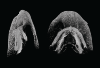
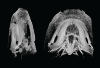
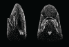
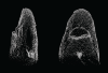
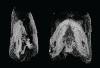
This contribution contains the 3D models described and figured in the following publication: Georgalis, G.L., G. Guinot, K.E. Kassegne, Y.Z. Amoudji, A.K.C. Johnson, H. Cappetta and L. Hautier. 2021. An assemblage of giant aquatic snakes (Serpentes, Palaeophiidae) from the Eocene of Togo. Swiss Journal of Palaeontology 140, https://doi.org/10.1186/s13358-021-00236-w
Palaeophis africanus UM KPO 21 View specimen
|
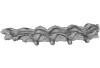
M3#821Trunk vertebra UM KPO 21 of Palaeophis africanus Type: "3D_surfaces"doi: 10.18563/m3.sf.821 state:published |
Download 3D surface file |
Palaeophis africanus UM KPO 22 View specimen
|
M3#822Trunk vertebra UM KPO 22 of Palaeophis africanus from the Eocene of Togo Type: "3D_surfaces"doi: 10.18563/m3.sf.822 state:published |
Download 3D surface file |
Palaeophis africanus UM KPO 23 View specimen
|
M3#823Trunk vertebra UM KPO 23 of Palaeophis africanus Type: "3D_surfaces"doi: 10.18563/m3.sf.823 state:published |
Download 3D surface file |
Palaeophis africanus UM KPO 24 View specimen
|
M3#824Trunk vertebra UM KPO 24 of Palaeophis africanus Type: "3D_surfaces"doi: 10.18563/m3.sf.824 state:published |
Download 3D surface file |
Palaeophis africanus UM KPO 25 View specimen
|
M3#825Trunk vertebra UM KPO 25 of Palaeophis africanus Type: "3D_surfaces"doi: 10.18563/m3.sf.825 state:published |
Download 3D surface file |
Palaeophis africanus UM KPO 26 View specimen
|
M3#826Trunk vertebra UM KPO 26 of Palaeophis africanus Type: "3D_surfaces"doi: 10.18563/m3.sf.826 state:published |
Download 3D surface file |
Palaeophis africanus UM KPO 27 View specimen
|
M3#827Trunk vertebra UM KPO 27 of Palaeophis africanus Type: "3D_surfaces"doi: 10.18563/m3.sf.827 state:published |
Download 3D surface file |
Palaeophis africanus UM KPO 28 View specimen
|
M3#828Trunk vertebra UM KPO 28 of Palaeophis africanus Type: "3D_surfaces"doi: 10.18563/m3.sf.828 state:published |
Download 3D surface file |
Palaeophis africanus UM KPO 29 View specimen
|
M3#829Trunk vertebra UM KPO 29 of Palaeophis africanus Type: "3D_surfaces"doi: 10.18563/m3.sf.829 state:published |
Download 3D surface file |
Palaeophis africanus UM KPO 30 View specimen
|
M3#830Trunk vertebra UM KPO 30 of Palaeophis africanus Type: "3D_surfaces"doi: 10.18563/m3.sf.830 state:published |
Download 3D surface file |
Palaeophis africanus UM KPO 31 View specimen
|
M3#831Trunk vertebra UM KPO 28 of Palaeophis africanus Type: "3D_surfaces"doi: 10.18563/m3.sf.831 state:published |
Download 3D surface file |
Palaeophis africanus UM KPO 32 View specimen
|
M3#832Trunk vertebra UM KPO 32 of Palaeophis africanus Type: "3D_surfaces"doi: 10.18563/m3.sf.832 state:published |
Download 3D surface file |
Palaeophis africanus UM KPO 33 View specimen
|
M3#833Trunk vertebra UM KPO 33 of Palaeophis africanus Type: "3D_surfaces"doi: 10.18563/m3.sf.833 state:published |
Download 3D surface file |
Palaeophis africanus UM KPO 34 View specimen

|
M3#839Trunk vertebra UM KPO 34 of Palaeophis africanus Type: "3D_surfaces"doi: 10.18563/m3.sf.839 state:published |
Download 3D surface file |
Palaeophis africanus UM KPO 35 View specimen
|
M3#840Trunk vertebra UM KPO 35 of Palaeophis africanus Type: "3D_surfaces"doi: 10.18563/m3.sf.840 state:published |
Download 3D surface file |
Palaeophis africanus UM KPO 36 View specimen
|
M3#841Trunk vertebra UM KPO 36 of Palaeophis africanus Type: "3D_surfaces"doi: 10.18563/m3.sf.841 state:published |
Download 3D surface file |
Palaeophis africanus UM KPO 37 View specimen
|
M3#842Trunk vertebra UM KPO 37 of Palaeophis africanus Type: "3D_surfaces"doi: 10.18563/m3.sf.842 state:published |
Download 3D surface file |
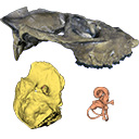
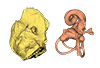
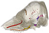
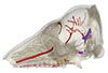
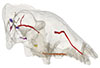
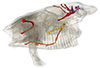
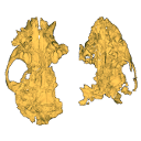
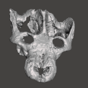
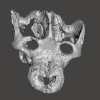
The present 3D Dataset contains the 3D models described in “Comparative masticatory myology in anteaters and its implications for interpreting morphological convergence in myrmecophagous placentals”.
Cyclopes didactylus M1571_JAG View specimen
|
M3#522Skull, mandible, and muscles of Cyclopes didactylus Type: "3D_surfaces"doi: 10.18563/m3.sf.522 state:published |
Download 3D surface file |
Tamandua tetradactyla M3075_JAG View specimen
|
M3#524Skull, left mandibles, and muscles of Tamandua tetradactyla. Type: "3D_surfaces"doi: 10.18563/m3.sf.524 state:published |
Download 3D surface file |
Myrmecophaga tridactyla M3023_JAG View specimen
|
M3#523Skull, left mandible and muscles of Myrmecophaga tridactyla. Type: "3D_surfaces"doi: 10.18563/m3.sf.523 state:published |
Download 3D surface file |
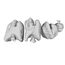
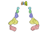
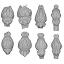
The present 3D Dataset contains the 3D models analyzed in ”Morphological features of tooth development and replacement in the rabbit Oryctolagus cuniculus”, Archives of Oral Biology, https://doi.org/10.1016/j.archoralbio.2019.104576
Oryctogalus cuniculus E14 View specimen
|
M3#390Right cheek teeth, Left and right incisors at 14 dpf Type: "3D_surfaces"doi: 10.18563/m3.sf.390 state:published |
Download 3D surface file |
Oryctogalus cuniculus E16 View specimen

|
M3#391Left cheek teeth, Left and right incisors at 16 dpf Type: "3D_surfaces"doi: 10.18563/m3.sf.391 state:published |
Download 3D surface file |
Oryctogalus cuniculus E18 View specimen
|
M3#392Left cheek teeth and incisors at 18 dpf Type: "3D_surfaces"doi: 10.18563/m3.sf.392 state:published |
Download 3D surface file |
Oryctogalus cuniculus E20 View specimen
|
M3#393Left cheek teeth and incisors at 20 dpf Type: "3D_surfaces"doi: 10.18563/m3.sf.393 state:published |
Download 3D surface file |
Oryctogalus cuniculus E22 View specimen
|
M3#394Left lower cheek teeth and incisors, right upper cheek teeth and incisors at 22 dpf Type: "3D_surfaces"doi: 10.18563/m3.sf.394 state:published |
Download 3D surface file |
Oryctogalus cuniculus E24 View specimen
|
M3#395Left cheek teeth and incisors at 24 dpf Type: "3D_surfaces"doi: 10.18563/m3.sf.395 state:published |
Download 3D surface file |
Oryctogalus cuniculus E28 View specimen
|
M3#396Right cheek teeth and incisors at 28 dpf Type: "3D_surfaces"doi: 10.18563/m3.sf.396 state:published |
Download 3D surface file |
Oryctogalus cuniculus E26 View specimen
|
M3#397Right cheek teeth and incisors at 26 dpf Type: "3D_surfaces"doi: 10.18563/m3.sf.397 state:published |
Download 3D surface file |
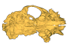
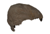
This contribution contains the 3D model described and figured in the following publication: New turtles from the Late Cretaceous of Monte Alto-SP, Brazil, including cranial osteology, neuroanatomy and phylogenetic position of a new taxon. PalZ. https://doi.org/10.1007/s12542-017-0397-x
Yuraramirim montealtensis 04-0008/89 View specimen
|
M3#2783D surfaces related to specimen MPMA 04-0008/89. Type: "3D_surfaces"doi: 10.18563/m3.sf.278 state:published |
Download 3D surface file |
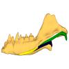
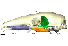
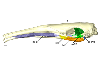
The present 3D Dataset contains the 3D model analyzed in The largest freshwater odontocete: a South Asian river dolphin relative from the Proto-Amazonia.
Pebanista yacuruna MUSM 4017 View specimen
|
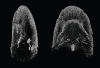
M3#1394Holotype skull of Pebanista yacuruna MUSM 4017 Type: "3D_surfaces"doi: 10.18563/m3.sf.1394 state:published |
Download 3D surface file |
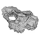
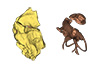
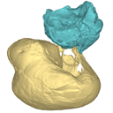
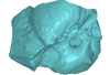
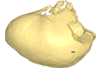
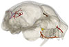
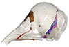
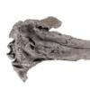
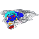
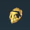
This contribution contains the 3D model of an endocranial cast analyzed in “A 10 ka intentionally deformed human skull from Northeast Asia”. There are many studies on the morphological characteristics of intentional cranial deformation (ICD), but few related 3D models were published. Here, we present the surface model of an intentionally deformed 10 ka human cranium for further research on ICD practice. The 3D model of the endocranial cast of this ICD cranium was discovered near Harbin City, Province Heilongjiang, Northeast China. The fossil preserved only the frontal, parietal, and occipital bones. To complete the endocast model of the specimen, we printed a 3D model and used modeling clay to reconstruct the missing part based on the general form of the modern human endocast morphology.
Homo sapiens IVPP-PA1616 View specimen
|
M3#972The frontal region of the endocast is flattened, probably formed by the constant pressure on the frontal bone during growth. There is a well-developed frontal crest on the endocranial surface. The endocast widens posteriorly from the frontal lobe. The widest point of the endocast is at the lateral border of the parietal lobe. The lower parietal areas display a marked lateral expansion. The overall shape of the endocast is asymmetrical, with the left side of the parietal lobe being more laterally expanded than the right side. Like the frontal lobe, the occipital lobe is also anteroposteriorly flattened. Type: "3D_surfaces"doi: 10.18563/m3.sf.972 state:published |
Download 3D surface file |
|
M3#976The original endocranial cast model (with texture) of IVPP-PA1616. It shows the original structures of the specimen, and was not altered in any way. Type: "3D_surfaces"doi: 10.18563/m3.sf.976 state:published |
Download 3D surface file |
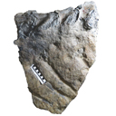
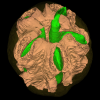
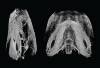
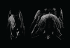
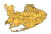
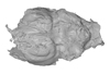
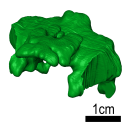
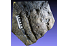
The present 3D Dataset contains the 3D model analyzed in Hendrickx, C. and Bell, P. R. 2021. The scaly skin of the abelisaurid Carnotaurus sastrei (Theropoda: Ceratosauria) from the Upper Cretaceous of Patagonia. Cretaceous Research. https://doi.org/10.1016/j.cretres.2021.104994
Carnotaurus sastrei MACN 894 View specimen
|
M3#8023D reconstruction of the biggest patch of skin (~1200 cm2) from the anterior tail region of the holotype of Carnotaurus, which is the largest single patch of squamous integument available for any saurischian. The skin consists of medium to large (up to 65 mm in diameter) conical feature scales surrounded by a network of low and small (< 14 mm) irregular basement scales separated by narrow interstitial tissue. Type: "3D_surfaces"doi: 10.18563/m3.sf.802 state:published |
Download 3D surface file |
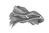
The present 3D Dataset contains the 3D models analyzed in: "a giant dapediid from the Late Triassic of Switzerland and insights into neopterygian phylogeny", Royal Society Open Science, https://doi.org/10.1098/rsos.180497
Scopulipiscis saxciput PIMUZ A/I 3026 View specimen

|
M3#1773D surfaces of the skull and endocranial spaces inside neurocranium, including the aortic canal, braincase, fossa bridgei, lateral cranial canal, nerves and other passageways, notochord, posterior myodome, and right semicircular canals. Type: "3D_surfaces"doi: 10.18563/m3.sf.177 state:published |
Download 3D surface file |

|
M3#178Scan of the neurocranium of PIMUZ A/I 3026 Type: "3D_CT"doi: 10.18563/m3.sf.178 state:published |
Download CT data |
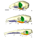
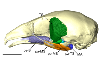
This contribution contains the 3D model described and figured in the following publication: Ramdarshan A., Orliac M.J., 2015. Endocranial morphology of Microchoerus erinaceus (Euprimates, Tarsiiformes) and early evolution of the Euprimates brain. American Journal of Physical Anthropology. doi: 10.1002/ajpa.22868
Microchoerus erinaceus UM-PRR1771 View specimen
|
M3#15Labelled 3D model of the endocranial cast and sinuse of Microchoerus erinaceus. Type: "3D_surfaces"doi: 10.18563/m3.sf15 state:published |
Download 3D surface file |
|
M3#130350µm voxel size µCT scan of the cranium of UM PRR1771 Type: "3D_CT"doi: 10.18563/m3.sf.1303 state:published |
Download CT data |
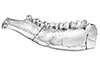
The present 3D Dataset contains the 3D models of a skull and lower jaw of the holotype of Santagnathus mariensis, described in “Old fossil findings in the Upper Triassic rocks of southern Brazil improve diversity of traversodontid cynodonts (Therapsida, Cynodontia)”
Santagnathus mariensis UFRGS-PV-1419-T View specimen
|
M3#1157Skull Type: "3D_surfaces"doi: 10.18563/m3.sf.1157 state:published |
Download 3D surface file |
|
M3#1158Lower jaw Type: "3D_surfaces"doi: 10.18563/m3.sf.1158 state:published |
Download 3D surface file |