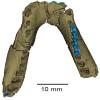
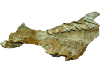
3D models of Kalakocetus, the earliest Cetacea
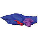
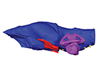
The specimens of Speothos pacivorus
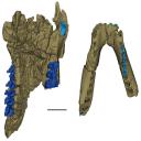
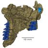
3D models related to the publication: Hidden diversity of Palaeogene metatherians: a new family of polydolopimorphian marsupials from Peruvian Amazonia
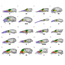
3D GM dataset of bird skeletal variation
Skeletal embryonic development in the catshark
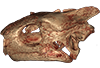
Bony connexions of the petrosal bone of extant hippos
bony labyrinth (14) , inner ear (11) , Eocene (11) , geometric morphometrics (10) , CT-scan (10) , Oligocene (9) , Micro-CT (9)
Maëva Judith Orliac (24) , Lionel Hautier (24) , Laurent Marivaux (18) , Renaud Lebrun (15) , Rodolphe Tabuce (14) , Pierre-Olivier Antoine (13) , Bastien Mennecart (13)
|
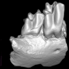
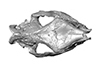
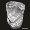
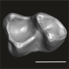
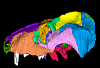
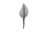
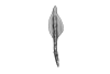
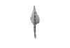
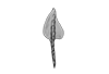
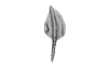
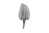
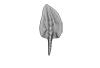
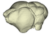
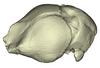
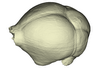
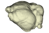
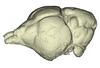
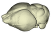
Brain damage: the endocranial cast of Mixtotherium cuspidatum (Mammalia, Artiodactyla) from the Victor Brun Museum (Montauban, France)Maëva J. Orliac
Published online: 25/11/2021 |
|
M3#857endocast of the brain cavity Type: "3D_surfaces"doi: 10.18563/m3.sf.857 state:published |
Download 3D surface file |
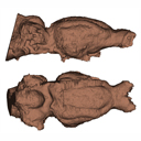
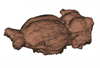
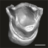
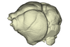
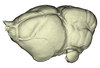
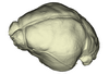
This contribution provides for the first time the 3D model of the type specimen of Molassitherium delemontense (Mammalia, Rhinocerotidae) described in the following publication: Becker et al. (2013), Journal of Systematic Palaeontology, Vol. 11, Issue 8, 947–972, https://doi.org/10.1080/14772019.2012.699007. Conservation issues of the specimen and solutions using 3D model and 3D prints are detailed.
Molassitherium delemontense MJSN POI007–245 View specimen
|
M3#384Skull of Molassitherium delemontense Becker and Antoine, 2013 (in Becker et al. 2013): holotype Type: "3D_surfaces"doi: 10.18563/m3.sf.384 state:published |
Download 3D surface file |
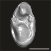
Current knowledge on the skeletogenesis of Chondrichthyes is scarce compared with their extant sister group, the bony fishes. Most of the previously described developmental tables in Chondrichthyes have focused on embryonic external morphology only. Due to its small body size and relative simplicity to raise eggs in laboratory conditions, the small-spotted catshark Scyliorhinus canicula has emerged as a reference species to describe developmental mechanisms in the Chondrichthyes lineage. Here we investigate the dynamic of mineralization in a set of six embryonic specimens using X-ray microtomography and describe the developing units of both the dermal skeleton (teeth and dermal scales) and endoskeleton (vertebral axis). This preliminary data on skeletogenesis in the catshark sets the first bases to a more complete investigation of the skeletal developmental in Chondrichthyes. It should provide comparison points with data known in osteichthyans and could thus be used in the broader context of gnathostome skeletal evolution.
Scyliorhinus canicula SC6_2_2015_03_20 View specimen

|
M3#50Mineralized skeleton of a 6,2 cm long embryo of Scyliorhinus canicula Type: "3D_surfaces"doi: 10.18563/m3.sf.50 state:published |
Download 3D surface file |
Scyliorhinus canicula SC6_7_2015_03_20 View specimen

|
M3#51Mineralized skeleton of a 6,7 cm long embryo of Scyliorhinus canicula Type: "3D_surfaces"doi: 10.18563/m3.sf.51 state:published |
Download 3D surface file |
Scyliorhinus canicula SC7_1_2015_04_03 View specimen

|
M3#52Mineralized skeleton of a 7,1 cm long embryo of Scyliorhinus canicula Type: "3D_surfaces"doi: 10.18563/m3.sf.52 state:published |
Download 3D surface file |
Scyliorhinus canicula SC7_5_2015_03_13 View specimen

|
M3#53Mineralized skeleton of a 7,5 cm long embryo of Scyliorhinus canicula Type: "3D_surfaces"doi: 10.18563/m3.sf.53 state:published |
Download 3D surface file |
Scyliorhinus canicula SC8_2015_03_20 View specimen

|
M3#54Mineralized skeleton of a 8 cm long embryo of Scyliorhinus canicula Type: "3D_surfaces"doi: 10.18563/m3.sf.54 state:published |
Download 3D surface file |
Scyliorhinus canicula SC10_2015_02_27 View specimen

|
M3#55Mineralized skeleton of a 10 cm long embryo of Scyliorhinus canicula Type: "3D_surfaces"doi: 10.18563/m3.sf.55 state:published |
Download 3D surface file |
This contribution contains 3D models of mandibles of Cypriot mice (Mus cypriacus) and house mice (Mus musculus domesticus) from the island of Cyprus. The niche partitioning of the two species was investigated using isotopic ecology, geometric morphometrics and biomechanics. Both species displayed generalist feeding behavior, modulated by fine-tuned adaptation to their feeding habits. The house mouse mandible, with a relatively large masseter area and an optimization for incisor biting, appears as an all-rounder tool for foraging on diverse non-natural items.
These models are analyzed in the following publication: Renaud et al 2024, “Trophic differentiation between the endemic Cypriot mouse and the house mouse: a study coupling stable isotopes and morphometrics”, https://doi.org/10.1007/s10914-024-09740-5
Mus cypriacus Cypriacus_5GE View specimen
|
M3#15843D model of the right mandible Type: "3D_surfaces"doi: 10.18563/m3.sf.1584 state:published |
Download 3D surface file |
Mus cypriacus Cypriacus_BET2 View specimen

|
M3#15853D model of the right mandible Type: "3D_surfaces"doi: 10.18563/m3.sf.1585 state:published |
Download 3D surface file |
Mus cypriacus Cypriacus_FON1 View specimen
|
M3#15863D model of the right mandible Type: "3D_surfaces"doi: 10.18563/m3.sf.1586 state:published |
Download 3D surface file |
Mus cypriacus Cypriacus_FON2 View specimen
|
M3#15873D model of the right mandible Type: "3D_surfaces"doi: 10.18563/m3.sf.1587 state:published |
Download 3D surface file |
Mus cypriacus Cypriacus_KOU1 View specimen
|
M3#15883D model of the right mandible Type: "3D_surfaces"doi: 10.18563/m3.sf.1588 state:published |
Download 3D surface file |
Mus musculus Cyprus_dom_KOF1 View specimen
|
M3#15893D model of the right mandible Type: "3D_surfaces"doi: 10.18563/m3.sf.1589 state:published |
Download 3D surface file |
Mus musculus Cyprus_dom_LEF1 View specimen
|
M3#15903D model of the right mandible Type: "3D_surfaces"doi: 10.18563/m3.sf.1590 state:published |
Download 3D surface file |
Mus musculus Cyprus_dom_MEN1 View specimen

|
M3#15913D model of the right mandible Type: "3D_surfaces"doi: 10.18563/m3.sf.1591 state:published |
Download 3D surface file |
Mus musculus Cyprus_dom_TSE2 View specimen
|
M3#15923D model of the mirrored left mandible Type: "3D_surfaces"doi: 10.18563/m3.sf.1592 state:published |
Download 3D surface file |
Mus musculus Cyprus_dom_XYL5 View specimen
|
M3#15933D model of the right mandible Type: "3D_surfaces"doi: 10.18563/m3.sf.1593 state:published |
Download 3D surface file |
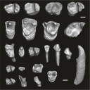
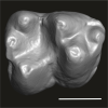
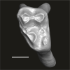
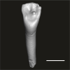
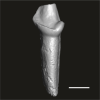
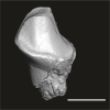
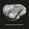
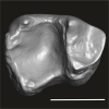
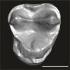
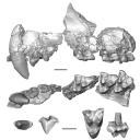
This contribution contains the three-dimensional digital models of the dental fossil material of anthropoid and strepsirrhine primates, discovered in Lower Oligocene detrital deposits outcropping in the Porto Rico and El Argoub areas, east of the Dakhla peninsula region (Atlantic Sahara; in the south of Morocco, near the northern border of Mauritania). These fossils were described, figured and discussed in the following publication: Marivaux et al. (2024), A new primate community from the earliest Oligocene of the Atlantic margin of Northwest Africa: Systematic, paleobiogeographic and paleoenvironmental implications. Journal of Human Evolution. https://doi.org/10.1016/j.jhevol.2024.103548
Catopithecus aff. browni DAK-Arg-087 View specimen

|
M3#1211Isolated right lower m3 (worn) Type: "3D_surfaces"doi: 10.18563/m3.sf.1211 state:published |
Download 3D surface file |
Catopithecus aff. browni DAK-Arg-088 View specimen
|
M3#1212Isolated right lower m2 (abraded/corroded) Type: "3D_surfaces"doi: 10.18563/m3.sf.1212 state:published |
Download 3D surface file |
Catopithecus aff. browni DAK-Arg-089 View specimen
|
M3#1213Isolated left lower m1 (worn) Type: "3D_surfaces"doi: 10.18563/m3.sf.1213 state:published |
Download 3D surface file |
Catopithecus aff. browni DAK-Pto-052 View specimen
|
M3#1214Isolated right lower m1 (pristine but lacking the mesiobuccal region) Type: "3D_surfaces"doi: 10.18563/m3.sf.1214 state:published |
Download 3D surface file |
Catopithecus aff. browni DAK-Arg-090 View specimen
|
M3#1215Isolated left upper P4 Type: "3D_surfaces"doi: 10.18563/m3.sf.1215 state:published |
Download 3D surface file |
Catopithecus aff. browni DAK-Arg-091 View specimen
|
M3#1216Isolated left upper M2 (worn and corroded) Type: "3D_surfaces"doi: 10.18563/m3.sf.1216 state:published |
Download 3D surface file |
Catopithecus aff. browni DAK-Pto-053 View specimen
|
M3#1217Isolated right upper M1 (lacking the buccal region) Type: "3D_surfaces"doi: 10.18563/m3.sf.1217 state:published |
Download 3D surface file |
Abuqatrania cf. basiodontos DAK-Arg-092 View specimen
|
M3#1218Isolated left lower c1 Type: "3D_surfaces"doi: 10.18563/m3.sf.1218 state:published |
Download 3D surface file |
?Propliopithecus sp. DAK-Pto-056 View specimen
|
M3#1219Isolated right lower m3 (fragment of talonid of a germ) Type: "3D_surfaces"doi: 10.18563/m3.sf.1219 state:published |
Download 3D surface file |
Abuqatrania cf. basiodontos DAK-Arg-093 View specimen
|
M3#1469Isolated right lower m1 Type: "3D_surfaces"doi: 10.18563/m3.sf.1469 state:published |
Download 3D surface file |
Abuqatrania cf. basiodontos DAK-Arg-094 View specimen
|
M3#1221Isolated left upper M1 or M2 (corroded, lacking the enamel cap [exposed dentine]) Type: "3D_surfaces"doi: 10.18563/m3.sf.1221 state:published |
Download 3D surface file |
Abuqatrania cf. basiodontos DAK-Arg-095 View specimen
|
M3#1222Isolated right lower i1 or i2 Type: "3D_surfaces"doi: 10.18563/m3.sf.1222 state:published |
Download 3D surface file |
Abuqatrania cf. basiodontos DAK-Arg-096 View specimen
|
M3#1223Isolated right lower p2 (worn apex) Type: "3D_surfaces"doi: 10.18563/m3.sf.1223 state:published |
Download 3D surface file |
Abuqatrania cf. basiodontos DAK-Arg-097 View specimen
|
M3#1224Isolated right lower p2 (worn apex and broken root) Type: "3D_surfaces"doi: 10.18563/m3.sf.1224 state:published |
Download 3D surface file |
Afrotarsius sp. DAK-Arg-098 View specimen
|
M3#1225Isolated left lower p3 Type: "3D_surfaces"doi: 10.18563/m3.sf.1225 state:published |
Download 3D surface file |
Afrotarsius sp. DAK-Pto-054 View specimen
|
M3#1226Isolated right lower m1 (abraded/corroded) Type: "3D_surfaces"doi: 10.18563/m3.sf.1226 state:published |
Download 3D surface file |
Orolemur mermozi DAK-Pto-055 View specimen
|
M3#1227Isolated right upper M1 or M2 (pristine, Holotype) Type: "3D_surfaces"doi: 10.18563/m3.sf.1227 state:published |
Download 3D surface file |
Wadilemur cf. elegans DAK-Arg-099 View specimen
|
M3#1228Isolated right lower m2 Type: "3D_surfaces"doi: 10.18563/m3.sf.1228 state:published |
Download 3D surface file |
cf. 'Anchomomys' milleri DAK-Arg-100 View specimen

|
M3#1229Isolated right lower c1 Type: "3D_surfaces"doi: 10.18563/m3.sf.1229 state:published |
Download 3D surface file |
Abuqatrania cf. basiodontos DAK-Arg-101 View specimen
|
M3#1396Isolated left upper M3 (abraded) Type: "3D_surfaces"doi: 10.18563/m3.sf.1396 state:published |
Download 3D surface file |
Orogalago saintexuperyi DAK-Arg-102 View specimen
|
M3#1397Isolated left lower m2 Type: "3D_surfaces"doi: 10.18563/m3.sf.1397 state:published |
Download 3D surface file |
Wadilemur cf. elegans DAK-Arg-103 View specimen

|
M3#1473Isolated right upper M1 or M2 (lacking the mesial and buccal regions) Type: "3D_surfaces"doi: 10.18563/m3.sf.1473 state:published |
Download 3D surface file |
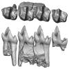
The present 3D Dataset contains 3D models of the holotypes described in Aiglstorfer et al. (2023a). Miocene Moschidae (Mammalia, Ruminantia) from the Linxia Basin (China) connect Europe and Asia and show early evolutionary diversity of a today monogeneric family. Palaeogeography, Palaeoclimatology, Palaeoecology.
Micromeryx? caoi CUGB GV 87045 View specimen
|
M3#11123D models of the holotype of “Micromeryx” caoi (CUGB GV87045) including the models of the teeth, the mandibule, and the sediment. Type: "3D_surfaces"doi: 10.18563/m3.sf.1112 state:published |
Download 3D surface file |
Hispanomeryx linxiaensis IVPP V28596 View specimen
|
M3#11133D models of the holotype of Hispanomeryx linxiaensis (IVPP V28596) including the models of the teeth, the mandibule, and the sediment. Type: "3D_surfaces"doi: 10.18563/m3.sf.1113 state:published |
Download 3D surface file |
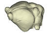
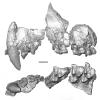
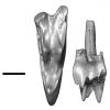
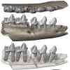
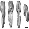
This contribution contains the 3D models described and figured in the following publication: Gaetano, L. C., Abdala, F., Mancuso, C, and Vega N.2025. New traversodontid cynodont from the Late Triassic Chañares Formation. Publicación Electrónica de la Asociación Paleontológica Argentina.
Pontognathus ignotus PULR-V 287 View specimen
|
M3#1647partial snout preserving the lateralmost incisor, the base of the canine, and several postcanines Type: "3D_surfaces"doi: 10.18563/m3.sf.1647 state:published |
Download 3D surface file |
Massetognathus pascuali PULR-V 289 View specimen
|
M3#1646partial lower jaw Type: "3D_surfaces"doi: 10.18563/m3.sf.1646 state:published |
Download 3D surface file |
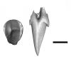
This contribution contains the three-dimensional models of the turbinal complex of 10 myrmecophagous and 10 non-myrmecophagous placental species. These specimens were analyzed and discussed in: Wright et. al (2024), Sniffing out morphological convergence in the turbinal complex of myrmecophagous placentals. https://doi.org/10.1002/ar.25603
Priodontes maximus NHMUK 732-a View specimen

|
M3#1536Turbinals of Priodontes maximus Type: "3D_surfaces"doi: 10.18563/m3.sf.1536 state:published |
Download 3D surface file |
Dasypus pilosus NHMUK 94-10-1-13 View specimen
|
M3#1537Turbinals of Dasypus pilosus Type: "3D_surfaces"doi: 10.18563/m3.sf.1537 state:published |
Download 3D surface file |
Dasypus novemcinctus AMNH 263287 View specimen
|
M3#1538Turbinals of Dasypus novemcinctus Type: "3D_surfaces"doi: 10.18563/m3.sf.1538 state:published |
Download 3D surface file |
Bradypus tridactylus UM 789N View specimen
|
M3#1539Turbinals of Bradypus tridactylus Type: "3D_surfaces"doi: 10.18563/m3.sf.1539 state:published |
Download 3D surface file |
Choloepus didactylus UM 767V View specimen
|
M3#1540Turbinals of Choloepus didactylus Type: "3D_surfaces"doi: 10.18563/m3.sf.1540 state:published |
Download 3D surface file |
Cyclopes didactylus NHMUK 88-8-8-14 View specimen

|
M3#1541Turbinals of Cyclopes didactylus Type: "3D_surfaces"doi: 10.18563/m3.sf.1541 state:published |
Download 3D surface file |
Myrmecophaga tridactyla UM 065V View specimen
|
M3#1542Turbinals of Myrmecophaga tridactyla Type: "3D_surfaces"doi: 10.18563/m3.sf.1542 state:published |
Download 3D surface file |
Tamandua tetradactyla NHMUK 3-7-7-135 View specimen
|
M3#1543Turbinals of Tamandua tetradactyla Type: "3D_surfaces"doi: 10.18563/m3.sf.1543 state:published |
Download 3D surface file |
Tamandua mexicana NHMUK 79-1-6-1 View specimen
|
M3#1544Turbinals of Tamandua mexicana Type: "3D_surfaces"doi: 10.18563/m3.sf.1544 state:published |
Download 3D surface file |
Orycteropus afer NHMUK 2-9-9-58 View specimen
|
M3#1545Turbinals of Orycteropus afer Type: "3D_surfaces"doi: 10.18563/m3.sf.1545 state:published |
Download 3D surface file |
Tenrec eucaudatus UM N439 View specimen
|
M3#1546Turbinals of Tenrec eucaudatus Type: "3D_surfaces"doi: 10.18563/m3.sf.1546 state:published |
Download 3D surface file |
Elephantulus rozeti UM N227 View specimen
|
M3#1547Turbinals of Elephantulus rozeti Type: "3D_surfaces"doi: 10.18563/m3.sf.1547 state:published |
Download 3D surface file |
Phataginus tetradactyla NHMUK 1-11-21-35 View specimen

|
M3#1548Turbinals of Phataginus tetradactyla Type: "3D_surfaces"doi: 10.18563/m3.sf.1548 state:published |
Download 3D surface file |
Smutsia gigantea KMMA 25479 View specimen

|
M3#1549Turbinals of Smutsia gigantea Type: "3D_surfaces"doi: 10.18563/m3.sf.1549 state:published |
Download 3D surface file |
Manis culionensis MNHN ZM-MO 1884-1822 View specimen

|
M3#1550Turbinals of Manis culionensis Type: "3D_surfaces"doi: 10.18563/m3.sf.1550 state:published |
Download 3D surface file |
Vulpes vulpes UM N140 View specimen
|
M3#1551Turbinals of Vulpes vulpes Type: "3D_surfaces"doi: 10.18563/m3.sf.1551 state:published |
Download 3D surface file |
Alopex lagopus UM N110 View specimen

|
M3#1552Turbinals of Alopex lagopus Type: "3D_surfaces"doi: 10.18563/m3.sf.1552 state:published |
Download 3D surface file |
Felis silvestris UM N149 View specimen
|
M3#1553Turbinals of Felis sylvestris Type: "3D_surfaces"doi: 10.18563/m3.sf.1553 state:published |
Download 3D surface file |
Hyaena hyaena UM N109 View specimen
|
M3#1554Turbinals of Hyaena hyaena Type: "3D_surfaces"doi: 10.18563/m3.sf.1554 state:published |
Download 3D surface file |
Proteles cristata NHMUK 4-3-1-58 View specimen

|
M3#1555Turbinals of Proteles cristata Type: "3D_surfaces"doi: 10.18563/m3.sf.1555 state:published |
Download 3D surface file |
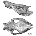
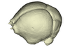
The present 3D Dataset contains the 3D model analyzed in the following publication: Carolina A. Hoffmann, A. G. Martinelli & M. B. Andrade. 2023. Anatomy of the holotype of “Probelesodon” kitchingi revisited, a chiniquodontid cynodont (Synapsida, Probainognathia) from the early Late Triassic of southern Brazil, Journal of Paleontology
Probelesodon kitchingi MCP 1600 PV View specimen
|
M3#11513D models of the skull with segmented bones and without the segmentation. colormap and orientation files also added. Type: "3D_surfaces"doi: 10.18563/m3.sf.1151 state:published |
Download 3D surface file |
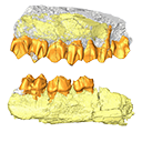
This contribution contains the 3D models of the set of Famennian conodont elements belonging to the species Polygnathus glaber and Polygnathus communis analyzed in the following publication: Renaud et al. 2021: Patterns of bilateral asymmetry and allometry in Late Devonian Polygnathus. Palaeontology. https://doi.org/10.1111/pala.12513
Polygnathus glaber UM BUS 001 View specimen
|
M3#574right P1 element Type: "3D_surfaces"doi: 10.18563/m3.sf.574 state:published |
Download 3D surface file |
Polygnathus glaber UM BUS 002 View specimen
|
M3#575right P1 element Type: "3D_surfaces"doi: 10.18563/m3.sf.575 state:published |
Download 3D surface file |
Polygnathus glaber UM BUS 003 View specimen
|
M3#576right P1 element Type: "3D_surfaces"doi: 10.18563/m3.sf.576 state:published |
Download 3D surface file |
Polygnathus glaber UM BUS 004 View specimen
|
M3#577left P1 element Type: "3D_surfaces"doi: 10.18563/m3.sf.577 state:published |
Download 3D surface file |
Polygnathus glaber UM BUS 005 View specimen

|
M3#578left P1 element Type: "3D_surfaces"doi: 10.18563/m3.sf.578 state:published |
Download 3D surface file |
Polygnathus glaber UM BUS 006 View specimen
|
M3#579right P1 element Type: "3D_surfaces"doi: 10.18563/m3.sf.579 state:published |
Download 3D surface file |
Polygnathus glaber UM BUS 007 View specimen
|
M3#580right P1 element Type: "3D_surfaces"doi: 10.18563/m3.sf.580 state:published |
Download 3D surface file |
Polygnathus glaber UM BUS 008 View specimen
|
M3#581left P1 element Type: "3D_surfaces"doi: 10.18563/m3.sf.581 state:published |
Download 3D surface file |
Polygnathus glaber UM BUS 009 View specimen
|
M3#582left P1 element Type: "3D_surfaces"doi: 10.18563/m3.sf.582 state:published |
Download 3D surface file |
Polygnathus glaber UM BUS 010 View specimen
|
M3#583right P1 element Type: "3D_surfaces"doi: 10.18563/m3.sf.583 state:published |
Download 3D surface file |
Polygnathus glaber UM BUS 011 View specimen

|
M3#584right P1 element Type: "3D_surfaces"doi: 10.18563/m3.sf.584 state:published |
Download 3D surface file |
Polygnathus glaber UM BUS 012 View specimen
|
M3#585right P1 element Type: "3D_surfaces"doi: 10.18563/m3.sf.585 state:published |
Download 3D surface file |
Polygnathus glaber UM BUS 013 View specimen

|
M3#586left P1 element Type: "3D_surfaces"doi: 10.18563/m3.sf.586 state:published |
Download 3D surface file |
Polygnathus glaber UM BUS 014 View specimen
|
M3#587left P1 element Type: "3D_surfaces"doi: 10.18563/m3.sf.587 state:published |
Download 3D surface file |
Polygnathus glaber UM BUS 015 View specimen

|
M3#588left P1 element Type: "3D_surfaces"doi: 10.18563/m3.sf.588 state:published |
Download 3D surface file |
Polygnathus glaber UM BUS 016 View specimen
|
M3#589right P1 element Type: "3D_surfaces"doi: 10.18563/m3.sf.589 state:published |
Download 3D surface file |
Polygnathus glaber UM BUS 017 View specimen

|
M3#590left P1 element Type: "3D_surfaces"doi: 10.18563/m3.sf.590 state:published |
Download 3D surface file |
Polygnathus glaber UM BUS 018 View specimen

|
M3#591left P1 element Type: "3D_surfaces"doi: 10.18563/m3.sf.591 state:published |
Download 3D surface file |
Polygnathus glaber UM BUS 019 View specimen
|
M3#592left P1 element Type: "3D_surfaces"doi: 10.18563/m3.sf.592 state:published |
Download 3D surface file |
Polygnathus glaber UM BUS 020 View specimen

|
M3#593left P1 element Type: "3D_surfaces"doi: 10.18563/m3.sf.593 state:published |
Download 3D surface file |
Polygnathus glaber UM BUS 021 View specimen
|
M3#594right P1 element Type: "3D_surfaces"doi: 10.18563/m3.sf.594 state:published |
Download 3D surface file |
Polygnathus glaber UM BUS 022 View specimen
|
M3#595left P1 element Type: "3D_surfaces"doi: 10.18563/m3.sf.595 state:published |
Download 3D surface file |
Polygnathus glaber UM BUS 023 View specimen

|
M3#596left P1 element Type: "3D_surfaces"doi: 10.18563/m3.sf.596 state:published |
Download 3D surface file |
Polygnathus glaber UM BUS 024 View specimen
|
M3#597left P1 element Type: "3D_surfaces"doi: 10.18563/m3.sf.597 state:published |
Download 3D surface file |
Polygnathus glaber UM BUS 025 View specimen

|
M3#598left P1 element Type: "3D_surfaces"doi: 10.18563/m3.sf.598 state:published |
Download 3D surface file |
Polygnathus glaber UM BUS 026 View specimen

|
M3#599left P1 element Type: "3D_surfaces"doi: 10.18563/m3.sf.599 state:published |
Download 3D surface file |
Polygnathus glaber UM BUS 027 View specimen

|
M3#600right P1 element Type: "3D_surfaces"doi: 10.18563/m3.sf.600 state:published |
Download 3D surface file |
Polygnathus glaber UM BUS 028 View specimen
|
M3#601right P1 element Type: "3D_surfaces"doi: 10.18563/m3.sf.601 state:published |
Download 3D surface file |
Polygnathus glaber UM BUS 029 View specimen

|
M3#602right P1 element Type: "3D_surfaces"doi: 10.18563/m3.sf.602 state:published |
Download 3D surface file |
Polygnathus glaber UM BUS 030 View specimen
|
M3#603right P1 element Type: "3D_surfaces"doi: 10.18563/m3.sf.603 state:published |
Download 3D surface file |
Polygnathus communis UM CTB 001 View specimen
|
M3#604right P1 element Type: "3D_surfaces"doi: 10.18563/m3.sf.604 state:published |
Download 3D surface file |
Polygnathus communis UM CTB 002 View specimen

|
M3#605right P1 element Type: "3D_surfaces"doi: 10.18563/m3.sf.605 state:published |
Download 3D surface file |
Polygnathus communis UM CTB 003 View specimen
|
M3#606right P1 element Type: "3D_surfaces"doi: 10.18563/m3.sf.606 state:published |
Download 3D surface file |
Polygnathus communis UM CTB 004 View specimen

|
M3#607right P1 element Type: "3D_surfaces"doi: 10.18563/m3.sf.607 state:published |
Download 3D surface file |
Polygnathus communis UM CTB 005 View specimen
|
M3#608left P1 element Type: "3D_surfaces"doi: 10.18563/m3.sf.608 state:published |
Download 3D surface file |
Polygnathus communis UM CTB 006 View specimen
|
M3#609left P1 element Type: "3D_surfaces"doi: 10.18563/m3.sf.609 state:published |
Download 3D surface file |
Polygnathus communis UM CTB 007 View specimen

|
M3#610left P1 element Type: "3D_surfaces"doi: 10.18563/m3.sf.610 state:published |
Download 3D surface file |
Polygnathus communis UM CTB 008 View specimen
|
M3#611left P1 element Type: "3D_surfaces"doi: 10.18563/m3.sf.611 state:published |
Download 3D surface file |
Polygnathus communis UM CTB 009 View specimen
|
M3#612right P1 element Type: "3D_surfaces"doi: 10.18563/m3.sf.612 state:published |
Download 3D surface file |
Polygnathus communis UM CTB 010 View specimen

|
M3#613left P1 element Type: "3D_surfaces"doi: 10.18563/m3.sf.613 state:published |
Download 3D surface file |
Polygnathus communis UM CTB 011 View specimen

|
M3#614right P1 element Type: "3D_surfaces"doi: 10.18563/m3.sf.614 state:published |
Download 3D surface file |
Polygnathus communis UM CTB 012 View specimen

|
M3#615right P1 element Type: "3D_surfaces"doi: 10.18563/m3.sf.615 state:published |
Download 3D surface file |
Polygnathus communis UM CTB 013 View specimen

|
M3#616right P1 element Type: "3D_surfaces"doi: 10.18563/m3.sf.616 state:published |
Download 3D surface file |
Polygnathus communis UM CTB 014 View specimen

|
M3#617right P1 element Type: "3D_surfaces"doi: 10.18563/m3.sf.617 state:published |
Download 3D surface file |
Polygnathus communis UM CTB 015 View specimen
|
M3#618right P1 element Type: "3D_surfaces"doi: 10.18563/m3.sf.618 state:published |
Download 3D surface file |
Polygnathus communis UM CTB 016 View specimen

|
M3#619left P1 element Type: "3D_surfaces"doi: 10.18563/m3.sf.619 state:published |
Download 3D surface file |
Polygnathus communis UM CTB 017 View specimen

|
M3#620right P1 element Type: "3D_surfaces"doi: 10.18563/m3.sf.620 state:published |
Download 3D surface file |
Polygnathus communis UM CTB 018 View specimen

|
M3#621right P1 element Type: "3D_surfaces"doi: 10.18563/m3.sf.621 state:published |
Download 3D surface file |
Polygnathus communis UM CTB 019 View specimen

|
M3#622right P1 element Type: "3D_surfaces"doi: 10.18563/m3.sf.622 state:published |
Download 3D surface file |
Polygnathus communis UM CTB 020 View specimen

|
M3#623right P1 element Type: "3D_surfaces"doi: 10.18563/m3.sf.623 state:published |
Download 3D surface file |
Polygnathus communis UM CTB 021 View specimen

|
M3#624left P1 element Type: "3D_surfaces"doi: 10.18563/m3.sf.624 state:published |
Download 3D surface file |
Polygnathus communis UM CTB 022 View specimen

|
M3#625left element Type: "3D_surfaces"doi: 10.18563/m3.sf.625 state:published |
Download 3D surface file |
Polygnathus communis UM CTB 023 View specimen

|
M3#626left P1 element Type: "3D_surfaces"doi: 10.18563/m3.sf.626 state:published |
Download 3D surface file |
Polygnathus communis UM CTB 024 View specimen

|
M3#627left P1 element Type: "3D_surfaces"doi: 10.18563/m3.sf.627 state:published |
Download 3D surface file |
Polygnathus communis UM CTB 025 View specimen

|
M3#628left P1 element Type: "3D_surfaces"doi: 10.18563/m3.sf.628 state:published |
Download 3D surface file |
Polygnathus communis UM CTB 026 View specimen
|
M3#629left P1 element Type: "3D_surfaces"doi: 10.18563/m3.sf.629 state:published |
Download 3D surface file |
Polygnathus communis UM CTB 027 View specimen

|
M3#630left P1 element Type: "3D_surfaces"doi: 10.18563/m3.sf.630 state:published |
Download 3D surface file |
Polygnathus communis UM CTB 028 View specimen

|
M3#631left P1 element Type: "3D_surfaces"doi: 10.18563/m3.sf.631 state:published |
Download 3D surface file |
Polygnathus communis UM CTB 029 View specimen
|
M3#632left P1 element Type: "3D_surfaces"doi: 10.18563/m3.sf.632 state:published |
Download 3D surface file |
Polygnathus communis UM CTB 030 View specimen

|
M3#633left P1 element Type: "3D_surfaces"doi: 10.18563/m3.sf.633 state:published |
Download 3D surface file |
Polygnathus communis UM CTB 031 View specimen
|
M3#634left P1 element Type: "3D_surfaces"doi: 10.18563/m3.sf.634 state:published |
Download 3D surface file |
Polygnathus communis UM CTB 032 View specimen
|
M3#635left P1 element Type: "3D_surfaces"doi: 10.18563/m3.sf.635 state:published |
Download 3D surface file |
Polygnathus communis UM CTB 033 View specimen

|
M3#636left P1 element Type: "3D_surfaces"doi: 10.18563/m3.sf.636 state:published |
Download 3D surface file |
Polygnathus communis UM CTB 034 View specimen

|
M3#637right P1 element Type: "3D_surfaces"doi: 10.18563/m3.sf.637 state:published |
Download 3D surface file |
The present 3D Dataset contains the 3D model analyzed in Wazir, W. A., Sehgal, R. K., Čerňanský, A., Patnaik, R., Kumar, N., Singh, A. P. and Singh, N. P. 2022. A find from the Ladakh Himalaya reveals a survival of madtsoiid snakes (Serpentes, Madtsoiidae) in India through the late Oligocene. Journal of Vertebrate Paleontology, 41(6), e2058401. https://doi.org/10.1080/02724634.2021.2058401
indet. indet. WIMF/A 4816 View specimen

|
M3#1754Vertebra Type: "3D_surfaces"doi: 10.18563/m3.sf.1754 state:published |
Download 3D surface file |
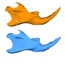
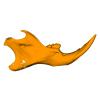
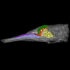
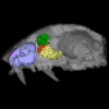
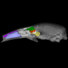
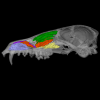
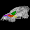
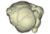
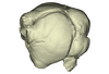
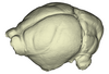
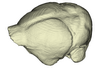
The present 3D Dataset contains the 3D models of extant Chiropteran endocranial casts, documenting 16 of the 19 extant bat families. They are used by Maugoust & Orliac (2023) to assess the correspondences between the brain and brain-surrounding tissues (i.e., neural tissues, blood vessels, meninges) and their imprint on the braincase, allowing for eventually proposing a Chiroptera-scale nomenclature of the endocast.
Balantiopteryx plicata UMMZ 102659 View specimen
|
M3#1132Endocranial cast of the corresponding cranium of Balantiopteryx plicata Type: "3D_surfaces"doi: 10.18563/m3.sf.1132 state:published |
Download 3D surface file |
Idiurus macrotis AMNH M-187705 View specimen
|
M3#1133Endocranial cast of the corresponding cranium of Nycteris macrotis Type: "3D_surfaces"doi: 10.18563/m3.sf.1133 state:published |
Download 3D surface file |
Thyroptera tricolor UMMZ 53240 View specimen
|
M3#1134Endocranial cast of the corresponding cranium of Thyroptera tricolor Type: "3D_surfaces"doi: 10.18563/m3.sf.1134 state:published |
Download 3D surface file |
Noctilio albiventris UMMZ 105827 View specimen
|
M3#1135Endocranial cast of the corresponding cranium of Noctilio albiventris Type: "3D_surfaces"doi: 10.18563/m3.sf.1135 state:published |
Download 3D surface file |
Mormoops blainvillii AMNH M-271513 View specimen
|
M3#1136Endocranial cast of the corresponding cranium of Mormoops blainvillii Type: "3D_surfaces"doi: 10.18563/m3.sf.1136 state:published |
Download 3D surface file |
Macrotus waterhousii UMMZ 95718 View specimen
|
M3#1137Endocranial cast of the corresponding cranium of Macrotus waterhousii Type: "3D_surfaces"doi: 10.18563/m3.sf.1137 state:published |
Download 3D surface file |
Nyctiellus lepidus UMMZ 105767 View specimen
|
M3#1138Endocranial cast of the corresponding cranium of Nyctiellus lepidus Type: "3D_surfaces"doi: 10.18563/m3.sf.1138 state:published |
Download 3D surface file |
Cheiromeles torquatus AMNH M-247585 View specimen
|
M3#1139Endocranial cast of the corresponding cranium of Cheiromeles torquatus Type: "3D_surfaces"doi: 10.18563/m3.sf.1139 state:published |
Download 3D surface file |
Miniopterus schreibersii UMMZ 156998 View specimen
|
M3#1140Endocranial cast of the corresponding cranium of Miniopterus schreibersii Type: "3D_surfaces"doi: 10.18563/m3.sf.1140 state:published |
Download 3D surface file |
Kerivoula pellucida UMMZ 161396 View specimen
|
M3#1141Endocranial cast of the corresponding cranium of Kerivoula pellucida Type: "3D_surfaces"doi: 10.18563/m3.sf.1141 state:published |
Download 3D surface file |
Scotophilus kuhlii UMMZ 157013 View specimen
|
M3#1142Endocranial cast of the corresponding cranium of Scotophilus kuhlii Type: "3D_surfaces"doi: 10.18563/m3.sf.1142 state:published |
Download 3D surface file |
Rhinolophus luctus MNHN CG-2006-87 View specimen
|
M3#1143Endocranial cast of the corresponding cranium of Rhinolophus luctus Type: "3D_surfaces"doi: 10.18563/m3.sf.1143 state:published |
Download 3D surface file |
Triaenops persicus AM RG-38552 View specimen
|
M3#1144Endocranial cast of the corresponding cranium of Triaenops persicus Type: "3D_surfaces"doi: 10.18563/m3.sf.1144 state:published |
Download 3D surface file |
Hipposideros armiger UM ZOOL-762-V View specimen
|
M3#1145Endocranial cast of the corresponding cranium of Hipposideros armiger Type: "3D_surfaces"doi: 10.18563/m3.sf.1145 state:published |
Download 3D surface file |
Lavia frons AM RG-12268 View specimen
|
M3#1146Endocranial cast of the corresponding cranium of Lavia frons Type: "3D_surfaces"doi: 10.18563/m3.sf.1146 state:published |
Download 3D surface file |
Rhinopoma hardwickii AM RG-M31166 View specimen
|
M3#1147Endocranial cast of the corresponding cranium of Rhinopoma hardwickii Type: "3D_surfaces"doi: 10.18563/m3.sf.1147 state:published |
Download 3D surface file |
Sphaerias blanfordi AMNH M-274330 View specimen
|
M3#1148Endocranial cast of the corresponding cranium of Sphaerias blanfordi Type: "3D_surfaces"doi: 10.18563/m3.sf.1148 state:published |
Download 3D surface file |
Rousettus aegyptiacus UMMZ 161026 View specimen
|
M3#1149Endocranial cast of the corresponding cranium of Rousettus aegyptiacus Type: "3D_surfaces"doi: 10.18563/m3.sf.1149 state:published |
Download 3D surface file |
Pteropus pumilus UMMZ 162253 View specimen
|
M3#1150Endocranial cast of the corresponding cranium of Pteropus pumilus Type: "3D_surfaces"doi: 10.18563/m3.sf.1150 state:published |
Download 3D surface file |
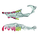
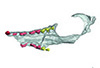
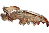
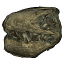
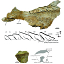
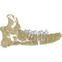
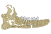
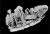
This contribution contains the 3D models described and figured in the following publication: Kassegne K. E., Mourlam M. J., Guinot G., Amoudji Y. Z., Martin J. E., Togbe K. A., Johnson A. K., Hautier L. 2021. First partial cranium of Togocetus from Kpogamé (Togo) and the protocetid diversity in the Togolese phosphate basin. Annales de Paléontologie, Issue 2, April–June 2021, 102488. https://doi.org/10.1016/j.annpal.2021.102488
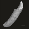
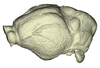
Togocetus cf. traversei ULDG-KPO1 View specimen
|
M3#768The specimen consists of a partial cranium prepared out of a calcareous phosphate matrix. The partial cranium lacks the anterior part of the rostrum, the cranial roof, and most of the basicranium apart from the left zygomatic process of the squamosal. The maxilla, nasal, palatine, pterygoid, alisphenoid, and squamosal bones are preserved, as well as two incomplete dental rows described hereafter. Type: "3D_surfaces"doi: 10.18563/m3.sf.768 state:published |
Download 3D surface file |
|
M3#770µCT . Resolution: 0.3156mm. This scan can easily be opened with Fiji, MorphoDig, 3DSlicer, or any software that reads .MHD file format. Also, the .RAW file can be opened easily with other software such as Avizo/Amira when providing the correct dimensions (which are enclosed within the file name) Type: "3D_CT"doi: 10.18563/m3.sf.770 state:published |
Download CT data |
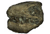
The present 3D Dataset contains the 3D model used in in the following publication: Interacting with the inaccessible: utilization of multimedia-based visual contents of Japan’s National Monument, the Taniwhasaurus mikasaensis (Mosasauridae) holotype for educational workshops at Mikasa City Museum.
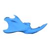
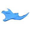
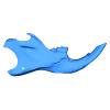
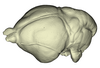
Taniwhasaurus mikasaensis MCM.M0009 View specimen
|
M3#499Taniwhasaurus mikasaensis, Caldwell et al. 2008 Type: "3D_surfaces"doi: 10.18563/m3.sf.499 state:published |
Download 3D surface file |
This contribution contains the three-dimensional models of the most complete and/or informative fossil materials attributed to Peradectes crocheti Gernelle, 2024, the earliest peradectid metatherian species of Europe, from its type locality (Palette, Provence, ~55 Ma). These specimens were analyzed and discussed in: Gernelle et al. (2024), Taxonomy and evolutionary history of peradectids (Metatheria): new data from the early Eocene of France. https://doi.org/10.1007/s10914-024-09724-5
Peradectes crocheti MHN.AIX.PV.2018.26.14 View specimen
|
M3#14993D surface model of MHN.AIX.PV.2018.26.14, fragmentary left maxilla with C-P1, anterior root of P2, and M1-M3 Type: "3D_surfaces"doi: 10.18563/m3.sf.1499 state:published |
Download 3D surface file |
Peradectes crocheti MHN.AIX.PV.2017.6.6 View specimen
|
M3#15003D surface model of MHN.AIX.PV.2017.6.6, left P2 Type: "3D_surfaces"doi: 10.18563/m3.sf.1500 state:published |
Download 3D surface file |
Peradectes crocheti MHN.AIX.PV.2017.6.7 View specimen
|
M3#15013D surface model of MHN.AIX.PV.2017.6.7, left M3 Type: "3D_surfaces"doi: 10.18563/m3.sf.1501 state:published |
Download 3D surface file |
Peradectes crocheti MHN.AIX.PV.2017.6.8 View specimen
|
M3#15023D surface model of MHN.AIX.PV.2017.6.8, right hemi-mandible fragment with canine alveolus, posterior root of p1, partial p2, p3, partial m1, and m2-m3 Type: "3D_surfaces"doi: 10.18563/m3.sf.1502 state:published |
Download 3D surface file |
Peradectes crocheti MHN.AIX.PV.2017.6.9 View specimen
|
M3#15033D surface model of MHN.AIX.PV.2017.6.9, leftm1-m4 row with fragments of dentary Type: "3D_surfaces"doi: 10.18563/m3.sf.1503 state:published |
Download 3D surface file |
Peradectes crocheti MHN.AIX.PV.2017.6.14 View specimen
|
M3#15043D surface model of MHN.AIX.PV.2017.6.14, right astragalus Type: "3D_surfaces"doi: 10.18563/m3.sf.1504 state:published |
Download 3D surface file |
This contribution contains the 3D models described and figured in: The Neogene record of northern South American native ungulates. Smithsonian Contributions to Paleobiology. Doi: 10.5479/si.1943-6688.101
Hilarcotherium miyou IGMp 881327 View specimen
|
M3#318Right upper M2 Type: "3D_surfaces"doi: 10.18563/m3.sf.318 state:published |
Download 3D surface file |
Hilarcotherium miyou MUN-STRI 34216 View specimen
|
M3#319Right upper P4 Type: "3D_surfaces"doi: 10.18563/m3.sf.319 state:published |
Download 3D surface file |
|
M3#320Right upper M2 Type: "3D_surfaces"doi: 10.18563/m3.sf.320 state:published |
Download 3D surface file |
Falcontoxodon aguilerai AMU-CURS 585 View specimen
|
M3#321Maxilla with left M3-P2 and right I2 Type: "3D_surfaces"doi: 10.18563/m3.sf.321 state:published |
Download 3D surface file |
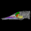
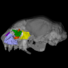
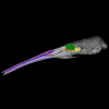
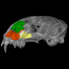
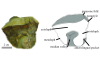
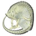
The present publication contains the µCT dataset and the 3D models analyzed in the following publication: Mautner, A.-K., A. E. Latimer, U. Fritz, and T. M. Scheyer. An updated description of the osteology of the pancake tortoise Malacochersus tornieri (Testudines: Testudinidae) with special focus on intraspecific variation. Journal of Morphology. https://doi.org/10.1002/jmor.20640
Malacochersus tornieri ZM 100.102 View specimen
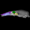
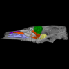
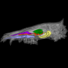
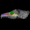
|
M3#129Virtual brain and inner ear endocast of Malacochersus tornieri (ZM 100.102; Zoological Museum of The University of Zurich). This virtual model is accompanied by the 3D dataset. Blue, endocranium; red, blood vessels; purple, semicircular canals; yellow, cranial nerves. Type: "3D_surfaces"doi: 10.18563/m3.sf.129 state:published |
Download 3D surface file |
|
M3#1303D dataset of skull of Malacochersus tornieri (ZM 100.102) Type: "3D_CT"doi: 10.18563/m3.sf.130 state:published |
Download CT data |
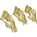
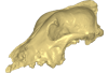
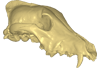
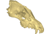
Archaeozoological studies are increasingly using new methods and approaches to explore questions about domestication. Here, we provide 3D models of three archaeological Canis lupus skulls from Belgium originating from the sites of Goyet (31,680±250BP; 31,890+240/-220BP), Trou des Nutons (21,810±90BP) and Trou Balleux (postglacial). Since their identification as either wolves or early dogs is still debated, we present these models as additional tools for further investigating their evolutionary history and the history of dog domestication.
Canis lupus Goyet 2860 View specimen
|
M3#213D surface model of the cranium of the Late Pleistocene Canis lupus "Goyet 2860" from the Royal Belgian Institute of Natural Sciences. Type: "3D_surfaces"doi: 10.18563/m3.sf21 state:published |
Download 3D surface file |
Canis lupus Trou Balleux no-nr View specimen
|
M3#223D surface model of the cranium of the Late Pleistocene Canis lupus "Trou Balleux no-nr" from the University of Liège, Belgium Type: "3D_surfaces"doi: 10.18563/m3.sf22 state:published |
Download 3D surface file |
Canis lupus Trou des Nutons 2559-1 View specimen
|
M3#233D surface model of the cranium of the Late Pleistocene Canis lupus "Trou des Nutons 2559-1" from the Royal Belgian Institute of Natural Sciences. Type: "3D_surfaces"doi: 10.18563/m3.sf23 state:published |
Download 3D surface file |
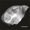
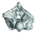
This project presents a µCT dataset and an associated 3D surface model of the holotype of Donrussellia magna (UM PAT 17; Primates, Adapiformes). UM PAT17 is the only known specimen for the species and consists of a well-preserved left lower jaw with p4-m3. It documents one of the oldest European primates, eventually dated near the Paleocene Eocene Thermal Maximum.
Donrussellia magna UM PAT 17 View specimen
|
M3#173D surface file model of UM PAT 17 (type specimen of Donrussellia magna), which is a well preserved left lower jaw with p4-m3. The teeth (and roots) were manually segmented. Type: "3D_surfaces"doi: 10.18563/m3.sf17 state:published |
Download 3D surface file |
|
M3#18CT Scan Data of Donrussellia magna UM PAT 17. Voxel size (in µm): 36µm (isotropic voxels). Dimensions in x,y,z : 594 pixels, 294 pixels, 1038 pixels. Image type : 8-bit voxels. Image format : raw data format (no header). Type: "3D_CT"doi: 10.18563/m3.sf18 state:published |
Download CT data |
This contribution contains 3D models of the holotype of a new species of long-nosed armadillos, the Guianan long-nosed armadillo (Dasypus guianensis) described in the following publication: Barthe M., Rancilhac L., Arteaga M. C., Feijó A., Tilak M.-K., Justy F., Loughry W. J., McDonough C. M., de Thoisy B., Catzeflis F., Billet G., Hautier L., Nabholz B., and Delsuc F. 2024. Exon capture museomics deciphers the nine-banded armadillo species complex and identifies a new species endemic to the Guiana Shield. Systematic Biology, syae027. https://doi.org/10.1093/sysbio/syae027
Dasypus guianensis MNHN-ZM-MO-2001-1317 View specimen
|
M3#1200Skeleton and carapace Type: "3D_surfaces"doi: 10.18563/m3.sf.1200 state:published |
Download 3D surface file |
|
M3#1201Frontal sinuses Type: "3D_surfaces"doi: 10.18563/m3.sf.1201 state:published |
Download 3D surface file |