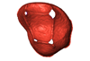
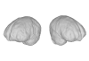
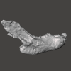
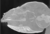
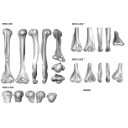
3D models of Kalakocetus, the earliest Cetacea
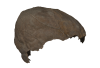
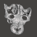
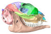
Explodable 3D Dog Skull for Veterinary Education
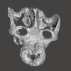
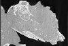
3D models related to the publication: Hidden diversity of Palaeogene metatherians: a new family of polydolopimorphian marsupials from Peruvian Amazonia
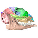
3D GM dataset of bird skeletal variation
Skeletal embryonic development in the catshark
Bony connexions of the petrosal bone of extant hippos
bony labyrinth (14) , inner ear (11) , Eocene (11) , geometric morphometrics (10) , CT-scan (10) , Oligocene (9) , Micro-CT (9)
Maëva Judith Orliac (24) , Lionel Hautier (24) , Laurent Marivaux (18) , Renaud Lebrun (15) , Rodolphe Tabuce (14) , Pierre-Olivier Antoine (13) , Bastien Mennecart (13)
|
3D models related to the publication: “The functional significance of aberrant cervical counts in sloths: insights from automated exhaustive analysis of cervical range of motion”Luisa J. Merten
Published online: 04/11/2023 |
|
M3#1260cervical vertebral series (7 vertebrae) Type: "3D_surfaces"doi: 10.18563/m3.sf.1260 state:published |
Download 3D surface file |
Bradypus variegatus ZMB_Mam_91345 View specimen
|
M3#1261cervical vertebral series (8 vertebrae) + first thoracic vertebra Type: "3D_surfaces"doi: 10.18563/m3.sf.1261 state:published |
Download 3D surface file |
Bradypus variegatus ZMB_Mam_35824 View specimen

|
M3#1262cervical vertebral series (8 vertebrae) + first & second thoracic vertebra Type: "3D_surfaces"doi: 10.18563/m3.sf.1262 state:published |
Download 3D surface file |
Choloepus didactylus ZMB_Mam_38388 View specimen
|
M3#1263cervical vertebral series (7 vertebrae) Type: "3D_surfaces"doi: 10.18563/m3.sf.1263 state:published |
Download 3D surface file |
Choloepus didactylus ZMB_Mam_102634 View specimen
|
M3#1264cervical vertebral series (6 vertebrae) + first thoracic vertebra Type: "3D_surfaces"doi: 10.18563/m3.sf.1264 state:published |
Download 3D surface file |
Tamandua tetradactyla ZMB_Mam_91288 View specimen
|
M3#1266cervical vertebral series (7 vertebrae) + first thoracic vertebra Type: "3D_surfaces"doi: 10.18563/m3.sf.1266 state:published |
Download 3D surface file |
Glossotherium robustum MNHN_n/n View specimen
|
M3#1267cervical vertebral series (7 vertebrae) + first thoracic vertebra Type: "3D_surfaces"doi: 10.18563/m3.sf.1267 state:published |
Download 3D surface file |
This contribution contains the three-dimensional models of the inner ear of the hetaxodontid rodents Amblyrhiza, Clidomys and Elasmodontomys from the West Indies. These specimens were analyzed and discussed in : The inner ear of caviomorph rodents: phylogenetic implications and application to extinct West Indian taxa.
Amblyrhiza inundata 11842 View specimen
|
M3#11543D surface of the left-oriented inner ear of Amblyrhiza. Type: "3D_surfaces"doi: 10.18563/m3.sf.1154 state:published |
Download 3D surface file |
Clidomys sp NA View specimen
|
M3#11553D surface of the left-oriented inner ear of Clidomys sp. Type: "3D_surfaces"doi: 10.18563/m3.sf.1155 state:published |
Download 3D surface file |
Elasmodontomys obliquus 17127 View specimen
|
M3#11563D surface of the left-oriented inner ear of Elasmodontomys obliquus. Type: "3D_surfaces"doi: 10.18563/m3.sf.1156 state:published |
Download 3D surface file |
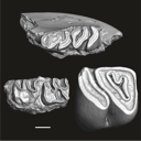
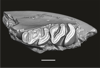
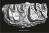
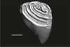
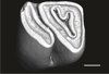
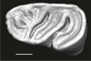
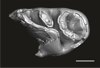
This contribution contains the three-dimensional digital models of a part of the dental fossil material (the large specimens) of caviomorph rodents, discovered in late middle Miocene detrital deposits of the TAR-31 locality in Peruvian Amazonia (San Martín, Peru). These fossils were described, figured and discussed in the following publication: Boivin, Marivaux et al. (2021), Late middle Miocene caviomorph rodents from Tarapoto, Peruvian Amazonia. PLoS ONE 16(11): e0258455. https://doi.org/10.1371/journal.pone.0258455
Microscleromys paradoxalis MUSM 4643 View specimen
|
M3#1115Fragment of left mandibule preserving dp4, m1 and a portion of incisor Type: "3D_surfaces"doi: 10.18563/m3.sf.1115 state:published |
Download 3D surface file |
Ricardomys longidens MUSM 4375 View specimen
|
M3#1116Fragment of left maxillary preserving DP4 and M1 (or M1 and M2) Type: "3D_surfaces"doi: 10.18563/m3.sf.1116 state:published |
Download 3D surface file |
"Scleromys" sp. MUSM 4272 View specimen
|
M3#1117Isolated left upper molar Type: "3D_surfaces"doi: 10.18563/m3.sf.1117 state:published |
Download 3D surface file |
"Scleromys" sp. MUSM 4275 View specimen
|
M3#1118Isolated right upper molar Type: "3D_surfaces"doi: 10.18563/m3.sf.1118 state:published |
Download 3D surface file |
"Scleromys" sp. MUSM 4273 View specimen
|
M3#1119Isolated left upper molar Type: "3D_surfaces"doi: 10.18563/m3.sf.1119 state:published |
Download 3D surface file |
"Scleromys" sp. MUSM 4276 View specimen
|
M3#1120Isolated right upper molar Type: "3D_surfaces"doi: 10.18563/m3.sf.1120 state:published |
Download 3D surface file |
"Scleromys" sp. MUSM 4282 View specimen
|
M3#1121Isolated right lower molar Type: "3D_surfaces"doi: 10.18563/m3.sf.1121 state:published |
Download 3D surface file |
"Scleromys" sp. MUSM 4281 View specimen
|
M3#1122Isolated right lower molar Type: "3D_surfaces"doi: 10.18563/m3.sf.1122 state:published |
Download 3D surface file |
"Scleromys" sp. MUSM 4280 View specimen
|
M3#1123Isolated left p4 Type: "3D_surfaces"doi: 10.18563/m3.sf.1123 state:published |
Download 3D surface file |
"Scleromys" sp. MUSM 4277 View specimen
|
M3#1124Isolated left lower dp4 Type: "3D_surfaces"doi: 10.18563/m3.sf.1124 state:published |
Download 3D surface file |
"Scleromys" sp. MUSM 4279 View specimen
|
M3#1125Isolated right lower dp4 (mesial fragment) Type: "3D_surfaces"doi: 10.18563/m3.sf.1125 state:published |
Download 3D surface file |
gen.indet sp. indet MUSM 4283 View specimen
|
M3#1126Isolated right lower p4 Type: "3D_surfaces"doi: 10.18563/m3.sf.1126 state:published |
Download 3D surface file |
Microscleromys sp. MUSM 4658 View specimen
|
M3#1127Isolated left tarsal bone (astragalus) Type: "3D_surfaces"doi: 10.18563/m3.sf.1127 state:published |
Download 3D surface file |
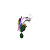
This contribution includes the 3D models of the reconstructed ossicular chain of the cainotheriid Caenomeryx filholi from the late Oligocene locality of Pech Desse (MP28, Quercy, France) described and figured in the publication of Assemat et al. (2020). It represents the oldest ossicular chain reconstruction for a Paleogene terrestrial artiodactyl species.
Caenomeryx filholi UM PDS 3353 View specimen
|
M3#508reconstruction of the middle ear with petrosal, bulla, stapes, incus, malleus Type: "3D_surfaces"doi: 10.18563/m3.sf.508 state:published |
Download 3D surface file |
The present 3D Dataset contains the 3D models of the enamel-dentine junctions of upper third molars and of the bony labyrinths of the extant cercopithecoid specimens analyzed in the following publication: Beaudet, A., Dumoncel, J., Thackeray, J.F., Bruxelles, L., Duployer, B., Tenailleau, C., Bam, L., Hoffman, J., de Beer, F., Braga, J.: Upper third molar internal structural organization and semicircular canal morphology in Plio-Pleistocene South African cercopithecoids. Journal of Human Evolution 95, 104-120. https://doi.org/10.1016/j.jhevol.2016.04.004
Cercocebus atys 81.007-M-0041 View specimen
|
M3#4453D model of the enamel-dentine junction of the right upper third molar. Type: "3D_surfaces"doi: 10.18563/m3.sf.445 state:published |
Download 3D surface file |
Cercocebus torquatus 73.018-M-0359 View specimen
|
M3#4463D model of the enamel-dentine junction of the right upper third molar. Type: "3D_surfaces"doi: 10.18563/m3.sf.446 state:published |
Download 3D surface file |

|
M3#4963D model of the left bony labyrinth. Type: "3D_surfaces"doi: 10.18563/m3.sf.496 state:published |
Download 3D surface file |
Mandrillus leucophaeus 73.029-M-0106 View specimen
|
M3#4473D model of the enamel-dentine junction of the right upper third molar. Type: "3D_surfaces"doi: 10.18563/m3.sf.447 state:published |
Download 3D surface file |

|
M3#4703D model of the right bony labyrinth. Type: "3D_surfaces"doi: 10.18563/m3.sf.470 state:published |
Download 3D surface file |
Lophocebus albigena 73.029-M-0109 View specimen
|
M3#4483D model of the enamel-dentine junction of the right upper third molar. Type: "3D_surfaces"doi: 10.18563/m3.sf.448 state:published |
Download 3D surface file |
|
M3#4713D model of the right bony labyrinth. Type: "3D_surfaces"doi: 10.18563/m3.sf.471 state:published |
Download 3D surface file |
Piliocolobus foai 91.060-M-0071 View specimen
|
M3#4493D model of the enamel-dentine junction of the right upper third molar. Type: "3D_surfaces"doi: 10.18563/m3.sf.449 state:published |
Download 3D surface file |

|
M3#4723D model of the right bony labyrinth. Type: "3D_surfaces"doi: 10.18563/m3.sf.472 state:published |
Download 3D surface file |
Colobus guereza 1215 View specimen
|
M3#4503D model of the enamel-dentine junction of the right upper third molar. Type: "3D_surfaces"doi: 10.18563/m3.sf.450 state:published |
Download 3D surface file |

|
M3#4733D model of the right bony labyrinth. Type: "3D_surfaces"doi: 10.18563/m3.sf.473 state:published |
Download 3D surface file |
Colobus guereza 2800 View specimen
|
M3#4513D model of the enamel-dentine junction of the right upper third molar. Type: "3D_surfaces"doi: 10.18563/m3.sf.451 state:published |
Download 3D surface file |

|
M3#4743D model of the right bony labyrinth. Type: "3D_surfaces"doi: 10.18563/m3.sf.474 state:published |
Download 3D surface file |
Papio cynocephalus kindae 3503 View specimen
|
M3#4523D model of the enamel-dentine junction of the right upper third molar. Type: "3D_surfaces"doi: 10.18563/m3.sf.452 state:published |
Download 3D surface file |

|
M3#4753D model of the right bony labyrinth. Type: "3D_surfaces"doi: 10.18563/m3.sf.475 state:published |
Download 3D surface file |
Erythrocebus patas 8452 View specimen
|
M3#4533D model of the enamel-dentine junction of the right upper third molar. Type: "3D_surfaces"doi: 10.18563/m3.sf.453 state:published |
Download 3D surface file |

|
M3#4763D model of the right bony labyrinth. Type: "3D_surfaces"doi: 10.18563/m3.sf.476 state:published |
Download 3D surface file |
Papio cynocephalus kindae 17979 View specimen
|
M3#4543D model of the enamel-dentine junction of the right upper third molar. Type: "3D_surfaces"doi: 10.18563/m3.sf.454 state:published |
Download 3D surface file |

|
M3#4773D model of the right bony labyrinth. Type: "3D_surfaces"doi: 10.18563/m3.sf.477 state:published |
Download 3D surface file |
Colobus angolensis 25456 View specimen
|
M3#4553D model of the enamel-dentine junction of the right upper third molar. Type: "3D_surfaces"doi: 10.18563/m3.sf.455 state:published |
Download 3D surface file |

|
M3#4783D model of the right bony labyrinth. Type: "3D_surfaces"doi: 10.18563/m3.sf.478 state:published |
Download 3D surface file |
Chlorocebus pygerythrus 37477 View specimen
|
M3#4563D model of the enamel-dentine junction of the right upper third molar. Type: "3D_surfaces"doi: 10.18563/m3.sf.456 state:published |
Download 3D surface file |
|
M3#4813D model of the right bony labyrinth. Type: "3D_surfaces"doi: 10.18563/m3.sf.481 state:published |
Download 3D surface file |
Chlorocebus pygerythrus 37478 View specimen
|
M3#4573D model of the enamel-dentine junction of the right upper third molar. Type: "3D_surfaces"doi: 10.18563/m3.sf.457 state:published |
Download 3D surface file |

|
M3#4823D model of the right bony labyrinth. Type: "3D_surfaces"doi: 10.18563/m3.sf.482 state:published |
Download 3D surface file |
Lophocebus albigena 37572 View specimen
|
M3#4583D model of the enamel-dentine junction of the right upper third molar. Type: "3D_surfaces"doi: 10.18563/m3.sf.458 state:published |
Download 3D surface file |

|
M3#4833D model of the right bony labyrinth. Type: "3D_surfaces"doi: 10.18563/m3.sf.483 state:published |
Download 3D surface file |
Lophocebus albigena 37579 View specimen
|
M3#4593D model of the enamel-dentine junction of the right upper third molar. Type: "3D_surfaces"doi: 10.18563/m3.sf.459 state:published |
Download 3D surface file |
Erythrocebus patas OST.2002-26 View specimen
|
M3#4603D model of the enamel-dentine junction of the right upper third molar. Type: "3D_surfaces"doi: 10.18563/m3.sf.460 state:published |
Download 3D surface file |
|
M3#4843D model of the right bony labyrinth. Type: "3D_surfaces"doi: 10.18563/m3.sf.484 state:published |
Download 3D surface file |
Mandrillus sphinx OST.AC.488 View specimen
|
M3#4613D model of the enamel-dentine junction of the right upper third molar. Type: "3D_surfaces"doi: 10.18563/m3.sf.461 state:published |
Download 3D surface file |
|
M3#4853D model of the left bony labyrinth. Type: "3D_surfaces"doi: 10.18563/m3.sf.485 state:published |
Download 3D surface file |
Macaca mulatta OST.AC.492 View specimen
|
M3#4623D model of the enamel-dentine junction of the right upper third molar. Type: "3D_surfaces"doi: 10.18563/m3.sf.462 state:published |
Download 3D surface file |

|
M3#4863D model of the right bony labyrinth. Type: "3D_surfaces"doi: 10.18563/m3.sf.486 state:published |
Download 3D surface file |
Chlorocebus aethiops OST.AC.523 View specimen
|
M3#4633D model of the enamel-dentine junction of the right upper third molar. Type: "3D_surfaces"doi: 10.18563/m3.sf.463 state:published |
Download 3D surface file |

|
M3#4913D model of the right bony labyrinth. Type: "3D_surfaces"doi: 10.18563/m3.sf.491 state:published |
Download 3D surface file |
Cercopithecus cephus OST.AC.533 View specimen
|
M3#4643D model of the enamel-dentine junction of the right upper third molar. Type: "3D_surfaces"doi: 10.18563/m3.sf.464 state:published |
Download 3D surface file |
|
M3#4933D model of the right bony labyrinth. Type: "3D_surfaces"doi: 10.18563/m3.sf.493 state:published |
Download 3D surface file |
Chlorocebus aethiops OST.AC.540 View specimen
|
M3#4653D model of the enamel-dentine junction of the right upper third molar. Type: "3D_surfaces"doi: 10.18563/m3.sf.465 state:published |
Download 3D surface file |

|
M3#4943D model of the right bony labyrinth. Type: "3D_surfaces"doi: 10.18563/m3.sf.494 state:published |
Download 3D surface file |
Mandrillus sphinx OST.AC.543 View specimen
|
M3#4663D model of the enamel-dentine junction of the right upper third molar. Type: "3D_surfaces"doi: 10.18563/m3.sf.466 state:published |
Download 3D surface file |
|
M3#4953D model of the right bony labyrinth. Type: "3D_surfaces"doi: 10.18563/m3.sf.495 state:published |
Download 3D surface file |
Cercocebus torquatus 73.018-M-389 View specimen

|
M3#4683D model of the right bony labyrinth. Type: "3D_surfaces"doi: 10.18563/m3.sf.468 state:published |
Download 3D surface file |
Mandrillus leucophaeus 73.029-M-0105 View specimen
|
M3#4693D model of the right bony labyrinth. Type: "3D_surfaces"doi: 10.18563/m3.sf.469 state:published |
Download 3D surface file |
Mandrillus leucophaeus 28425 View specimen

|
M3#4793D model of the right bony labyrinth. Type: "3D_surfaces"doi: 10.18563/m3.sf.479 state:published |
Download 3D surface file |
Cercocebus atys 28998 View specimen

|
M3#4803D model of the right bony labyrinth. Type: "3D_surfaces"doi: 10.18563/m3.sf.480 state:published |
Download 3D surface file |
Macaca sylvanus OST.AC.493 View specimen

|
M3#4873D model of the right bony labyrinth. Type: "3D_surfaces"doi: 10.18563/m3.sf.487 state:published |
Download 3D surface file |
Chlorocebus aethiops OST.AC.508 View specimen

|
M3#4883D model of the left bony labyrinth. Type: "3D_surfaces"doi: 10.18563/m3.sf.488 state:published |
Download 3D surface file |
Cercopithecus cephus OST.AC.515 View specimen

|
M3#4893D model of the right bony labyrinth. Type: "3D_surfaces"doi: 10.18563/m3.sf.489 state:published |
Download 3D surface file |
Colobus guereza OST.AC.519 View specimen

|
M3#4903D model of the right bony labyrinth. Type: "3D_surfaces"doi: 10.18563/m3.sf.490 state:published |
Download 3D surface file |
Macaca sp. OST.AC.532 View specimen

|
M3#4923D model of the left bony labyrinth. Type: "3D_surfaces"doi: 10.18563/m3.sf.492 state:published |
Download 3D surface file |
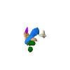
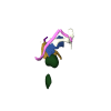
This project presents the 3D models of two isolated petrosals from the Oligocene locality of Pech de Fraysse (Quercy, France) here attributed to the genus Prodremotherium Filhol, 1877. Our aim is to describe the petrosal morphology of this Oligocene “early ruminant” as only few data are available in the literature for Oligocene taxa.
Prodremotherium sp. UM PFY 4053 View specimen

|
M3#7Labelled 3D model of right isolated petrosal of Prodremotherium sp. from Pech de Fraysse (Quercy, MP 28) Type: "3D_surfaces"doi: 10.18563/m3.sf7 state:published |
Download 3D surface file |
Prodremotherium sp. UM PFY 4054 View specimen

|
M3#8Labelled 3D model of right isolated petrosal of Prodremotherium sp. from Pech de Fraysse (Quercy, MP 28) Type: "3D_surfaces"doi: 10.18563/m3.sf8 state:published |
Download 3D surface file |
The present 3D Dataset contains the 3D models analyzed in Brualla et al., 2024: Comparative anatomy of the vocal apparatus in bats and implication for the diversity of laryngeal echolocation. Zoological Journal of the Linnean Society, vol. zlad180. (https://doi.org/10.1093/zoolinnean/zlad180). Bat larynges are understudied in the previous anatomical studies. The description and comparison of the different morphological traits might provide important proxies to investigate the evolutionary origin of laryngeal echolocation in bats.
Eonycteris spelaea VN18-026 View specimen

|
M3#1305Laryngeal cartilages and muscles of the cave nectar bat Type: "3D_surfaces"doi: 10.18563/m3.sf.1305 state:published |
Download 3D surface file |
Macroglossus sobrinus VN15-017 View specimen
|
M3#1306Laryngeal anatomy of Macroglossus sobrinus Type: "3D_surfaces"doi: 10.18563/m3.sf.1306 state:published |
Download 3D surface file |
Aselliscus dongbacana VTTu15-013 View specimen
|
M3#1307Laryngeal anatomy of Aselliscus dongbacana Type: "3D_surfaces"doi: 10.18563/m3.sf.1307 state:published |
Download 3D surface file |
Coelops frithii VN19-196 View specimen
|
M3#1308Laryngeal anatomy of Coelops frithii Type: "3D_surfaces"doi: 10.18563/m3.sf.1308 state:published |
Download 3D surface file |
Hipposideros larvatus VN18-209 View specimen
|
M3#1309Laryngeal anatomy of Hipposideros larvatus Type: "3D_surfaces"doi: 10.18563/m3.sf.1309 state:published |
Download 3D surface file |
Rhinolophus cornutus JP21-025 View specimen
|
M3#14763D surfaces of Rhinolophus cornutus Type: "3D_surfaces"doi: 10.18563/m3.sf.1476 state:published |
Download 3D surface file |
Rhinolophus macrotis VN11-089 View specimen
|
M3#1477Laryngeal cartilages and muscles of Rhinolophus macrotis Type: "3D_surfaces"doi: 10.18563/m3.sf.1477 state:published |
Download 3D surface file |
Lyroderma lyra VN17-535 View specimen
|
M3#1312Laryngeal anatomy of Lyroderma lyra Type: "3D_surfaces"doi: 10.18563/m3.sf.1312 state:published |
Download 3D surface file |
Saccolaimus mixtus A3257 View specimen
|
M3#1478Laryngeal components of Saccolaimus mixtus Type: "3D_surfaces"doi: 10.18563/m3.sf.1478 state:published |
Download 3D surface file |
Taphozous melanopogon VN17-0252 View specimen
|
M3#1479Laryngeal cartilages and muscles of Taphozous melanopogon Type: "3D_surfaces"doi: 10.18563/m3.sf.1479 state:published |
Download 3D surface file |
Artibeus jamaicensis AJ001 View specimen
|
M3#1316Laryngeal anatomy of Artibeus jamaicensis Type: "3D_surfaces"doi: 10.18563/m3.sf.1316 state:published |
Download 3D surface file |
Kerivoula hardwickii VN11-0043 View specimen
|
M3#1317Laryngeal anatomy of Kerivoula hardwickii Type: "3D_surfaces"doi: 10.18563/m3.sf.1317 state:published |
Download 3D surface file |
Myotis ater VN19-016 View specimen
|
M3#1318Laryngeal anatomy of Myotis ater Type: "3D_surfaces"doi: 10.18563/m3.sf.1318 state:published |
Download 3D surface file |
Myotis siligorensis VTTu14-018 View specimen
|
M3#1319Laryngeal anatomy of Myotis siligorensis Type: "3D_surfaces"doi: 10.18563/m3.sf.1319 state:published |
Download 3D surface file |
Suncus murinus KATS_835A View specimen
|
M3#1395Laryngeal anatomy of Suncus murinus Type: "3D_surfaces"doi: 10.18563/m3.sf.1395 state:published |
Download 3D surface file |
The present 3D Dataset contains the 3D models analyzed in 3D Finite Element Analysis and Geometric Morphometrics of Sloths (Xenarthra, Folivora) Mandibles Show Insights on the Dietary Specializations of Fossil Taxa. Journal of South American Earth Sciences. https://doi.org/10.1016/j.jsames.2023.104445
Mylodon darwinii CAV 379 View specimen
|
M3#1159Right hemimandible Type: "3D_surfaces"doi: 10.18563/m3.sf.1159 state:published |
Download 3D surface file |
Scelidotherium leptocephalum MNHN-M 137,722 View specimen
|
M3#1160Mandible Type: "3D_surfaces"doi: 10.18563/m3.sf.1160 state:published |
Download 3D surface file |
Glossotherium robustum MNHN-M 914 View specimen
|
M3#1161Mandible Type: "3D_surfaces"doi: 10.18563/m3.sf.1161 state:published |
Download 3D surface file |
Lestodon armatus MPAC 899 View specimen
|
M3#1162Mandible Type: "3D_surfaces"doi: 10.18563/m3.sf.1162 state:published |
Download 3D surface file |
Valgipes bucklandi NHMD.Z.M.K. 1/1845:3540 View specimen
|
M3#1163Mandible Type: "3D_surfaces"doi: 10.18563/m3.sf.1163 state:published |
Download 3D surface file |
The present 3D Dataset contains the 3D models analyzed in the following publication: Le Verger K., González Ruiz L.R., Billet G. 2021. Comparative anatomy and phylogenetic contribution of intracranial osseous canals and cavities in armadillos and glyptodonts (Xenarthra, Cingulata). Journal of Anatomy 00: 1-30 p. https://doi.org/10.1111/joa.13512
Bradypus tridactylus MNHN ZM-MO-1999-1065 View specimen
|
M3#844Bradypus tridactylus MNHN ZM-MO-1999-1065: cranium, cranial canals & alveolar cavities. Type: "3D_surfaces"doi: 10.18563/m3.sf.844 state:published |
Download 3D surface file |
Tamandua tetradactyla NHMUK ZD-1903.7.7.135 View specimen
|
M3#845Tamandua tetradactyla NHMUK ZD-1903.7.7.135: cranium, cranial canals & alveolar cavities. Type: "3D_surfaces"doi: 10.18563/m3.sf.845 state:published |
Download 3D surface file |
Dasypus novemcinctus AMNH 33150 View specimen
|
M3#846Dasypus novemcinctus AMNH 33150: cranium, cranial canals & alveolar cavities. Type: "3D_surfaces"doi: 10.18563/m3.sf.846 state:published |
Download 3D surface file |
Dasypus novemcinctus AMNH 133261 View specimen
|
M3#847Dasypus novemcinctus AMNH 133261: cranium, cranial canals & alveolar cavities. Type: "3D_surfaces"doi: 10.18563/m3.sf.847 state:published |
Download 3D surface file |
Dasypus novemcinctus AMNH 133328 View specimen
|
M3#848Dasypus novemcinctus AMNH 133328: cranium, cranial canals & alveolar cavities. Type: "3D_surfaces"doi: 10.18563/m3.sf.848 state:published |
Download 3D surface file |
Zaedyus pichiy ZMB-MAM-49039 View specimen
|
M3#849Zaedyus pichiy ZMB-MAM-49039: cranium, cranial canals & alveolar cavities. Type: "3D_surfaces"doi: 10.18563/m3.sf.849 state:published |
Download 3D surface file |
Zaedyus pichiy MHNG 1627.053 View specimen

|
M3#850Zaedyus pichiy MHNG 1627.053: cranium, cranial canals & alveolar cavities. Type: "3D_surfaces"doi: 10.18563/m3.sf.850 state:published |
Download 3D surface file |
Zaedyus pichiy MHNG 1276.076 View specimen
|
M3#851Zaedyus pichiy MHNG 1276.076: cranium, cranial canals & alveolar cavities. Type: "3D_surfaces"doi: 10.18563/m3.sf.851 state:published |
Download 3D surface file |
Cabassous unicinctus NBC_ZMA.MAM.26326.a View specimen
|
M3#852Cabassous unicinctus NBC ZMA.MAM.26326.a: cranium, cranial canals & alveolar cavities. Type: "3D_surfaces"doi: 10.18563/m3.sf.852 state:published |
Download 3D surface file |
Cabassous unicinctus MNHN-CG-1999-1044 View specimen
|
M3#853Cabassous unicinctus MNHN-CG-1999-1044: cranium, cranial canals & alveolar cavities. Type: "3D_surfaces"doi: 10.18563/m3.sf.853 state:published |
Download 3D surface file |
Vassallia maxima FMNH P14424 View specimen
|
M3#854Vassallia maxima FMNH P14424: cranium, cranial canals & alveolar cavities. Type: "3D_surfaces"doi: 10.18563/m3.sf.854 state:published |
Download 3D surface file |
Glyptodon sp. MNHN-F-PAM-759 View specimen
|
M3#855Glyptodon sp. MNHN-F-PAM-759: cranium, cranial canals & alveolar cavities. Type: "3D_surfaces"doi: 10.18563/m3.sf.855 state:published |
Download 3D surface file |
Glyptodon sp. MNHN-F-PAM-760 View specimen

|
M3#856Glyptodon sp. MNHN-F-PAM-760: cranium, cranial canals & alveolar cavities. Type: "3D_surfaces"doi: 10.18563/m3.sf.856 state:published |
Download 3D surface file |
The present 3D Dataset contains the 3D model analyzed in Hendrickx, C. and Bell, P. R. 2021. The scaly skin of the abelisaurid Carnotaurus sastrei (Theropoda: Ceratosauria) from the Upper Cretaceous of Patagonia. Cretaceous Research. https://doi.org/10.1016/j.cretres.2021.104994
Carnotaurus sastrei MACN 894 View specimen
|
M3#8023D reconstruction of the biggest patch of skin (~1200 cm2) from the anterior tail region of the holotype of Carnotaurus, which is the largest single patch of squamous integument available for any saurischian. The skin consists of medium to large (up to 65 mm in diameter) conical feature scales surrounded by a network of low and small (< 14 mm) irregular basement scales separated by narrow interstitial tissue. Type: "3D_surfaces"doi: 10.18563/m3.sf.802 state:published |
Download 3D surface file |
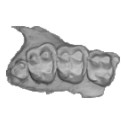
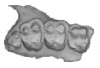
This contribution contains the 3D models of the fossil remains (maxilla, dentary, and talus) attributed to Djebelemur martinezi, a ca. 50 Ma primate from Tunisia (Djebel Chambi), described and figured in the following publication: Marivaux et al. (2013), Djebelemur, a tiny pre-tooth-combed primate from the Eocene of Tunisia: a glimpse into the origin of crown strepsirhines. PLoS ONE. https://doi.org/10.1371/journal.pone.0080778
Djebelemur martinezi CBI-1-544 View specimen
|
M3#365CBI-1-544, left maxilla preserving P3-M3 and alveoli for P2 and C1 Type: "3D_surfaces"doi: 10.18563/m3.sf.365 state:published |
Download 3D surface file |
Djebelemur martinezi CBI-1-567 View specimen
|
M3#363Isolated left upper P4 Type: "3D_surfaces"doi: 10.18563/m3.sf.363 state:published |
Download 3D surface file |
Djebelemur martinezi CBI-1-565-577-587-580 View specimen
|
M3#366- CBI-1-565, a damaged right mandible, which consists of three isolated pieces found together and reassembled here: the anterior part of the dentary bears the p3 and m1, and alveoli for p4, p2 and c, while the posterior part preserves m3 and a portion of the ascending ramus; the m2 was found isolated but in the same small calcareous block treated by acid processing. - CBI-1-577, isolated right lower p4. - CBI-1-587, isolated left lower p2 (reversed). - CBI-1-580, isolated left lower canine (reversed). Type: "3D_surfaces"doi: 10.18563/m3.sf.366 state:published |
Download 3D surface file |
Djebelemur martinezi CBI-1-545 View specimen
|
M3#364Right Talus Type: "3D_surfaces"doi: 10.18563/m3.sf.364 state:published |
Download 3D surface file |
This contribution contains the 3D model of an endocranial cast analyzed in “A 10 ka intentionally deformed human skull from Northeast Asia”. There are many studies on the morphological characteristics of intentional cranial deformation (ICD), but few related 3D models were published. Here, we present the surface model of an intentionally deformed 10 ka human cranium for further research on ICD practice. The 3D model of the endocranial cast of this ICD cranium was discovered near Harbin City, Province Heilongjiang, Northeast China. The fossil preserved only the frontal, parietal, and occipital bones. To complete the endocast model of the specimen, we printed a 3D model and used modeling clay to reconstruct the missing part based on the general form of the modern human endocast morphology.
Homo sapiens IVPP-PA1616 View specimen
|
M3#972The frontal region of the endocast is flattened, probably formed by the constant pressure on the frontal bone during growth. There is a well-developed frontal crest on the endocranial surface. The endocast widens posteriorly from the frontal lobe. The widest point of the endocast is at the lateral border of the parietal lobe. The lower parietal areas display a marked lateral expansion. The overall shape of the endocast is asymmetrical, with the left side of the parietal lobe being more laterally expanded than the right side. Like the frontal lobe, the occipital lobe is also anteroposteriorly flattened. Type: "3D_surfaces"doi: 10.18563/m3.sf.972 state:published |
Download 3D surface file |
|
M3#976The original endocranial cast model (with texture) of IVPP-PA1616. It shows the original structures of the specimen, and was not altered in any way. Type: "3D_surfaces"doi: 10.18563/m3.sf.976 state:published |
Download 3D surface file |
Considerable morphological variations are found in the middle ear among mammals. Here I present a three-dimensional atlas of the middle ear ossicles of eulipotyphlan mammals. This group has radiated into various environments as terrestrial, aquatic, and subterranean habitats independently in multiple lineages. Therefore, eulipotyphlans are an ideal group to explore the form-function relationship of the middle ear ossicles. This comparative atlas of hedgehogs, true shrews, water shrews, mole shrews, true moles, and shrew moles encourages future studies of the middle ear morphology of this diverse group.
Erinaceus europaeus DK2331 View specimen
|
M3#151Left middle ear ossicles Type: "3D_surfaces"doi: 10.18563/m3.sf.151 state:published |
Download 3D surface file |
Anourosorex yamashinai SIK_yamashinai View specimen
|
M3#152Left middle ear ossicles Type: "3D_surfaces"doi: 10.18563/m3.sf.152 state:published |
Download 3D surface file |
Blarina brevicauda M8003 View specimen
|
M3#153Right middle ear ossicles Type: "3D_surfaces"doi: 10.18563/m3.sf.153 state:published |
Download 3D surface file |
Chimarrogale platycephala DK5481 View specimen
|
M3#162Left middle ear ossicles Type: "3D_surfaces"doi: 10.18563/m3.sf.162 state:published |
Download 3D surface file |
Suncus murinus DK1227 View specimen
|
M3#155Left middle ear ossicles Type: "3D_surfaces"doi: 10.18563/m3.sf.155 state:published |
Download 3D surface file |
Condylura cristata SIK0050 View specimen
|
M3#156Right middle ear ossicles Type: "3D_surfaces"doi: 10.18563/m3.sf.156 state:published |
Download 3D surface file |
Euroscaptor klossi SIK0673 View specimen
|
M3#163Left middle ear ossicles Type: "3D_surfaces"doi: 10.18563/m3.sf.163 state:published |
Download 3D surface file |
Euroscaptor malayana SIK_malayana View specimen
|
M3#164Left middle ear ossicles Type: "3D_surfaces"doi: 10.18563/m3.sf.164 state:published |
Download 3D surface file |
Mogera wogura DK2551 View specimen
|
M3#159Left middle ear ossicles Type: "3D_surfaces"doi: 10.18563/m3.sf.159 state:published |
Download 3D surface file |
Talpa altaica SIK_altaica View specimen
|
M3#161Right middle ear ossicles Type: "3D_surfaces"doi: 10.18563/m3.sf.161 state:published |
Download 3D surface file |
Urotrichus talpoides DK0887 View specimen
|
M3#165Left middle ear ossicles Type: "3D_surfaces"doi: 10.18563/m3.sf.165 state:published |
Download 3D surface file |
Oreoscaptor mizura DK6545 View specimen
|
M3#166Left middle ear ossicles Type: "3D_surfaces"doi: 10.18563/m3.sf.166 state:published |
Download 3D surface file |
Scalopus aquaticus SIK_aquaticus View specimen
|
M3#167Left middle ear ossicles Type: "3D_surfaces"doi: 10.18563/m3.sf.167 state:published |
Download 3D surface file |
Scapanus orarius SIK_orarius View specimen
|
M3#168Left middle ear ossicles Type: "3D_surfaces"doi: 10.18563/m3.sf.168 state:published |
Download 3D surface file |
Neurotrichus gibbsii SIK_gibbsii View specimen
|
M3#169Left middle ear ossicles Type: "3D_surfaces"doi: 10.18563/m3.sf.169 state:published |
Download 3D surface file |
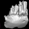
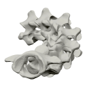
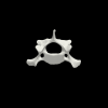
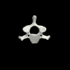
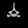
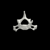
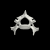
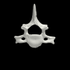
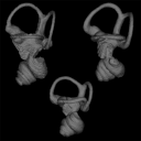
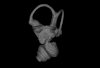
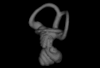
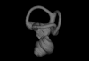
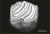
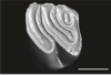
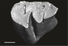
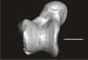
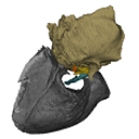
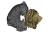
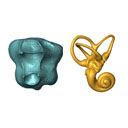
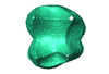
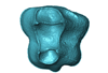
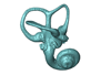
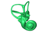
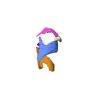
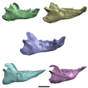
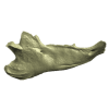
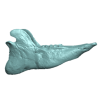
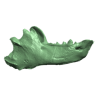
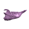
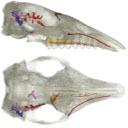
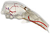
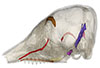
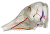
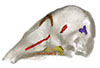
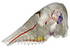
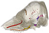
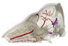
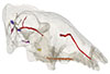
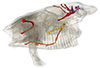
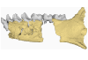
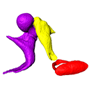
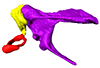
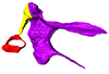
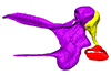
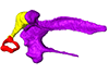
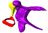
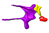
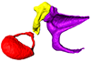
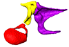
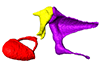
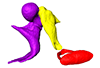
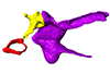
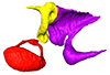
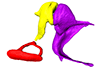
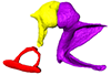
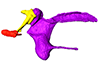
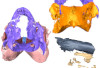
This project presents the osteological connexions of the petrosal bone of the extant Hippopotamidae Hippopotamus amphibius and Choeropsis liberiensis by a virtual osteological dissection of the ear region. The petrosal, the bulla, the sinuses and the major morphological features surrounding the petrosal bone are labelled, both in situ and in an exploded model presenting disassembly views. The directional underwater hearing mode of Hippopotamidae is discussed based on the new observations.
Choeropsis liberiensis UPPal-M09-5-005a View specimen
|
M3#1Labelled compact model of the right ear region of Choeropsis liberiensis (UPPal-M09-5-005a) Type: "3D_surfaces"doi: 10.18563/m3.sf1 state:published |
Download 3D surface file |

|
M3#2Labelled exploded model of the right ear region of Choeropsis liberiensis (UPPal-M09-5-005a) Type: "3D_surfaces"doi: 10.18563/m3.sf2 state:published |
Download 3D surface file |
Hippopotamus amphibius UM N179 View specimen

|
M3#3Labelled compact model of the right ear region of Hippopotamus amphibius (UM N 179) Type: "3D_surfaces"doi: 10.18563/m3.sf3 state:published |
Download 3D surface file |

|
M3#4Labelled exploded model of the right ear region of Hippopotamus amphibius (UM N 179) Type: "3D_surfaces"doi: 10.18563/m3.sf4 state:published |
Download 3D surface file |
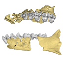
The present 3D Dataset contains the 3D models of a skull and lower jaw of the holotype of Santagnathus mariensis, described in “Old fossil findings in the Upper Triassic rocks of southern Brazil improve diversity of traversodontid cynodonts (Therapsida, Cynodontia)”
Santagnathus mariensis UFRGS-PV-1419-T View specimen
|
M3#1157Skull Type: "3D_surfaces"doi: 10.18563/m3.sf.1157 state:published |
Download 3D surface file |
|
M3#1158Lower jaw Type: "3D_surfaces"doi: 10.18563/m3.sf.1158 state:published |
Download 3D surface file |
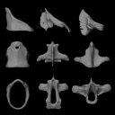
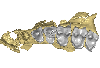
The present 3D Dataset contains the 3D models analyzed in Keppeler, H., Schultz, J. A., Ruf, I., & Martin, T., 2023. Cranial anatomy of Hypisodus minimus (Artiodactyla: Ruminantia) from the Oligocene Brule Formation of North America. Palaeontographica Abteilung A.
Hypisodus minimus SMNK-PAL 27212 View specimen
|
M3#1031CT image stack of a skull of Hypisodus minimus. Also includes a lumbar vertebra and a probable proximal phalanx of digit III or IV. Type: "3D_CT"doi: 10.18563/m3.sf.1031 state:published |
Download CT data |
|
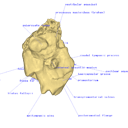
M3#10363D surface models of a skull of Hypisodus minimus (SMNK-PAL27212). The data includes a surface model for: basisphenoid, tympanic bullae, ethmoid (lamina perpendicularis), frontals, jugal (left), jugal (right), lacrimals, lower dentition, mandibles, mastoid processes, maxillaries, maxilloturbinals, nasals, occipital, palatine, parietals, petrosals, presphenoid, squamosals, turbinates, upper dentition, and the vomer. Type: "3D_surfaces"doi: 10.18563/m3.sf.1036 state:published |
Download 3D surface file |
Hypisodus minimus SMNK-PAL 27213 View specimen
|
M3#1033CT image stack of a skull of Hypisodus minimus. Also shows numerous postcranial material including an atlas articulated with the occipital bone, the distal part of a left humerus articulated to radius and ulna, a part of a femur, a part of a tibia and fibula, unidentifiable tarsal bones, parts of the metatarsals II, III, IV and V and their phalanges, a proximal phalanx of digit III or IV, a middle phalanx of digit III or IV, a possible patella and calcaneus, as well as numerous unidentifiable broken bony fragments. Type: "3D_CT"doi: 10.18563/m3.sf.1033 state:published |
Download CT data |
|
M3#10353D surface models of a skull of Hypisodus minimus (SMNK-PAL27213). The data includes a surface model for: atlas, basisphenoid, tympanic bullae, nasals, occipital, the petrosals, and the inner ear. Type: "3D_surfaces"doi: 10.18563/m3.sf.1035 state:published |
Download 3D surface file |
The present 3D Dataset contains the 3D models analyzed in the publication “Systematic and locomotor diversification of the Adapis group (Primates, Adapiformes) in the late Eocene of the Quercy (Southwest France), revealed by humeral remains”. In this paper, twenty humeral specimens from the old and new Quercy collections attributed to the fossil primates Adapis and Palaeolemur are described and analysed together. In this dataset only the scans of the fossils belonging to the collections of Université de Montpellier are provided.
In our paper (Marigó et al., 2019) we provide a qualitative and quantitative analysis of the different humeri, revealing that high variability is present within the “Adapis group” sample. Six different morphotypes are identified, confirming that what has often been called “Adapis parisiensis” is a mix of different species that present different locomotor adaptations.
Adapis sp. UM ROS 2-95 View specimen
|
M3#356Complete right humerus ROS 2-95 attributed to the Adapis group Type: "3D_surfaces"doi: 10.18563/m3.sf.356 state:published |
Download 3D surface file |
Adapis sp. UM ROS 2-536 View specimen
|
M3#357Proximal end of the right humerus ROS 2-536 attributed to the Adapis group Type: "3D_surfaces"doi: 10.18563/m3.sf.357 state:published |
Download 3D surface file |
Adapis sp. UM ROS 2-534 View specimen
|
M3#358Distal end of the left humerus ROS 2-534 attributed to the Adapis group Type: "3D_surfaces"doi: 10.18563/m3.sf.358 state:published |
Download 3D surface file |
Adapis sp. UM ROS 2-535 View specimen
|
M3#359Distal end of the left humerus ROS 2-535 attributed to the Adapis group Type: "3D_surfaces"doi: 10.18563/m3.sf.359 state:published |
Download 3D surface file |
Adapis sp. UM ROS 2-80 View specimen
|
M3#360Proximal end of the right humerus ROS 2-80 attributed to the Adapis group Type: "3D_surfaces"doi: 10.18563/m3.sf.360 state:published |
Download 3D surface file |
Adapis sp. UM ROS 2-79 View specimen
|
M3#361Distal end of the right humerus ROS 2-79 attributed to the Adapis group Type: "3D_surfaces"doi: 10.18563/m3.sf.361 state:published |
Download 3D surface file |
Adapis sp. UM ECA 1364 View specimen
|
M3#362Distal end of the left humerus ECA 1364 attributed to the Adapis group Type: "3D_surfaces"doi: 10.18563/m3.sf.362 state:published |
Download 3D surface file |
Adapis sp. UM ACQ-262 View specimen

|
M3#3733D model of ACQ 262. Humerus Type: "3D_surfaces"doi: 10.18563/m3.sf373 state:published |
Download 3D surface file |
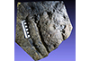
This contribution contains the 3D model of the holotype of Chambius kasserinensis, the basalmost ‘elephant-shrew’ figured in the following publication: New remains of Chambius kasserinensis from the Eocene of Tunisia and evaluation of proposed affinities for Macroscelidea (Mammalia, Afrotheria). https://doi.org/10.1080/08912963.2017.1297433
Chambius kasserinensis CBI-1-06 View specimen
|
M3#1463D model of the holotype maxilla of Chambius kasserinensis. The 3D surface was extracted manually from the limestone matrix within AVIZO 9.2 Type: "3D_surfaces"doi: 10.18563/m3.sf.146 state:published |
Download 3D surface file |
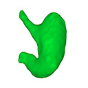
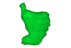
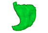
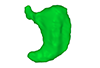
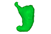
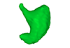
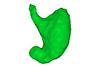
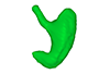
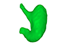
The present 3D Dataset contains the 3D models analyzed in: Kaigai N et al. Morphogenesis and three-dimensional movement of the stomach during the human embryonic period, Anat Rec (Hoboken). 2014 May;297(5):791-797. doi: 10.1002/ar.22833.
Homo sapiens KC-CS16STM27159 View specimen
|
M3#56computationally reconstructed stomach of the human embryo (M3#56_KC-CS16STM27159) at Carnegie Stage 16 (Crown Rump Length= 9.9mm). Type: "3D_surfaces"doi: 10.18563/m3.sf56 state:published |
Download 3D surface file |
Homo sapiens KC-CS17STM20383 View specimen
|
M3#57computationally reconstructed stomach of the human embryo (M3#57_KC-CS17STM20383) at Carnegie Stage 17 (Crown Rump Length= 12.3mm). Type: "3D_surfaces"doi: 10.18563/m3.sf57 state:published |
Download 3D surface file |
Homo sapiens KC-CS18STM21807 View specimen
|
M3#58computationally reconstructed stomach of the human embryo (M3#58_KC-CS18STM21807) at Carnegie Stage 18 (Crown Rump Length= 14.7mm). Type: "3D_surfaces"doi: 10.18563/m3.sf58 state:published |
Download 3D surface file |
Homo sapiens KC-CS19STM17998 View specimen
|
M3#59computationally reconstructed stomach of the human embryo (M3#59_KC-CS19STM17998) at Carnegie Stage 19 (Crown Rump Length was unmeasured ). Type: "3D_surfaces"doi: 10.18563/m3.sf59 state:published |
Download 3D surface file |
Homo sapiens KC-CS20STM20785 View specimen
|
M3#60computationally reconstructed stomach of the human embryo (M3#60_KC-CS20STM20785) at Carnegie Stage 20 (Crown Rump Length= 18.7 mm). Type: "3D_surfaces"doi: 10.18563/m3.sf60 state:published |
Download 3D surface file |
Homo sapiens KC-CS21STM24728 View specimen
|
M3#61computationally reconstructed stomach of the human embryo (M3#61_KC-CS21STM24728) at Carnegie Stage 21 (Crown Rump Length= 20.9 mm). Type: "3D_surfaces"doi: 10.18563/m3.sf61 state:published |
Download 3D surface file |
Homo sapiens KC-CS22STM26438 View specimen
|
M3#62computationally reconstructed stomach of the human embryo (M3#62_KC-CS22STM26438) at Carnegie Stage 22 (Crown Rump Length= 21.5 mm). Type: "3D_surfaces"doi: 10.18563/m3.sf62 state:published |
Download 3D surface file |
Homo sapiens KC-CS23STM20018 View specimen
|
M3#63computationally reconstructed stomach of the human embryo (M3#63_KC-CS23STM20018) at Carnegie Stage 23 (Crown Rump Length= 23.1 mm). Type: "3D_surfaces"doi: 10.18563/m3.sf63 state:published |
Download 3D surface file |
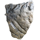
This contribution contains the 3D model described and figured in the following publication: Hautier L, Sarr R, Lihoreau F, Tabuce R, Marwan Hameh P. 2014. First record of the family Protocetidae in the Lutetian of Senegal (West Africa). Palaeovertebrata 38(2)-e2
indet. indet. SN103 View specimen

|
M3#5SN103, partial left innominate. Age and occurrence – Taïba Formation, Lutetian of the near Taïba Ndiaye, quarry of the Industries Chimiques du Sénégal (ICS) Type: "3D_surfaces"doi: 10.18563/m3.sf5 state:published |
Download 3D surface file |