Explodable 3D Dog Skull for Veterinary Education
3D models of a Sheep and Goat Skull and Inner ear
3D models of anteaters snout morphology
3D GM dataset of bird skeletal variation
Skeletal embryonic development in the catshark
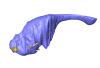
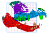
Bony connexions of the petrosal bone of extant hippos
bony labyrinth (11) , inner ear (10) , Eocene (8) , South America (8) , Paleobiogeography (7) , skull (7) , phylogeny (6)
Lionel Hautier (23) , Maëva Judith Orliac (22) , Laurent Marivaux (17) , Rodolphe Tabuce (14) , Renaud Lebrun (13) , Bastien Mennecart (13) , Pierre-Olivier Antoine (12)
|
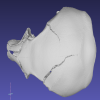
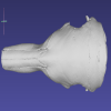
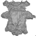
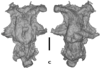
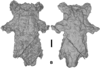
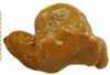
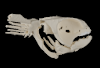
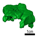
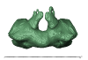
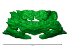
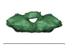
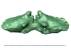
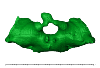
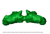
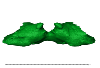
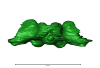
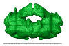
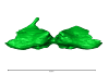
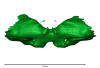
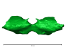
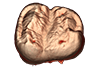
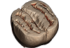
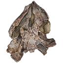
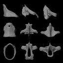
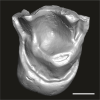
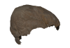
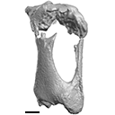
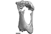
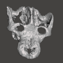
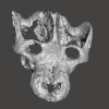
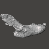
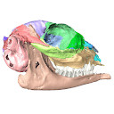
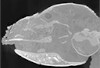
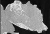
3D models related to the publication: Phylogenetic implications of the systematic reassessment of Xenacanthiformes and ‘Ctenacanthiformes’ (Chondrichthyes) neurocrania from the Carboniferous-Permian Autun Basin (France)Vincent Luccisano
Published online: 20/10/2021 |
|
M3#834MHNH.F.AUT811 (isolated neurocranium) in dorsal view. Type: "3D_surfaces"doi: 10.18563/m3.sf.834 state:published |
Download 3D surface file |
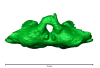
indet indet MNHN.F.AUT812 View specimen
|
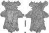
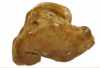
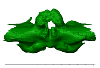
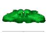
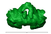
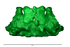
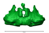
M3#835MHNH.F.AUT812 (isolated neurocranium) in dorsal view. Type: "3D_surfaces"doi: 10.18563/m3.sf.835 state:published |
Download 3D surface file |
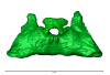
indet indet MNHN.F.AUT813 View specimen
|
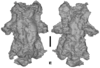
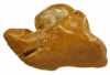
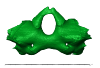
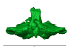
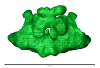
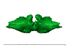
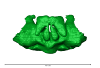
M3#836MHNH.F.AUT813 (isolated neurocranium) in dorsal view. Type: "3D_surfaces"doi: 10.18563/m3.sf.836 state:published |
Download 3D surface file |
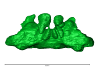
cf. Triodus sp MNHN.F.AUT814 View specimen
|
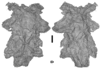
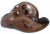
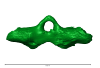
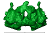
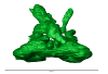
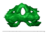
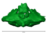
M3#837MHNH.F.AUT814 (isolated neurocranium) in dorsal view. Type: "3D_surfaces"doi: 10.18563/m3.sf.837 state:published |
Download 3D surface file |
cf. Triodus sp MHNE.2021.9.1 View specimen
|
M3#838MHNE.2021.9.1 (isolated neurocranium) in dorsal view. Type: "3D_surfaces"doi: 10.18563/m3.sf.838 state:published |
Download 3D surface file |
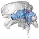
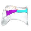
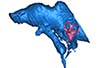
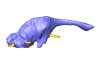
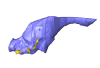
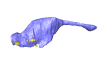
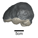
This contribution contains the 3D models described and figured in the following publication: Paulina-Carabajal A and Calvo JO 2021. Re-description of the braincase of the rebbachisaurid sauropod Limaysaurus tessonei and novel endocranial information based on CT scans. Anais da Academia Brasileira de Ciências 93(Suppl. 2): e20200762 https://doi.org/10.1590/0001-3765202120200762
Limaysaurus tessonei MUCPv-205 View specimen
|
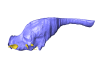
M3#700Renderings of the virtually isolate braincase, brain, and right inner ear. Type: "3D_surfaces"doi: 10.18563/m3.sf.700 state:published |
Download 3D surface file |
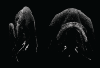
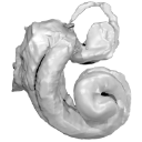
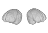
The present 3D Dataset contains the 3D model of the brain endocast of Neoepiblema acreensis analyzed in “Small within the largest: Brain size and anatomy of the extinct Neoepiblema acreensis, a giant rodent from the Neotropics”. The 3D model was generated using CT-Scanning and techniques of virtual reconstruction.
Neoepiblema acreensis UFAC 4515 View specimen
|
M3#502Brain endocast of Neoepiblema acreensis Type: "3D_surfaces"doi: 10.18563/m3.sf.502 state:published |
Download 3D surface file |
The present 3D Dataset contains the 3D models analyzed in the following manuscript: L. Roese-Miron, M.E.H. Jones, J.D. Ferreira and A.S. Hsiou., 2023. Virtual endocasts of Clevosaurus brasiliensis and the tuatara: Rhynchocephalian neuroanatomy and the oldest endocranial record for Lepidosauria.
Sphenodon punctatus CM 30660 View specimen
|
M3#10993D surface model of the cranial endocast of specimen CM 30660 (Sphenodon punctatus). Type: "3D_surfaces"doi: 10.18563/m3.sf.1099 state:published |
Download 3D surface file |
Sphenodon punctatus KCLZJ 001 View specimen
|
M3#11003D surface models of the cranial endocast and the initial trunks of the cranial nerves of specimen KCLZJ 001 (Sphenodon punctatus). Type: "3D_surfaces"doi: 10.18563/m3.sf.1100 state:published |
Download 3D surface file |
Sphenodon punctatus LDUCZ x0036 View specimen
|
M3#11013D surface models of the cranial endocast and the initial trunks of the cranial nerves of specimen LDUCZ x0036 (Sphenodon punctatus). Type: "3D_surfaces"doi: 10.18563/m3.sf.1101 state:published |
Download 3D surface file |
Sphenodon punctatus LDUCZ x1126 View specimen
|
M3#11023D surface model of the cranial endocast of specimen LDUCZ x1126 (Sphenodon punctatus). Type: "3D_surfaces"doi: 10.18563/m3.sf.1102 state:published |
Download 3D surface file |
Clevosaurus brasiliensis MCN PV 2852 View specimen
|
M3#11033D surface model of the cranial endocast of specimen MCN PV 2852 (Clevosaurus brasiliensis). Type: "3D_surfaces"doi: 10.18563/m3.sf.1103 state:published |
Download 3D surface file |
Sphenodon punctatus SAMA 70524 View specimen
|
M3#11043D surface models of the cranial endocast, brain, endosseous labyrinth and initial trunks of the cranial nerves of specimen SAMA 70524 (Sphenodon punctatus). Type: "3D_surfaces"doi: 10.18563/m3.sf.1104 state:published |
Download 3D surface file |
Sphenodon punctatus SU1 View specimen
|
M3#11053D surface models of the cranial endocast and the initial trunks of the cranial nerves of specimen SU1 (Sphenodon punctatus). Type: "3D_surfaces"doi: 10.18563/m3.sf.1105 state:published |
Download 3D surface file |
Sphenodon punctatus YPM HERR 009194 View specimen
|
M3#11063D surface models of the cranial endocast and the initial trunks of the cranial nerves of specimen YPM HERR 009194 (Sphenodon punctatus). Type: "3D_surfaces"doi: 10.18563/m3.sf.1106 state:published |
Download 3D surface file |
This contribution contains the 3D models described and figured in the following publication: Aguirre-Fernández G, Jost J, and Hilfiker S. 2022. First records of extinct kentriodontid and squalodelphinid dolphins from the Upper Marine Molasse (Burdigalian age) of Switzerland and a reappraisal of the Swiss cetacean fauna.
Kentriodon sp. NMBE 5023944 View specimen
|
M3#8583D models of left periotic and bony labyrinth of NMBE 5023944 (Kentriodon sp.) Type: "3D_surfaces"doi: 10.18563/m3.sf.858 state:published |
Download 3D surface file |
Kentriodon sp. NMBE 5023945 View specimen
|
M3#8593D models of right periotic and bony labyrinth of NMBE 5023945 (Kentriodontidae indet.) Type: "3D_surfaces"doi: 10.18563/m3.sf.859 state:published |
Download 3D surface file |
Kentriodon sp. NMBE 5023946 View specimen
|
M3#8603D models of left periotic and bony labyrinth of NMBE 5023946 (Kentriodon sp.) Type: "3D_surfaces"doi: 10.18563/m3.sf.860 state:published |
Download 3D surface file |
Kentriodon sp. NMBE 5036436 View specimen
|
M3#8613D models of right periotic and bony labyrinth of NMBE 5036436 (Kentriodontidae indet.) Type: "3D_surfaces"doi: 10.18563/m3.sf.861 state:published |
Download 3D surface file |
indet. indet. NMBE 5023942 View specimen
|
M3#8623D models of right periotic and bony labyrinth of NMBE 5023942 (Squalodelphinidae indet.) Type: "3D_surfaces"doi: 10.18563/m3.sf.862 state:published |
Download 3D surface file |
indet. indet. NMBE 5023943 View specimen
|
M3#8633D models of left periotic and bony labyrinth of NMBE 5023943 (Squalodelphinidae indet.) Type: "3D_surfaces"doi: 10.18563/m3.sf.863 state:published |
Download 3D surface file |
indet. indet. NMBE 5036437 View specimen
|
M3#8643D models of left periotic and bony labyrinth of NMBE 5036437 (Physeteridae indet.) Type: "3D_surfaces"doi: 10.18563/m3.sf.864 state:published |
Download 3D surface file |
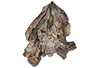
The present 3D Dataset contains 3D models of the cranial, visceral, and pectoral endoskeleton of Iniopera, an iniopterygian stem-group holocephalan from the Pennsylvanian of the USA. These data formed the basis for the analyses carried out in Dearden et al. (2023) “Evidence for high-performance suction feeding in the Pennsylvanian stem-group holocephalan Iniopera” PNAS.
Iniopera sp. KUNHM 22060, 158289 View specimen
|
M3#1034plys of the head endoskeleton of Iniopera sp. Type: "3D_surfaces"doi: 10.18563/m3.sf.1034 state:published |
Download 3D surface file |
The present 3D Dataset contains the 3D models analyzed in: Perrichon et al. 2023. Ontogenetic variability of the intertympanic sinus distinguishes lineages within Crocodylia.
Mecistops sp. ag SVSTUA 022001 View specimen
|
M3#980Intertympanic sinus system (expressed in meters) Type: "3D_surfaces"doi: 10.18563/m3.sf.980 state:published |
Download 3D surface file |
Crocodylus niloticus ag SVSTUA 022002 View specimen
|
M3#981Intertympanic sinus system Type: "3D_surfaces"doi: 10.18563/m3.sf.981 state:published |
Download 3D surface file |
Mecistops sp. AMU Zoo 04721 View specimen
|
M3#982Intertympanic sinus system Type: "3D_surfaces"doi: 10.18563/m3.sf.982 state:published |
Download 3D surface file |
Crocodylus sp. MHNL QV14 View specimen
|
M3#983Intertympanic sinus system Type: "3D_surfaces"doi: 10.18563/m3.sf.983 state:published |
Download 3D surface file |
Crocodylus rhombifer MHNL 42006506 View specimen

|
M3#1008Intertympanic sinus system (incomplete) Type: "3D_surfaces"doi: 10.18563/m3.sf.1008 state:published |
Download 3D surface file |
Crocodylus rhombifer MHNL 42006507 View specimen
|
M3#1007Intertympanic sinus system (incomplete) Type: "3D_surfaces"doi: 10.18563/m3.sf.1007 state:published |
Download 3D surface file |
Crocodylus niloticus MHNL 50001387 View specimen
|
M3#1006Intertympanic sinus system Type: "3D_surfaces"doi: 10.18563/m3.sf.1006 state:published |
Download 3D surface file |
Crocodylus palustris MHNL 50001388 View specimen
|
M3#1004Intertympanic sinus system Type: "3D_surfaces"doi: 10.18563/m3.sf.1004 state:published |
Download 3D surface file |
Crocodylus porosus MHNL 50001389 View specimen
|
M3#1009Intertympanic sinus system Type: "3D_surfaces"doi: 10.18563/m3.sf.1009 state:published |
Download 3D surface file |
Mecistops sp. MHNL 50001393 View specimen
|
M3#1010Intertympanic sinus system Type: "3D_surfaces"doi: 10.18563/m3.sf.1010 state:published |
Download 3D surface file |
Crocodylus niloticus MHNL 50001397 View specimen
|
M3#1018Intertympanic sinus system Type: "3D_surfaces"doi: 10.18563/m3.sf.1018 state:published |
Download 3D surface file |
Crocodylus porosus MHNL 50001398 View specimen
|
M3#1017Intertympanic sinus system Type: "3D_surfaces"doi: 10.18563/m3.sf.1017 state:published |
Download 3D surface file |
Crocodylus niloticus MHNL 50001405 View specimen
|
M3#1016Intertympanic sinus system Type: "3D_surfaces"doi: 10.18563/m3.sf.1016 state:published |
Download 3D surface file |
Gavialis gangeticus MHNL 50001407 View specimen
|
M3#1015Intertympanic sinus system Type: "3D_surfaces"doi: 10.18563/m3.sf.1015 state:published |
Download 3D surface file |
Crocodylus niloticus MHNL 90001850 View specimen
|
M3#1014Intertympanic sinus system Type: "3D_surfaces"doi: 10.18563/m3.sf.1014 state:published |
Download 3D surface file |
Crocodylus niloticus MHNL 90001851 View specimen
|
M3#1013Intertympanic sinus system Type: "3D_surfaces"doi: 10.18563/m3.sf.1013 state:published |
Download 3D surface file |
Crocodylus niloticus MHNL 90001855 View specimen
|
M3#1012Intertympanic sinus system Type: "3D_surfaces"doi: 10.18563/m3.sf.1012 state:published |
Download 3D surface file |
Osteolaemus tetraspis MHNM.9095.0 View specimen
|
M3#1011Intertympanic sinus system Type: "3D_surfaces"doi: 10.18563/m3.sf.1011 state:published |
Download 3D surface file |
Voay robustus MNHN F.1908-5 View specimen
|
M3#1003Intertympanic sinus system Type: "3D_surfaces"doi: 10.18563/m3.sf.1003 state:published |
Download 3D surface file |
Crocodylus sp. MNHN-F.1908-5-2 View specimen
|
M3#1005Intertympanic sinus system Type: "3D_surfaces"doi: 10.18563/m3.sf.1005 state:published |
Download 3D surface file |
Osteolaemus tetraspis MZS Cro 040 View specimen
|
M3#1002Intertympanic sinus system Type: "3D_surfaces"doi: 10.18563/m3.sf.1002 state:published |
Download 3D surface file |
Crocodylus acutus MZS Cro 055 View specimen
|
M3#991Intertympanic sinus system Type: "3D_surfaces"doi: 10.18563/m3.sf.991 state:published |
Download 3D surface file |
Melanosuchus niger MZS Cro 073 View specimen
|
M3#989Intertympanic sinus system Type: "3D_surfaces"doi: 10.18563/m3.sf.989 state:published |
Download 3D surface file |
Mecistops sp. MZS Cro 083 View specimen

|
M3#990Intertympanic sinus system Type: "3D_surfaces"doi: 10.18563/m3.sf.990 state:published |
Download 3D surface file |
Tomistoma schlegelii MZS Cro 094 View specimen
|
M3#988Intertympanic sinus system Type: "3D_surfaces"doi: 10.18563/m3.sf.988 state:published |
Download 3D surface file |
Gavialis gangeticus NHMUK 1846.1.7.3 View specimen
|
M3#987Intertympanic sinus system Type: "3D_surfaces"doi: 10.18563/m3.sf.987 state:published |
Download 3D surface file |
Osteolaemus tetraspis NHMUK 1862.6.30.5 View specimen
|
M3#986Intertympanic sinus system Type: "3D_surfaces"doi: 10.18563/m3.sf.986 state:published |
Download 3D surface file |
Gavialis gangeticus NHMUK 1873 View specimen
|
M3#985intertympanic sinus system Type: "3D_surfaces"doi: 10.18563/m3.sf.985 state:published |
Download 3D surface file |
Tomistoma schlegelii NHMUK 1893.3.6.14 View specimen
|
M3#984Intertympanic sinus system Type: "3D_surfaces"doi: 10.18563/m3.sf.984 state:published |
Download 3D surface file |
Mecistops sp. NHMUK 1924.5.10.1 View specimen
|
M3#992Intertympanic sinus system Type: "3D_surfaces"doi: 10.18563/m3.sf.992 state:published |
Download 3D surface file |
Voay robustus NHMUK PV R 36684 View specimen
|
M3#993Intertympanic sinus system Type: "3D_surfaces"doi: 10.18563/m3.sf.993 state:published |
Download 3D surface file |
Voay robustus NHMUK PV R 36685 View specimen
|
M3#1001Intertympanic sinus system Type: "3D_surfaces"doi: 10.18563/m3.sf.1001 state:published |
Download 3D surface file |
Crocodylus niloticus UCBL FSL 532077 View specimen

|
M3#1000Intertympanic sinus system Type: "3D_surfaces"doi: 10.18563/m3.sf.1000 state:published |
Download 3D surface file |
Crocodylus porosus/siamensis UCBLZ 2019-1-237 View specimen
|
M3#999Intertympanic sinus system Type: "3D_surfaces"doi: 10.18563/m3.sf.999 state:published |
Download 3D surface file |
Osteolaemus tetraspis UCBLZ 2019-1-236 View specimen
|
M3#998Intertympanic sinus system Type: "3D_surfaces"doi: 10.18563/m3.sf.998 state:published |
Download 3D surface file |
Alligator mississipiensis UCBLZ WB35 View specimen

|
M3#997Intertympanic sinus system Type: "3D_surfaces"doi: 10.18563/m3.sf.997 state:published |
Download 3D surface file |
Crocodylus siamensis UCBLZ WB41 View specimen
|
M3#996Intertympanic sinus system Type: "3D_surfaces"doi: 10.18563/m3.sf.996 state:published |
Download 3D surface file |
Tomistoma schlegelii UM 1097 View specimen
|
M3#995Intertympanic sinus system Type: "3D_surfaces"doi: 10.18563/m3.sf.995 state:published |
Download 3D surface file |
Crocodylus niloticus UM 2001-1756-1-434 NR View specimen
|
M3#994Intertympanic sinus system Type: "3D_surfaces"doi: 10.18563/m3.sf.994 state:published |
Download 3D surface file |
Mecistops sp. UM N89 View specimen
|
M3#1019Intertympanic sinus system Type: "3D_surfaces"doi: 10.18563/m3.sf.1019 state:published |
Download 3D surface file |
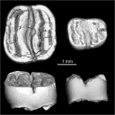
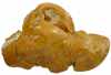
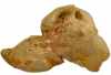
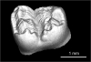
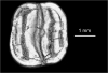
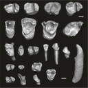
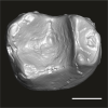
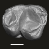
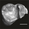
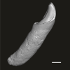
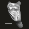
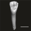
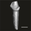
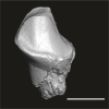
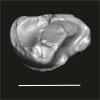
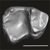
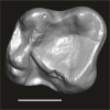
This contribution provides the raw files for the μCT-scan data and renderings of the three-dimensional digital models of two fossil teeth of a geomyin geomorph rodent (Caribeomys merzeraudi), discovered from lower Oligocene deposits of Puerto Rico, San Sebastian Formation (locality LACM Loc. 8060). These fossils were described, figured and discussed in the following publication: Marivaux et al. (2021), An unpredicted ancient colonization of the West Indies by North American rodents: dental evidence of a geomorph from the early Oligocene of Puerto Rico. Papers in Palaeontology. https://doi.org/10.1002/spp2.1388
Caribeomys merzeraudi LACM 162478 View specimen
|
M3#712Right lower dp4: isolated deciduous premolar. The specimen was scanned with a resolution of 5 µm using a μ-CT-scanning station EasyTom 150 / Rx Solutions (Montpellier RIO Imaging, ISE-M, Montpellier, France). AVIZO 7.1 (Visualization Sciences Group) software was used for visualization, segmentation, and 3D rendering. This isolated tooth was prepared within a “labelfield” module of AVIZO, using the segmentation threshold selection tool. Type: "3D_surfaces"doi: 10.18563/m3.sf.712 state:published |
Download 3D surface file |
|
M3#7145µm µCT data set . Right lower dp4: isolated deciduous premolar. The specimen was scanned with a resolution of 5 µm using a μ-CT-scanning station EasyTom 150 / Rx Solutions (Montpellier RIO Imaging, ISE-M, Montpellier, France). Type: "3D_CT"doi: 10.18563/m3.sf.714 state:published |
Download CT data |
Caribeomys merzeraudi LACM 162449 View specimen
|
M3#713Right lower molar (m1 or m2). The specimen was scanned with a resolution of 4.5 µm using a μ-CT-scanning station EasyTom 150 / Rx Solutions (Montpellier RIO Imaging, ISE-M, Montpellier, France). AVIZO 7.1 (Visualization Sciences Group) software was used for visualization, segmentation, and 3D rendering. This isolated tooth was prepared within a “labelfield” module of AVIZO, using the segmentation threshold selection tool. Type: "3D_surfaces"doi: 10.18563/m3.sf.713 state:published |
Download 3D surface file |
|
M3#715µCT data at 4.5µm Type: "3D_CT"doi: 10.18563/m3.sf.715 state:published |
Download CT data |
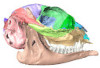
This contribution contains the 3D surface model of the holotype cranium of the Late Jurassic thalassochelydian turtle Solnhofia brachyrhyncha described and figured in the publication of Anquetin and Püntener (2020).
Solnhofia brachyrhyncha MJSN BAN001-2.1 View specimen
|
M3#536Textured 3D surface model of the holotype cranium of the Late Jurassic turtle Solnhofia brachyrhyncha Type: "3D_surfaces"doi: 10.18563/m3.sf.536 state:published |
Download 3D surface file |
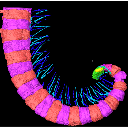
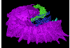
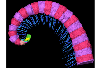
The present 3D Dataset contains the 3D models analyzed in the publication Fossils from the Montceau-les-Mines Lagerstätte (305 Ma) shed light on the anatomy, ecology and phylogeny of Carboniferous millipedes. Authors: Lheritier Mickael, Perroux Maëva, Vannier Jean, Escarguel Gilles, Wesener Thomas, Moritz Leif, Chabard Dominique, Adrien Jerome and Perrier Vincent. Journal of Systematics Palaeontology. https://doi.org/10.1080/14772019.2023.2169891
Amynilyspes fatimae MNHN.F.SOT.2134 View specimen

|
M3#1073Nearly complete specimen. Type: "3D_surfaces"doi: 10.18563/m3.sf.1073 state:published |
Download 3D surface file |
Amynilyspes fatimae MNHN.F.SOT.14983 View specimen
|
M3#1074Nearly complete specimen. Type: "3D_surfaces"doi: 10.18563/m3.sf.1074 state:published |
Download 3D surface file |
Amynilyspes fatimae MNHN.F.SOT.2129 View specimen

|
M3#1075Nearly complete specimen. Type: "3D_surfaces"doi: 10.18563/m3.sf.1075 state:published |
Download 3D surface file |
Blanzilius parriati MNHN.F.SOT.2114A View specimen
|
M3#1076Front part. Type: "3D_surfaces"doi: 10.18563/m3.sf.1076 state:published |
Download 3D surface file |
Blanzilius parriati MNHN.F.SOT.5148 View specimen
|
M3#1077Front part. Type: "3D_surfaces"doi: 10.18563/m3.sf.1077 state:published |
Download 3D surface file |
Blanzilius parriati MNHN.F.SOT.2113 View specimen
|
M3#1078Fragment with legs, sternites and possible tracheal openings. Type: "3D_surfaces"doi: 10.18563/m3.sf.1078 state:published |
Download 3D surface file |
Blanzilius parriati MNHN.F.SOT.81522 View specimen
|
M3#1079Nealry complete specimen. Type: "3D_surfaces"doi: 10.18563/m3.sf.1079 state:published |
Download 3D surface file |
The present 3D Dataset contains the 3D model analyzed in Gaetano, L. C., Abdala, F., Seoane, F. D., Tartaglione, A., Schulz, M., Otero, A., Leardi, J. M., Apaldetti, C., Krapovickas, V., and Steinbach, E. 2021. A new cynodont from the Upper Triassic Los Colorados Formation (Argentina, South America) reveals a novel paleobiogeographic context for mammalian ancestors. Scientific Reports.
Tessellatia bonapartei PULR-V121 View specimen
|
M3#9603D surface model of PULR-V121 Type: "3D_surfaces"doi: 10.18563/m3.sf.960 state:published |
Download 3D surface file |
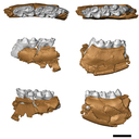
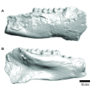
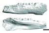
The present 3D Dataset contains the 3D models analyzed in Mennecart B., Wazir W.A., Sehgal R.K., Patnaik R., Singh N.P., Kumar N, and Nanda A.C. 2021. New remains of Nalamaeryx (Tragulidae, Mammalia) from the Ladakh Himalaya and their phylogenetical and palaeoenvironmental implications. Historical Biology. https://doi.org/10.1080/08912963.2021.2014479
Nalameryx savagei WIMF/A4801 View specimen
|
M3#766Nalameryx savagei, Partial lower right jaw preserving m2 and m3. Type: "3D_surfaces"doi: 10.18563/m3.sf.766 state:published |
Download 3D surface file |
Nalameryx savagei WIMF/A4802 View specimen
|
M3#767Nalameryx savagei, partial lower right jaw preserving m2 and m3 Type: "3D_surfaces"doi: 10.18563/m3.sf.767 state:published |
Download 3D surface file |
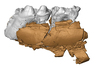
The present 3D Dataset contains the 3D model of a left dentary with m1-m3 analyzed in “A new fossil of Tayassuidae (Mammalia: Certartiodactyla) from the Pleistocene of northern Brazil”. The 3D model was generated using a laser scanning.
cf. Pecari tajacu UFSM 11606 View specimen
|
M3#498Left dentary with m1-m3 Type: "3D_surfaces"doi: 10.18563/m3.sf.498 state:published |
Download 3D surface file |
The present contribution contains the 3D virtual restoration of a Pliocene Lutrine right femur of Tobène, Senegal, described and figured in Lihoreau et al. (2021) : "A fossil terrestrial fauna from Tobène (Senegal) provides a unique early Pliocene window in Western Africa ". https://doi.org/10.1016/j.gr.2021.06.013
Indet indet SN-Tob-12-02 View specimen
|
M3#441Virtual restoration of SN-Tob-12-02 Type: "3D_surfaces"doi: 10.18563/m3.sf.441 state:published |
Download 3D surface file |
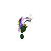
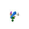
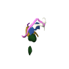
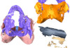
The present 3D Dataset contains the 3D models analyzed in Brualla et al., 2024: Comparative anatomy of the vocal apparatus in bats and implication for the diversity of laryngeal echolocation. Zoological Journal of the Linnean Society, vol. zlad180. (https://doi.org/10.1093/zoolinnean/zlad180). Bat larynges are understudied in the previous anatomical studies. The description and comparison of the different morphological traits might provide important proxies to investigate the evolutionary origin of laryngeal echolocation in bats.
Eonycteris spelaea VN18-026 View specimen

|
M3#1305Laryngeal cartilages and muscles of the cave nectar bat Type: "3D_surfaces"doi: 10.18563/m3.sf.1305 state:published |
Download 3D surface file |
Macroglossus sobrinus VN15-017 View specimen

|
M3#1306Laryngeal anatomy of Macroglossus sobrinus Type: "3D_surfaces"doi: 10.18563/m3.sf.1306 state:published |
Download 3D surface file |
Aselliscus dongbacana VTTu15-013 View specimen
|
M3#1307Laryngeal anatomy of Aselliscus dongbacana Type: "3D_surfaces"doi: 10.18563/m3.sf.1307 state:published |
Download 3D surface file |
Coelops frithii VN19-196 View specimen
|
M3#1308Laryngeal anatomy of Coelops frithii Type: "3D_surfaces"doi: 10.18563/m3.sf.1308 state:published |
Download 3D surface file |
Hipposideros larvatus VN18-209 View specimen
|
M3#1309Laryngeal anatomy of Hipposideros larvatus Type: "3D_surfaces"doi: 10.18563/m3.sf.1309 state:published |
Download 3D surface file |
Rhinolophus cornutus JP21-025 View specimen
|
M3#14763D surfaces of Rhinolophus cornutus Type: "3D_surfaces"doi: 10.18563/m3.sf.1476 state:published |
Download 3D surface file |
Rhinolophus macrotis VN11-089 View specimen
|
M3#1477Laryngeal cartilages and muscles of Rhinolophus macrotis Type: "3D_surfaces"doi: 10.18563/m3.sf.1477 state:published |
Download 3D surface file |
Lyroderma lyra VN17-535 View specimen
|
M3#1312Laryngeal anatomy of Lyroderma lyra Type: "3D_surfaces"doi: 10.18563/m3.sf.1312 state:published |
Download 3D surface file |
Saccolaimus mixtus A3257 View specimen
|
M3#1478Laryngeal components of Saccolaimus mixtus Type: "3D_surfaces"doi: 10.18563/m3.sf.1478 state:published |
Download 3D surface file |
Taphozous melanopogon VN17-0252 View specimen
|
M3#1479Laryngeal cartilages and muscles of Taphozous melanopogon Type: "3D_surfaces"doi: 10.18563/m3.sf.1479 state:published |
Download 3D surface file |
Artibeus jamaicensis AJ001 View specimen
|
M3#1316Laryngeal anatomy of Artibeus jamaicensis Type: "3D_surfaces"doi: 10.18563/m3.sf.1316 state:published |
Download 3D surface file |
Kerivoula hardwickii VN11-0043 View specimen
|
M3#1317Laryngeal anatomy of Kerivoula hardwickii Type: "3D_surfaces"doi: 10.18563/m3.sf.1317 state:published |
Download 3D surface file |
Myotis ater VN19-016 View specimen
|
M3#1318Laryngeal anatomy of Myotis ater Type: "3D_surfaces"doi: 10.18563/m3.sf.1318 state:published |
Download 3D surface file |
Myotis siligorensis VTTu14-018 View specimen
|
M3#1319Laryngeal anatomy of Myotis siligorensis Type: "3D_surfaces"doi: 10.18563/m3.sf.1319 state:published |
Download 3D surface file |
Suncus murinus KATS_835A View specimen
|
M3#1395Laryngeal anatomy of Suncus murinus Type: "3D_surfaces"doi: 10.18563/m3.sf.1395 state:published |
Download 3D surface file |
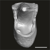
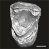
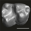
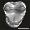
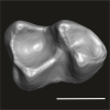
This contribution contains the three-dimensional digital models of the dental fossil material of anthropoid and strepsirrhine primates, discovered in Lower Oligocene detrital deposits outcropping in the Porto Rico and El Argoub areas, east of the Dakhla peninsula region (Atlantic Sahara; in the south of Morocco, near the northern border of Mauritania). These fossils were described, figured and discussed in the following publication: Marivaux et al. (2024), A new primate community from the earliest Oligocene of the Atlantic margin of Northwest Africa: Systematic, paleobiogeographic and paleoenvironmental implications. Journal of Human Evolution. https://doi.org/10.1016/j.jhevol.2024.103548
Catopithecus aff. browni DAK-Arg-087 View specimen

|
M3#1211Isolated right lower m3 (worn) Type: "3D_surfaces"doi: 10.18563/m3.sf.1211 state:published |
Download 3D surface file |
Catopithecus aff. browni DAK-Arg-088 View specimen
|
M3#1212Isolated right lower m2 (abraded/corroded) Type: "3D_surfaces"doi: 10.18563/m3.sf.1212 state:published |
Download 3D surface file |
Catopithecus aff. browni DAK-Arg-089 View specimen
|
M3#1213Isolated left lower m1 (worn) Type: "3D_surfaces"doi: 10.18563/m3.sf.1213 state:published |
Download 3D surface file |
Catopithecus aff. browni DAK-Pto-052 View specimen
|
M3#1214Isolated right lower m1 (pristine but lacking the mesiobuccal region) Type: "3D_surfaces"doi: 10.18563/m3.sf.1214 state:published |
Download 3D surface file |
Catopithecus aff. browni DAK-Arg-090 View specimen
|
M3#1215Isolated left upper P4 Type: "3D_surfaces"doi: 10.18563/m3.sf.1215 state:published |
Download 3D surface file |
Catopithecus aff. browni DAK-Arg-091 View specimen
|
M3#1216Isolated left upper M2 (worn and corroded) Type: "3D_surfaces"doi: 10.18563/m3.sf.1216 state:published |
Download 3D surface file |
Catopithecus aff. browni DAK-Pto-053 View specimen
|
M3#1217Isolated right upper M1 (lacking the buccal region) Type: "3D_surfaces"doi: 10.18563/m3.sf.1217 state:published |
Download 3D surface file |
Abuqatrania cf. basiodontos DAK-Arg-092 View specimen
|
M3#1218Isolated left lower c1 Type: "3D_surfaces"doi: 10.18563/m3.sf.1218 state:published |
Download 3D surface file |
?Propliopithecus sp. DAK-Pto-056 View specimen
|
M3#1219Isolated right lower m3 (fragment of talonid of a germ) Type: "3D_surfaces"doi: 10.18563/m3.sf.1219 state:published |
Download 3D surface file |
Abuqatrania cf. basiodontos DAK-Arg-093 View specimen
|
M3#1469Isolated right lower m1 Type: "3D_surfaces"doi: 10.18563/m3.sf.1469 state:published |
Download 3D surface file |
Abuqatrania cf. basiodontos DAK-Arg-094 View specimen
|
M3#1221Isolated left upper M1 or M2 (corroded, lacking the enamel cap [exposed dentine]) Type: "3D_surfaces"doi: 10.18563/m3.sf.1221 state:published |
Download 3D surface file |
Abuqatrania cf. basiodontos DAK-Arg-095 View specimen
|
M3#1222Isolated right lower i1 or i2 Type: "3D_surfaces"doi: 10.18563/m3.sf.1222 state:published |
Download 3D surface file |
Abuqatrania cf. basiodontos DAK-Arg-096 View specimen
|
M3#1223Isolated right lower p2 (worn apex) Type: "3D_surfaces"doi: 10.18563/m3.sf.1223 state:published |
Download 3D surface file |
Abuqatrania cf. basiodontos DAK-Arg-097 View specimen
|
M3#1224Isolated right lower p2 (worn apex and broken root) Type: "3D_surfaces"doi: 10.18563/m3.sf.1224 state:published |
Download 3D surface file |
Afrotarsius sp. DAK-Arg-098 View specimen
|
M3#1225Isolated left lower p3 Type: "3D_surfaces"doi: 10.18563/m3.sf.1225 state:published |
Download 3D surface file |
Afrotarsius sp. DAK-Pto-054 View specimen
|
M3#1226Isolated right lower m1 (abraded/corroded) Type: "3D_surfaces"doi: 10.18563/m3.sf.1226 state:published |
Download 3D surface file |
Orolemur mermozi DAK-Pto-055 View specimen
|
M3#1227Isolated right upper M1 or M2 (pristine, Holotype) Type: "3D_surfaces"doi: 10.18563/m3.sf.1227 state:published |
Download 3D surface file |
Wadilemur cf. elegans DAK-Arg-099 View specimen
|
M3#1228Isolated right lower m2 Type: "3D_surfaces"doi: 10.18563/m3.sf.1228 state:published |
Download 3D surface file |
cf. 'Anchomomys' milleri DAK-Arg-100 View specimen

|
M3#1229Isolated right lower c1 Type: "3D_surfaces"doi: 10.18563/m3.sf.1229 state:published |
Download 3D surface file |
Abuqatrania cf. basiodontos DAK-Arg-101 View specimen
|
M3#1396Isolated left upper M3 (abraded) Type: "3D_surfaces"doi: 10.18563/m3.sf.1396 state:published |
Download 3D surface file |
Orogalago saintexuperyi DAK-Arg-102 View specimen
|
M3#1397Isolated left lower m2 Type: "3D_surfaces"doi: 10.18563/m3.sf.1397 state:published |
Download 3D surface file |
Wadilemur cf. elegans DAK-Arg-103 View specimen

|
M3#1473Isolated right upper M1 or M2 (lacking the mesial and buccal regions) Type: "3D_surfaces"doi: 10.18563/m3.sf.1473 state:published |
Download 3D surface file |
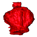
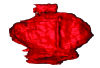
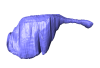
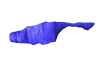
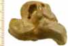
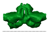
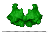
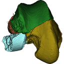
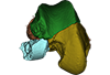
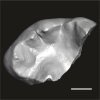
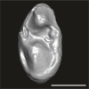
This contribution contains the 3D model of an endocranial cast analyzed in “A 10 ka intentionally deformed human skull from Northeast Asia”. There are many studies on the morphological characteristics of intentional cranial deformation (ICD), but few related 3D models were published. Here, we present the surface model of an intentionally deformed 10 ka human cranium for further research on ICD practice. The 3D model of the endocranial cast of this ICD cranium was discovered near Harbin City, Province Heilongjiang, Northeast China. The fossil preserved only the frontal, parietal, and occipital bones. To complete the endocast model of the specimen, we printed a 3D model and used modeling clay to reconstruct the missing part based on the general form of the modern human endocast morphology.
Homo sapiens IVPP-PA1616 View specimen
|
M3#972The frontal region of the endocast is flattened, probably formed by the constant pressure on the frontal bone during growth. There is a well-developed frontal crest on the endocranial surface. The endocast widens posteriorly from the frontal lobe. The widest point of the endocast is at the lateral border of the parietal lobe. The lower parietal areas display a marked lateral expansion. The overall shape of the endocast is asymmetrical, with the left side of the parietal lobe being more laterally expanded than the right side. Like the frontal lobe, the occipital lobe is also anteroposteriorly flattened. Type: "3D_surfaces"doi: 10.18563/m3.sf.972 state:published |
Download 3D surface file |
|
M3#976The original endocranial cast model (with texture) of IVPP-PA1616. It shows the original structures of the specimen, and was not altered in any way. Type: "3D_surfaces"doi: 10.18563/m3.sf.976 state:published |
Download 3D surface file |
The present 3D Dataset contains the 3D model analyzed in Presence of the ground sloth Valgipes bucklandi (Xenarthra, Folivora, Scelidotheriinae) in southern Uruguay during the Late Pleistocene: Ecological and biogeographical implications. Quaternary International. https://doi.org/10.1016/j.quaint.2021.06.011
Valgipes bucklandi CAV 1573 View specimen
|
M3#797Left tibia-fibula Type: "3D_surfaces"doi: 10.18563/m3.sf.797 state:published |
Download 3D surface file |
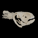
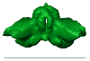
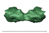
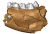
The present 3D Dataset contains the 3D models of a skull and lower jaw of the holotype of Santagnathus mariensis, described in “Old fossil findings in the Upper Triassic rocks of southern Brazil improve diversity of traversodontid cynodonts (Therapsida, Cynodontia)”
Santagnathus mariensis UFRGS-PV-1419-T View specimen
|
M3#1157Skull Type: "3D_surfaces"doi: 10.18563/m3.sf.1157 state:published |
Download 3D surface file |
|
M3#1158Lower jaw Type: "3D_surfaces"doi: 10.18563/m3.sf.1158 state:published |
Download 3D surface file |
The present 3D Dataset contains the 3D models analyzed in Keppeler, H., Schultz, J. A., Ruf, I., & Martin, T., 2023. Cranial anatomy of Hypisodus minimus (Artiodactyla: Ruminantia) from the Oligocene Brule Formation of North America. Palaeontographica Abteilung A.
Hypisodus minimus SMNK-PAL 27212 View specimen
|
M3#1031CT image stack of a skull of Hypisodus minimus. Also includes a lumbar vertebra and a probable proximal phalanx of digit III or IV. Type: "3D_CT"doi: 10.18563/m3.sf.1031 state:published |
Download CT data |
|
M3#10363D surface models of a skull of Hypisodus minimus (SMNK-PAL27212). The data includes a surface model for: basisphenoid, tympanic bullae, ethmoid (lamina perpendicularis), frontals, jugal (left), jugal (right), lacrimals, lower dentition, mandibles, mastoid processes, maxillaries, maxilloturbinals, nasals, occipital, palatine, parietals, petrosals, presphenoid, squamosals, turbinates, upper dentition, and the vomer. Type: "3D_surfaces"doi: 10.18563/m3.sf.1036 state:published |
Download 3D surface file |
Hypisodus minimus SMNK-PAL 27213 View specimen
|
M3#1033CT image stack of a skull of Hypisodus minimus. Also shows numerous postcranial material including an atlas articulated with the occipital bone, the distal part of a left humerus articulated to radius and ulna, a part of a femur, a part of a tibia and fibula, unidentifiable tarsal bones, parts of the metatarsals II, III, IV and V and their phalanges, a proximal phalanx of digit III or IV, a middle phalanx of digit III or IV, a possible patella and calcaneus, as well as numerous unidentifiable broken bony fragments. Type: "3D_CT"doi: 10.18563/m3.sf.1033 state:published |
Download CT data |
|
M3#10353D surface models of a skull of Hypisodus minimus (SMNK-PAL27213). The data includes a surface model for: atlas, basisphenoid, tympanic bullae, nasals, occipital, the petrosals, and the inner ear. Type: "3D_surfaces"doi: 10.18563/m3.sf.1035 state:published |
Download 3D surface file |