3D models of Cainotheriids Ossicular chain
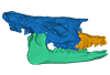
Explodable 3D Dog Skull for Veterinary Education
The specimens of Speothos pacivorus
3D GM dataset of bird skeletal variation
Skeletal embryonic development in the catshark
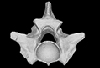
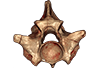
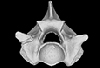
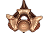
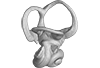
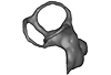
Bony connexions of the petrosal bone of extant hippos
bony labyrinth (14) , inner ear (11) , Eocene (11) , geometric morphometrics (10) , CT-scan (10) , Oligocene (9) , Micro-CT (9)
Lionel Hautier (25) , Maëva Judith Orliac (24) , Laurent Marivaux (18) , Renaud Lebrun (15) , Rodolphe Tabuce (15) , Pierre-Olivier Antoine (13) , Bastien Mennecart (13)
|
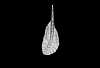
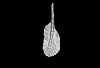
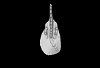
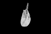
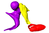
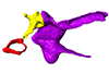
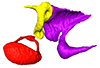
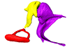
3D models related to the publication: Neogene sloth assemblages (Mammalia, Pilosa) of the Cocinetas Basin (La Guajira, Colombia): implications for the Great American Biotic InterchangeEli Amson
Published online: 08/06/2016 |
|
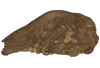
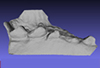
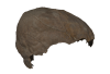
M3#106Fragmentary basicranium with posterior portion of the skull roof. Type: "3D_surfaces"doi: 10.18563/m3.sf.106 state:published |
Download 3D surface file |
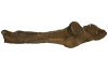
indet. indet. MUN STRI 16535 View specimen
|
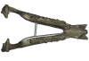
M3#107Complete left ulna of a Scelidotheriinae gen. et sp. indet. Type: "3D_surfaces"doi: 10.18563/m3.sf.107 state:published |
Download 3D surface file |
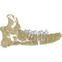
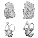
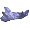
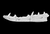
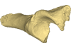
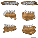
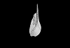
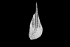
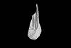
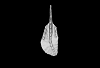
This project presents a µCT dataset and an associated 3D surface model of the holotype of Donrussellia magna (UM PAT 17; Primates, Adapiformes). UM PAT17 is the only known specimen for the species and consists of a well-preserved left lower jaw with p4-m3. It documents one of the oldest European primates, eventually dated near the Paleocene Eocene Thermal Maximum.
Donrussellia magna UM PAT 17 View specimen

|
M3#173D surface file model of UM PAT 17 (type specimen of Donrussellia magna), which is a well preserved left lower jaw with p4-m3. The teeth (and roots) were manually segmented. Type: "3D_surfaces"doi: 10.18563/m3.sf17 state:published |
Download 3D surface file |
|
M3#18CT Scan Data of Donrussellia magna UM PAT 17. Voxel size (in µm): 36µm (isotropic voxels). Dimensions in x,y,z : 594 pixels, 294 pixels, 1038 pixels. Image type : 8-bit voxels. Image format : raw data format (no header). Type: "3D_CT"doi: 10.18563/m3.sf18 state:published |
Download CT data |
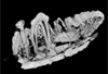
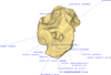
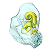
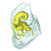
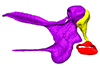
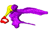
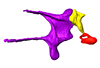
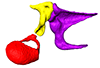
This project presents the 3D models of two isolated petrosals from the Oligocene locality of Pech de Fraysse (Quercy, France) here attributed to the genus Prodremotherium Filhol, 1877. Our aim is to describe the petrosal morphology of this Oligocene “early ruminant” as only few data are available in the literature for Oligocene taxa.
Prodremotherium sp. UM PFY 4053 View specimen

|
M3#7Labelled 3D model of right isolated petrosal of Prodremotherium sp. from Pech de Fraysse (Quercy, MP 28) Type: "3D_surfaces"doi: 10.18563/m3.sf7 state:published |
Download 3D surface file |
Prodremotherium sp. UM PFY 4054 View specimen
|
M3#8Labelled 3D model of right isolated petrosal of Prodremotherium sp. from Pech de Fraysse (Quercy, MP 28) Type: "3D_surfaces"doi: 10.18563/m3.sf8 state:published |
Download 3D surface file |
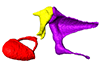
The present 3D Dataset contains the 3D models analyzed in Mennecart, B., Duranthon, F., & Costeur, L. 2024. Systematic contribution of the auditory region to the knowledge of the oldest European Bovidae (Mammalia, Ruminantia). Journal of Anatomy XXX. https://doi.org/10.1111/joa.14132
Pusillutragus montrealensis MHNT.PAL.2015.0.2261.4 View specimen
|
M3#1522Right petrosal, bony labyrinth, stapes Type: "3D_surfaces"doi: 10.18563/m3.sf.1522 state:published |
Download 3D surface file |
Pusillutragus montrealensis MHNT.PAL.2015.0.2261.9 View specimen
|
M3#1523Left petrosal and left bony labyrinth Type: "3D_surfaces"doi: 10.18563/m3.sf.1523 state:published |
Download 3D surface file |
Eotragus artenensis SMNS-P-41625 View specimen
|
M3#1524Petrosal (right), bony labyrinth (left) Type: "3D_surfaces"doi: 10.18563/m3.sf.1524 state:published |
Download 3D surface file |
Eotragus clavatus NMB San.15056 View specimen
|
M3#1528Right petrosal and right bony labyrinth Type: "3D_surfaces"doi: 10.18563/m3.sf.1528 state:published |
Download 3D surface file |
Eotragus clavatus NMB San.15055 View specimen
|
M3#1526Left Petrosal Type: "3D_surfaces"doi: 10.18563/m3.sf.1526 state:published |
Download 3D surface file |
The present 3D Dataset contains the 3D models analyzed in 3D Finite Element Analysis and Geometric Morphometrics of Sloths (Xenarthra, Folivora) Mandibles Show Insights on the Dietary Specializations of Fossil Taxa. Journal of South American Earth Sciences. https://doi.org/10.1016/j.jsames.2023.104445
Mylodon darwinii CAV 379 View specimen
|
M3#1159Right hemimandible Type: "3D_surfaces"doi: 10.18563/m3.sf.1159 state:published |
Download 3D surface file |
Scelidotherium leptocephalum MNHN-M 137,722 View specimen
|
M3#1160Mandible Type: "3D_surfaces"doi: 10.18563/m3.sf.1160 state:published |
Download 3D surface file |
Glossotherium robustum MNHN-M 914 View specimen
|
M3#1161Mandible Type: "3D_surfaces"doi: 10.18563/m3.sf.1161 state:published |
Download 3D surface file |
Lestodon armatus MPAC 899 View specimen
|
M3#1162Mandible Type: "3D_surfaces"doi: 10.18563/m3.sf.1162 state:published |
Download 3D surface file |
Valgipes bucklandi NHMD.Z.M.K. 1/1845:3540 View specimen
|
M3#1163Mandible Type: "3D_surfaces"doi: 10.18563/m3.sf.1163 state:published |
Download 3D surface file |
The present 3D Dataset contains the 3D models analyzed in Hendrickx, C., Gaetano, L. C., Choiniere, J., Mocke, H. and Abdala, F. in press. A new traversodontid cynodont with a peculiar postcanine dentition from the Middle/Late Triassic of Namibia and dental evolution in basal gomphodonts. Journal of Systematic Palaeontology.
Etjoia dentitransitus GSN F1591 View specimen
|
M3#557Surface model derived from µCT data of the holotype of Etjoia dentitransitus Type: "3D_surfaces"doi: 10.18563/m3.sf.557 state:published |
Download 3D surface file |
|
M3#558Photogrammetric 3D surface model of the postcanines of the Holotype of Etjoia dentitransitus Type: "3D_surfaces"doi: 10.18563/m3.sf.558 state:published |
Download 3D surface file |
|
M3#559Photogrammetric 3D surface model of the Holotype of Etjoia dentitransitus Type: "3D_surfaces"doi: 10.18563/m3.sf.559 state:published |
Download 3D surface file |
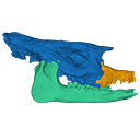
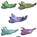
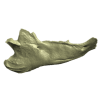
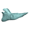
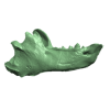
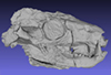
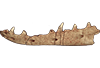
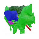
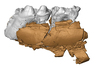
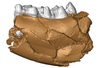
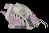
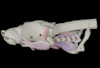
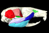
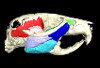
The present 3D Dataset contains two 3D models described in Tissier et al. (https://doi.org/10.1098/rsos.200633): the only known complete mandible of the early-branching rhinocerotoid Epiaceratherium magnum Uhlig, 1999, and a hypothetical reconstruction of the complete archetypic skull of Epiaceratherium Heissig, 1969, created by merging three cranial parts from three distinct Epiaceratherium species.
Epiaceratherium magnum NMB.O.B.928 View specimen
|
M3#5343D surface model of the mandible NMB.O.B.928 of Epiaceratherium magnum, with texture file. Type: "3D_surfaces"doi: 10.18563/m3.sf.534 state:published |
Download 3D surface file |
Epiaceratherium magnum NMB.O.B.928 + MJSN POI007–245 + NMB.I.O.43 View specimen
|
M3#535Archetypal reconstruction of the skull of Epiaceratherium, generated by 3D virtual association of the cranium of E. delemontense (MJSN POI007–245, in blue), mandible of E. magnum (NMB.O.B.928, green) and snout of E. bolcense (NMB.I.O.43, in orange). Type: "3D_surfaces"doi: 10.18563/m3.sf.535 state:published |
Download 3D surface file |
The present 3D Dataset contains the 3D models analyzed in the following publication: Georgalis, G. L., and T. M. Scheyer. A new species of Palaeopython (Serpentes) and other extinct squamates from the Eocene of Dielsdorf (Zurich, Switzerland). Swiss Journal of Geosciences (in press). https://doi.org/10.1007/s00015-019-00341-6
Palaeopython helveticus PIMUZ A/III 631 View specimen
|
M3#399ZIP file containing .ply of vertebra PIMUZ A/III 631 from Palaeopython helveticus n. sp. Type: "3D_surfaces"doi: 10.18563/m3.sf.399 state:published |
Download 3D surface file |
|
M3#403dataset of snake vertebra PIMUZ A/III 631 Type: "3D_CT"doi: 10.18563/m3.sf.403 state:published |
Download CT data |
Palaeopython helveticus PIMUZ A/III 634 View specimen
|
M3#400ZIP file containing .ply of vertebra PIMUZ A/III 634 from Palaeopython helveticus n. sp. (holotype) Type: "3D_surfaces"doi: 10.18563/m3.sf.400 state:published |
Download 3D surface file |
|
M3#404dataset of snake vertbra PIMUZ A/III 634 (holotype) Type: "3D_CT"doi: 10.18563/m3.sf.404 state:published |
Download CT data |
Palaeopython helveticus PIMUZ A/III 636 View specimen
|
M3#401ZIP file containing .ply of vertebra PIMUZ A/III 636 from Palaeopython helveticus n. sp. Type: "3D_surfaces"doi: 10.18563/m3.sf.401 state:published |
Download 3D surface file |
|
M3#406dataset of snake vertebra PIMUZ A/III 636 Type: "3D_CT"doi: 10.18563/m3.sf.406 state:published |
Download CT data |
Palaeovaranus sp. PIMUZ A/III 234 View specimen
|
M3#402ZIP file containing .ply of dentary PIMUZ A/III 234 of Palaeovaranus sp. Type: "3D_surfaces"doi: 10.18563/m3.sf.402 state:published |
Download 3D surface file |
|
M3#405dataset of dentary of Palaeovaranus sp. (PIMUZ A/III 234) Type: "3D_CT"doi: 10.18563/m3.sf.405 state:published |
Download CT data |

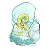
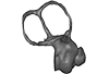
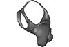
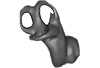
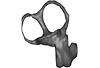
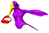
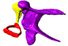
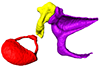
The present 3D Dataset contains the 3D models of the holotype (NMB Sth. 833) of the new species Micromeryx? eiselei analysed in the article Aiglstorfer, M., Costeur, L., Mennecart, B., Heizmann, E.P.J.. 2017. Micromeryx? eiselei - a new moschid species from Steinheim am Albuch, Germany, and the first comprehensive description of moschid cranial material from the Miocene of Central Europe. PlosOne https://doi.org/10.1371/journal.pone.0185679
Micromeryx? eiselei NMB Sth. 833 View specimen
|
M3#284The 3 D surfaces comprises the skull, petrosal, and bony labyrinth of NMB Sth.833, the holotype of Micromeryx? eiselei Type: "3D_surfaces"doi: 10.18563/m3.sf.284 state:published |
Download 3D surface file |
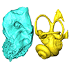
The present 3D Dataset contains the 3D models analyzed in Velazco P. M., Grohé C. 2017. Comparative anatomy of the bony labyrinth of the bats Platalina genovensium (Phyllostomidae, Lonchophyllinae) and Tomopeas ravus (Molossidae, Tomopeatinae). Biotempo 14(2).
Platalina genovensium 278520 View specimen

|
M3#276Right bony labyrinth surface positioned (.PLY) Labels associated (.FLG) Type: "3D_surfaces"doi: 10.18563/m3.sf.276 state:published |
Download 3D surface file |
Tomopeas ravus 278525 View specimen

|
M3#277Right bony labyrinth surface (.PLY) Labels associated (.FLG) Type: "3D_surfaces"doi: 10.18563/m3.sf.277 state:published |
Download 3D surface file |
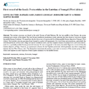
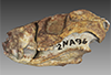
This contribution contains the 3D model described and figured in the following publication: Hautier L, Sarr R, Lihoreau F, Tabuce R, Marwan Hameh P. 2014. First record of the family Protocetidae in the Lutetian of Senegal (West Africa). Palaeovertebrata 38(2)-e2
indet. indet. SN103 View specimen
|
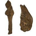
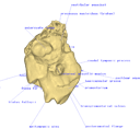
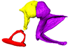
M3#5SN103, partial left innominate. Age and occurrence – Taïba Formation, Lutetian of the near Taïba Ndiaye, quarry of the Industries Chimiques du Sénégal (ICS) Type: "3D_surfaces"doi: 10.18563/m3.sf5 state:published |
Download 3D surface file |
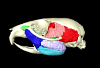
This contribution contains the 3D model of an endocranial cast analyzed in “A 10 ka intentionally deformed human skull from Northeast Asia”. There are many studies on the morphological characteristics of intentional cranial deformation (ICD), but few related 3D models were published. Here, we present the surface model of an intentionally deformed 10 ka human cranium for further research on ICD practice. The 3D model of the endocranial cast of this ICD cranium was discovered near Harbin City, Province Heilongjiang, Northeast China. The fossil preserved only the frontal, parietal, and occipital bones. To complete the endocast model of the specimen, we printed a 3D model and used modeling clay to reconstruct the missing part based on the general form of the modern human endocast morphology.
Homo sapiens IVPP-PA1616 View specimen
|
M3#972The frontal region of the endocast is flattened, probably formed by the constant pressure on the frontal bone during growth. There is a well-developed frontal crest on the endocranial surface. The endocast widens posteriorly from the frontal lobe. The widest point of the endocast is at the lateral border of the parietal lobe. The lower parietal areas display a marked lateral expansion. The overall shape of the endocast is asymmetrical, with the left side of the parietal lobe being more laterally expanded than the right side. Like the frontal lobe, the occipital lobe is also anteroposteriorly flattened. Type: "3D_surfaces"doi: 10.18563/m3.sf.972 state:published |
Download 3D surface file |
|
M3#976The original endocranial cast model (with texture) of IVPP-PA1616. It shows the original structures of the specimen, and was not altered in any way. Type: "3D_surfaces"doi: 10.18563/m3.sf.976 state:published |
Download 3D surface file |
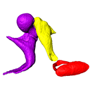
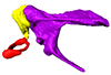
The present 3D Dataset contains the 3D models analyzed in Pochat-Cottilloux Y., Martin J.E., Jouve S., Perrichon G., Adrien J., Salaviale C., de Muizon C., Cespedes R. & Amiot R. (2021). The neuroanatomy of Zulmasuchus querejazus (Crocodylomorpha, Sebecidae) and its implications for the paleoecology of sebecosuchians. The Anatomical Record, https://doi.org/10.1002/ar.24826
Zulmasuchus querejazus MHNC 6672 View specimen

|
M3#798Left endosseous labyrinth of Z. querejazus (MHNC 6672). Type: "3D_surfaces"doi: 10.18563/m3.sf.798 state:published |
Download 3D surface file |
|
M3#799Reconstruction of the endocranial cavities of Z. querejazus (MHNC 6672). Type: "3D_surfaces"doi: 10.18563/m3.sf.799 state:published |
Download 3D surface file |
|
M3#800Three-dimensional reconstruction of the pneumatic cavities within the braincase of Z. querejazus (MHNC 6672) Type: "3D_surfaces"doi: 10.18563/m3.sf.800 state:published |
Download 3D surface file |
The present 3D Dataset contains the 3D models analyzed in Mennecart B., Wazir W.A., Sehgal R.K., Patnaik R., Singh N.P., Kumar N, and Nanda A.C. 2021. New remains of Nalamaeryx (Tragulidae, Mammalia) from the Ladakh Himalaya and their phylogenetical and palaeoenvironmental implications. Historical Biology. https://doi.org/10.1080/08912963.2021.2014479
Nalameryx savagei WIMF/A4801 View specimen
|
M3#766Nalameryx savagei, Partial lower right jaw preserving m2 and m3. Type: "3D_surfaces"doi: 10.18563/m3.sf.766 state:published |
Download 3D surface file |
Nalameryx savagei WIMF/A4802 View specimen
|
M3#767Nalameryx savagei, partial lower right jaw preserving m2 and m3 Type: "3D_surfaces"doi: 10.18563/m3.sf.767 state:published |
Download 3D surface file |
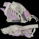
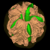
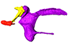
This contribution contains 3D models of the cranial skeleton and muscles in an elephantfish (Callorhinchus milii) and a catshark (Scyliorhinus canicula), based on synchrotron tomographic scans. These datasets were analyzed and described in Dearden et al. (2021) “The morphology and evolution of chondrichthyan cranial muscles: a digital dissection of the elephantfish Callorhinchus milii and the catshark Scyliorhinus canicula.” Journal of Anatomy.
Callorhinchus milii 001 View specimen
|
M3#7083D models of the cranial skeleton and muscles of Callorhinchus milii, created using Mimics. Type: "3D_surfaces"doi: 10.18563/m3.sf.708 state:published |
Download 3D surface file |
Scyliorhinus canicula 002 View specimen
|
M3#7093D models of the cranial skeleton and muscles of Scyliorhinus canicula, created using Mimics. Type: "3D_surfaces"doi: 10.18563/m3.sf.709 state:published |
Download 3D surface file |
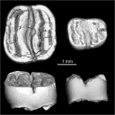
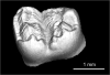
This contribution provides the raw files for the μCT-scan data and renderings of the three-dimensional digital models of two fossil teeth of a geomyin geomorph rodent (Caribeomys merzeraudi), discovered from lower Oligocene deposits of Puerto Rico, San Sebastian Formation (locality LACM Loc. 8060). These fossils were described, figured and discussed in the following publication: Marivaux et al. (2021), An unpredicted ancient colonization of the West Indies by North American rodents: dental evidence of a geomorph from the early Oligocene of Puerto Rico. Papers in Palaeontology. https://doi.org/10.1002/spp2.1388
Caribeomys merzeraudi LACM 162478 View specimen
|
M3#712Right lower dp4: isolated deciduous premolar. The specimen was scanned with a resolution of 5 µm using a μ-CT-scanning station EasyTom 150 / Rx Solutions (Montpellier RIO Imaging, ISE-M, Montpellier, France). AVIZO 7.1 (Visualization Sciences Group) software was used for visualization, segmentation, and 3D rendering. This isolated tooth was prepared within a “labelfield” module of AVIZO, using the segmentation threshold selection tool. Type: "3D_surfaces"doi: 10.18563/m3.sf.712 state:published |
Download 3D surface file |
|
M3#7145µm µCT data set . Right lower dp4: isolated deciduous premolar. The specimen was scanned with a resolution of 5 µm using a μ-CT-scanning station EasyTom 150 / Rx Solutions (Montpellier RIO Imaging, ISE-M, Montpellier, France). Type: "3D_CT"doi: 10.18563/m3.sf.714 state:published |
Download CT data |
Caribeomys merzeraudi LACM 162449 View specimen
|
M3#713Right lower molar (m1 or m2). The specimen was scanned with a resolution of 4.5 µm using a μ-CT-scanning station EasyTom 150 / Rx Solutions (Montpellier RIO Imaging, ISE-M, Montpellier, France). AVIZO 7.1 (Visualization Sciences Group) software was used for visualization, segmentation, and 3D rendering. This isolated tooth was prepared within a “labelfield” module of AVIZO, using the segmentation threshold selection tool. Type: "3D_surfaces"doi: 10.18563/m3.sf.713 state:published |
Download 3D surface file |
|
M3#715µCT data at 4.5µm Type: "3D_CT"doi: 10.18563/m3.sf.715 state:published |
Download CT data |
The present 3D Dataset contains the 3D models of the holotype and the paratypes of the new species Siphonodella leiosa described and analyzed in the following publication: L. Souquet, C. Corradini, C. Girard: Siphonodella leiosa (Conodonta), a new unornamented species from the Tournaisian (lower Carboniferous) of Puech de la Suque (Montagne Noire, France). Geobios, https://doi.org/10.1016/j.geobios.2020.06.004.
Siphonodella leiosa UM PSQ 1 View specimen
|
M3#525Siphonodella leiosa, paratype, dextral P1 element Type: "3D_surfaces"doi: 10.18563/m3.sf.525 state:published |
Download 3D surface file |
Siphonodella leiosa UM PSQ 2 View specimen
|
M3#526Siphonodella leiosa, holotype, dextral P1 element Type: "3D_surfaces"doi: 10.18563/m3.sf.526 state:published |
Download 3D surface file |
Siphonodella leiosa UM PSQ 3 View specimen

|
M3#527Siphonodella leiosa, paratype, dextral P1 element Type: "3D_surfaces"doi: 10.18563/m3.sf.527 state:published |
Download 3D surface file |
Siphonodella leiosa UM PSQ 4 View specimen
|
M3#528Siphonodella leiosa, paratype, dextral P1 element Type: "3D_surfaces"doi: 10.18563/m3.sf.528 state:published |
Download 3D surface file |
Siphonodella leiosa UM PSQ 5 View specimen
|
M3#529Siphonodella leiosa, paratype, sinistral P1 element Type: "3D_surfaces"doi: 10.18563/m3.sf.529 state:published |
Download 3D surface file |
Siphonodella leiosa UM PSQ 6 View specimen
|
M3#530Siphonodella leiosa, paratype, dextral P1 element Type: "3D_surfaces"doi: 10.18563/m3.sf.530 state:published |
Download 3D surface file |
Siphonodella leiosa UM PSQ 7 View specimen
|
M3#531Siphonodella leiosa, paratype, dextral P1 element Type: "3D_surfaces"doi: 10.18563/m3.sf.531 state:published |
Download 3D surface file |
Siphonodella leiosa UM PSQ 8 View specimen
|
M3#532Siphonodella leiosa, paratype, sinistral P1 element Type: "3D_surfaces"doi: 10.18563/m3.sf.532 state:published |
Download 3D surface file |
Siphonodella leiosa UM PSQ 9 View specimen
|
M3#533Siphonodella leiosa, paratype, dextral P1 element Type: "3D_surfaces"doi: 10.18563/m3.sf.533 state:published |
Download 3D surface file |
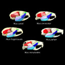
The present 3D Dataset contains the 3D models analyzed in the article entitled "One skull to rule them all? Descriptive and comparative anatomy of the masticatory apparatus in five mice species based on traditional and digital dissections" (Ginot et al. 2018, Journal of Morphology, https://doi.org/10.1002/jmor.20845).
Mus cervicolor R7314 View specimen
|
M3#343.ply surfaces of the skull and masticatory muscles of Mus cervicolor. Created with MorphoDig, .pos and .ntw files also included. Scans were obtained thanks to the Institut des Sciences de l'Evolution de Montpellier MRI platform. Type: "3D_surfaces"doi: 10.18563/m3.sf.343 state:published |
Download 3D surface file |
Mus caroli R7264 View specimen
|
M3#344.ply surfaces of the skull and masticatory muscles of Mus caroli. Created with MorphoDig, .pos and .ntw files also included. Scans were obtained thanks to the Institut des Sciences de l'Evolution de Montpellier MRI platform. Type: "3D_surfaces"doi: 10.18563/m3.sf.344 state:published |
Download 3D surface file |
Mus fragilicauda R7260 View specimen
|
M3#345.ply surfaces of the skull and masticatory muscles of Mus fragilicauda. Created with MorphoDig, .pos and .ntw files also included. Scans were obtained thanks to the Institut des Sciences de l'Evolution de Montpellier MRI platform. Type: "3D_surfaces"doi: 10.18563/m3.sf.345 state:published |
Download 3D surface file |
Mus pahari R7226 View specimen
|
M3#346.ply surfaces of the skull and masticatory muscles of Mus pahari. Created with MorphoDig, .pos and .ntw files also included. Scans were obtained thanks to the Institut des Sciences de l'Evolution de Montpellier MRI platform. Type: "3D_surfaces"doi: 10.18563/m3.sf.346 state:published |
Download 3D surface file |
Mus minutoides minutoides-1 View specimen
|
M3#347.ply surfaces of the skull and masticatory muscles of Mus minutoides. Created with MorphoDig, .pos and .ntw files also included. Scans were obtained thanks to the Institut des Sciences de l'Evolution de Montpellier MRI platform. Type: "3D_surfaces"doi: 10.18563/m3.sf.347 state:published |
Download 3D surface file |
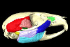
Here, the semicircular canals of the most aquatic seal, the rare Antarctic Ross Seal (Ommatophoca rossii), are presented for the first time, along with representatives of every species in the Lobodontini: the leopard seal (Hydrurga leptonyx), Weddell seal (Leptonychotes weddellii), and crabeater seal (Lobodon carcinophagus). Because encounters with wild Ross seal are rare, and few specimens are available in collections worldwide, this dataset increases accessibility to a rare species. For further comparison, we present the bony labyrinths of other carnivorans, the elephant seal (Mirounga leonina), harbor seal (Phoca vitulina), walrus (Odobenus rosmarus), South American sea lion (Otaria byronia).
Odobenus rosmarus MVZ 125566 View specimen
|
M3#173Surface of the semicircular canals and cochlea of the walrus, Odobenus rosmarus Type: "3D_surfaces"doi: 10.18563/m3.sf.173 state:published |
Download 3D surface file |
Phoca vitulina UZNH 17973 View specimen
|
M3#174Endocast surface of the semicircular canals and cochlea of the harbor seal, Phoca vitulina. Type: "3D_surfaces"doi: 10.18563/m3.sf.174 state:published |
Download 3D surface file |
Hydrurga leptonyx MLP 14.IV.48.11 View specimen
|
M3#285Endocast surface of the semicircular canals and cochlea of the leopard seal, Hydrurga leptonyx. Type: "3D_surfaces"doi: 10.18563/m3.sf.285 state:published |
Download 3D surface file |
Leptonychotes weddellii IAA 02-13 View specimen
|
M3#288Endocast surface of the semicircular canals and cochlea of the Weddell seal Leptonychotes weddellii. Type: "3D_surfaces"doi: 10.18563/m3.sf.288 state:published |
Download 3D surface file |
Lobodon carcinophagus IAA 530 View specimen
|
M3#286Endocast surface of the semicircular canals and cochlea of the crabeater seal, Lobodon carcinophagus. Type: "3D_surfaces"doi: 10.18563/m3.sf.286 state:published |
Download 3D surface file |
Ommatophoca rossii MACN 48259 View specimen
|
M3#176Endocast surface of the semicircular canals and cochlea of the Ross seal Ommatophoca rossii. Type: "3D_surfaces"doi: 10.18563/m3.sf.176 state:published |
Download 3D surface file |
Mirounga leonina IAA 03-5 View specimen
|
M3#287Right endocast surface of the semicircular canals and cochlea of the elephant seal, Mirounga leonina. Type: "3D_surfaces"doi: 10.18563/m3.sf.287 state:published |
Download 3D surface file |
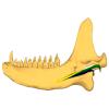
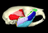
Considerable morphological variations are found in the middle ear among mammals. Here I present a three-dimensional atlas of the middle ear ossicles of eulipotyphlan mammals. This group has radiated into various environments as terrestrial, aquatic, and subterranean habitats independently in multiple lineages. Therefore, eulipotyphlans are an ideal group to explore the form-function relationship of the middle ear ossicles. This comparative atlas of hedgehogs, true shrews, water shrews, mole shrews, true moles, and shrew moles encourages future studies of the middle ear morphology of this diverse group.
Erinaceus europaeus DK2331 View specimen
|
M3#151Left middle ear ossicles Type: "3D_surfaces"doi: 10.18563/m3.sf.151 state:published |
Download 3D surface file |
Anourosorex yamashinai SIK_yamashinai View specimen
|
M3#152Left middle ear ossicles Type: "3D_surfaces"doi: 10.18563/m3.sf.152 state:published |
Download 3D surface file |
Blarina brevicauda M8003 View specimen
|
M3#153Right middle ear ossicles Type: "3D_surfaces"doi: 10.18563/m3.sf.153 state:published |
Download 3D surface file |
Chimarrogale platycephala DK5481 View specimen
|
M3#162Left middle ear ossicles Type: "3D_surfaces"doi: 10.18563/m3.sf.162 state:published |
Download 3D surface file |
Suncus murinus DK1227 View specimen
|
M3#155Left middle ear ossicles Type: "3D_surfaces"doi: 10.18563/m3.sf.155 state:published |
Download 3D surface file |
Condylura cristata SIK0050 View specimen
|
M3#156Right middle ear ossicles Type: "3D_surfaces"doi: 10.18563/m3.sf.156 state:published |
Download 3D surface file |
Euroscaptor klossi SIK0673 View specimen
|
M3#163Left middle ear ossicles Type: "3D_surfaces"doi: 10.18563/m3.sf.163 state:published |
Download 3D surface file |
Euroscaptor malayana SIK_malayana View specimen
|
M3#164Left middle ear ossicles Type: "3D_surfaces"doi: 10.18563/m3.sf.164 state:published |
Download 3D surface file |
Mogera wogura DK2551 View specimen
|
M3#159Left middle ear ossicles Type: "3D_surfaces"doi: 10.18563/m3.sf.159 state:published |
Download 3D surface file |
Talpa altaica SIK_altaica View specimen
|
M3#161Right middle ear ossicles Type: "3D_surfaces"doi: 10.18563/m3.sf.161 state:published |
Download 3D surface file |
Urotrichus talpoides DK0887 View specimen
|
M3#165Left middle ear ossicles Type: "3D_surfaces"doi: 10.18563/m3.sf.165 state:published |
Download 3D surface file |
Oreoscaptor mizura DK6545 View specimen
|
M3#166Left middle ear ossicles Type: "3D_surfaces"doi: 10.18563/m3.sf.166 state:published |
Download 3D surface file |
Scalopus aquaticus SIK_aquaticus View specimen
|
M3#167Left middle ear ossicles Type: "3D_surfaces"doi: 10.18563/m3.sf.167 state:published |
Download 3D surface file |
Scapanus orarius SIK_orarius View specimen
|
M3#168Left middle ear ossicles Type: "3D_surfaces"doi: 10.18563/m3.sf.168 state:published |
Download 3D surface file |
Neurotrichus gibbsii SIK_gibbsii View specimen
|
M3#169Left middle ear ossicles Type: "3D_surfaces"doi: 10.18563/m3.sf.169 state:published |
Download 3D surface file |