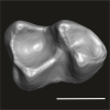
3D models of Ocnotherium skull
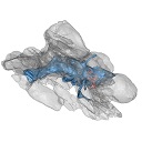
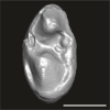
3D models of Kalakocetus, the earliest Cetacea
Explodable 3D Dog Skull for Veterinary Education
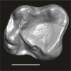
3D GM dataset of bird skeletal variation
Skeletal embryonic development in the catshark
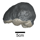
Bony connexions of the petrosal bone of extant hippos
bony labyrinth (14) , inner ear (11) , Eocene (11) , geometric morphometrics (10) , CT-scan (10) , Oligocene (9) , Micro-CT (9)
Maëva Judith Orliac (24) , Lionel Hautier (24) , Laurent Marivaux (18) , Renaud Lebrun (15) , Rodolphe Tabuce (14) , Pierre-Olivier Antoine (13) , Bastien Mennecart (13)
|
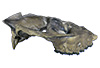
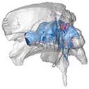
3D models related to the publication: A 50-million-year-old, three-dimensionally preserved bat skull supports an early origin for modern echolocationJacob Maugoust
Published online: 19/10/2023 |
|
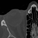
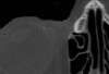
M3#1269External surface of the cranium Type: "3D_surfaces"doi: 10.18563/m3.sf.1269 state:published |
Download 3D surface file |
|
M3#1270Virtual endocast of the right bony labyrinth Type: "3D_surfaces"doi: 10.18563/m3.sf.1270 state:published |
Download 3D surface file |
The present Dataset contains the micro-CT scan of the head of an anonymous 54 year old female donor, at a voxel resolution of 145µm. The skin of the face has been masked in order to avoid the donor to be recognized.
Homo sapiens UM_HS_2018_09_13 View specimen
|
M3#1152Micro-ct data set Type: "3D_CT"doi: 10.18563/m3.sf.1152 state:published |
Download CT data |
The present 3D Dataset contains the 3D model of the skin of Allosaurus described in Hendrickx, C. et al. in press. Morphology and distribution of scales, dermal ossifications, and other non-feather integumentary structures in non-avialan theropod dinosaurs. Biological Reviews.
Allosaurus jimmadseni UMNH VP C481 View specimen
|
M3#902The material consists of a 3D reconstruction of the counterpart of a 30 cm2 patch of skin impression associated with the anterior dorsal ribs/pectoral region of the specimen of Allosaurus jimmadseni UMNH VP C481. The skin shows a semi-uniform basement of 1-2 mm diameter pebbles with a smaller number of slightly larger (up to 3 mm) ovoid scales. The irregular shape, distribution, and overall small size of these larger scales suggest that they are not classifiable as feature scales but rather as variations in the basement scales. Type: "3D_surfaces"doi: 10.18563/m3.sf.902 state:published |
Download 3D surface file |
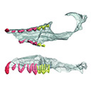
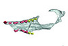
This contribution contains 3D models of the cranial skeleton and muscles in an elephantfish (Callorhinchus milii) and a catshark (Scyliorhinus canicula), based on synchrotron tomographic scans. These datasets were analyzed and described in Dearden et al. (2021) “The morphology and evolution of chondrichthyan cranial muscles: a digital dissection of the elephantfish Callorhinchus milii and the catshark Scyliorhinus canicula.” Journal of Anatomy.
Callorhinchus milii 001 View specimen
|
M3#7083D models of the cranial skeleton and muscles of Callorhinchus milii, created using Mimics. Type: "3D_surfaces"doi: 10.18563/m3.sf.708 state:published |
Download 3D surface file |
Scyliorhinus canicula 002 View specimen
|
M3#7093D models of the cranial skeleton and muscles of Scyliorhinus canicula, created using Mimics. Type: "3D_surfaces"doi: 10.18563/m3.sf.709 state:published |
Download 3D surface file |
The present 3D Dataset contains the 3D models analyzed in Mennecart B., Métais G., Costeur L., Ginsburg L, and Rössner G. 2021, Reassessment of the enigmatic ruminant Miocene genus Amphimoschus Bourgeois, 1873 (Mammalia, Artiodactyla, Pecora). PlosOne. https://doi.org/10.1371/journal.pone.0244661
Amphimoschus ponteleviensis MNHN.F.AR3266 View specimen
|
M3#701Surface scan of the cast of the skull of Amphimoschus ponteleviensis MNHN.F.AR3266 from Artenay (France) Type: "3D_surfaces"doi: 10.18563/m3.sf.701 state:published |
Download 3D surface file |
|
M3#702Right petrosal bone and bony labyrinth of the skull MNHN.F.AR3266 from Artenay (France) Type: "3D_surfaces"doi: 10.18563/m3.sf.702 state:published |
Download 3D surface file |
Amphimoschus ponteleviensis SMNS40693 View specimen
|
M3#704Left petrosal bone and bony labyrinth of the skull SMNS40693 from Langenau 1 (Germany) Type: "3D_surfaces"doi: 10.18563/m3.sf.704 state:published |
Download 3D surface file |
This contribution contains the 3D models described and figured in the following publication: Paulina-Carabajal A and Calvo JO 2021. Re-description of the braincase of the rebbachisaurid sauropod Limaysaurus tessonei and novel endocranial information based on CT scans. Anais da Academia Brasileira de Ciências 93(Suppl. 2): e20200762 https://doi.org/10.1590/0001-3765202120200762
Limaysaurus tessonei MUCPv-205 View specimen
|
M3#700Renderings of the virtually isolate braincase, brain, and right inner ear. Type: "3D_surfaces"doi: 10.18563/m3.sf.700 state:published |
Download 3D surface file |
This contribution contains the 3D model(s) described and figured in the following publication: The present 3D Dataset contains the 3D models and CT-Scan slices of the lower jaws and teeth analyzed in “A new prozostrodontian cynodont (Eucynodontia, Probainognathia) from the Upper Triassic of southern Brazil”. https://doi.org/10.1080/02724634.2020.1782415
Agudotherium gassenae CAPPA/UFSM 0262 View specimen
|
M3#546Left lower jaw and cheek teeth Type: "3D_surfaces"doi: 10.18563/m3.sf.546 state:published |
Download 3D surface file |
|
M3#5471578 slices Type: "3D_CT"doi: 10.18563/m3.sf.547 state:published |
Download CT data |
Agudotherium gassenae CAPPA/UFSM 0208 View specimen
|
M3#548right lower jaw Type: "3D_surfaces"doi: 10.18563/m3.sf.548 state:published |
Download 3D surface file |
|
M3#549CT data of CAPPA_UFSM_0208 Type: "3D_CT"doi: 10.18563/m3.sf.549 state:published |
Download CT data |
This contribution includes the 3D models of the reconstructed ossicular chain of the cainotheriid Caenomeryx filholi from the late Oligocene locality of Pech Desse (MP28, Quercy, France) described and figured in the publication of Assemat et al. (2020). It represents the oldest ossicular chain reconstruction for a Paleogene terrestrial artiodactyl species.
Caenomeryx filholi UM PDS 3353 View specimen
|
M3#508reconstruction of the middle ear with petrosal, bulla, stapes, incus, malleus Type: "3D_surfaces"doi: 10.18563/m3.sf.508 state:published |
Download 3D surface file |
This contribution contains the 3D models described and figured in the following publication: Paulina-Carabajal, A. and Nieto, M. N. In press. Brief comment on the brain and inner ear of Giganotosaurus carolinii (Dinosauria: Theropoda) based on CT scans. Ameghiniana. https://doi.org/10.5710/AMGH.25.10.2019.3237
Giganotosaurus carolinii MUCPv-CH-1 View specimen
|
M3#504The current file contents 3D models of the braincase, brain, left and right inner ears Type: "3D_surfaces"doi: 10.18563/m3.sf.504 state:published |
Download 3D surface file |
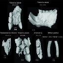
This contribution contains 3D models of extinct rodents Dinomyidae from Miocene and Quaternary of Brazil. The Miocene specimens that were digitalized include the holotypes of Potamarchus adamiae, Pseudopotamarchus villanuevai, and Ferigolomys pacarana collected in the Solimões Formation (Upper Miocene), northern Brazil. The Quaternary specimens are the holotype and paratype of Niedemys piauiensis, found in Upper Pleistocene deposits from northeast Brazil.
Potamarchus adamiae UFAC-CS 011 View specimen
|
M3#410UFAC-CS 011 – holotype, palatal region of the skull with cheek teeth Type: "3D_surfaces"doi: 10.18563/m3.sf.410 state:published |
Download 3D surface file |
Potamarchus adamiae UFAC-CS 043 View specimen
|
M3#411UFAC-CS 043, left dentary with cheek teeth Type: "3D_surfaces"doi: 10.18563/m3.sf.411 state:published |
Download 3D surface file |
Pseudopotamarchus villanuevai UFAC 4762 View specimen
|
M3#412UFAC 4762 – holotype, incomplete right maxilla with cheek teeth Type: "3D_surfaces"doi: 10.18563/m3.sf.412 state:published |
Download 3D surface file |
Ferigolomys pacarana UFAC 6460 View specimen
|
M3#413UFAC 6460 – holotype, palatal region of the skull with cheek teeth Type: "3D_surfaces"doi: 10.18563/m3.sf.413 state:published |
Download 3D surface file |
Drytomomys sp. UFAC 2742 View specimen

|
M3#414UFAC 2742, right dentary with cheek teeth Type: "3D_surfaces"doi: 10.18563/m3.sf.414 state:published |
Download 3D surface file |
Niedemys piauiensis FUMDHAM 113-146365-2 View specimen
|
M3#418FUMDHAM 113-146365-2 - holotype, upper right tooth Type: "3D_surfaces"doi: 10.18563/m3.sf.418 state:published |
Download 3D surface file |
Niedemys piauiensis FUMDHAM 113-145304-2 View specimen
|
M3#419FUMDHAM 113-145304-2 - paratype, left lower molar Type: "3D_surfaces"doi: 10.18563/m3.sf.419 state:published |
Download 3D surface file |
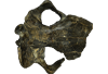
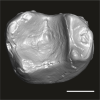
The present 3D Dataset contains the 3D model analyzed in the following publication: Solé et al. (2018), Niche partitioning of the European carnivorous mammals during the paleogene. Palaios. https://doi.org/10.2110/palo.2018.022
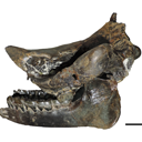
Hyaenodon leptorhynchus FSL848325 View specimen
|
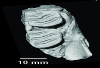
M3#336The specimen FSL848325 is separated in two fragments: the anterior part bears the incisors, the deciduous and permanent canines, while the posterior part bears the right P3, P4, M1 and M2. The P2 is isolated. When combined, the cranium length is approximatively 10.5 cm long. The anterior part is 6.9 cm long and 2.15 cm wide (taken at the level of the P1). The posterior part is 4.8 cm long. The anterior part of the cranium is very narrow. Type: "3D_surfaces"doi: 10.18563/m3.sf.336 state:published |
Download 3D surface file |
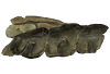
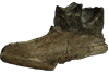
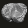
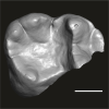
This contribution contains the 3D models described and figured in the following publication: Tissier et al. (in prep.).
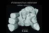
Sellamynodon zimborensis UBB MPS 15795 View specimen
|
M3#297Incomplete skull with left M3. Type: "3D_surfaces"doi: 10.18563/m3.sf.297 state:published |
Download 3D surface file |
Sellamynodon zimborensis UBB MPS 15795 View specimen

|
M3#298Mandible with complete molar and premolar rows, lacking symphysis. Type: "3D_surfaces"doi: 10.18563/m3.sf.298 state:published |
Download 3D surface file |
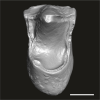
Amynodontopsis aff. bodei UBB MPS V545 View specimen
|
M3#299Maxillary fragment with M1-3. Type: "3D_surfaces"doi: 10.18563/m3.sf.299 state:published |
Download 3D surface file |
Amynodontopsis aff. bodei UBB MPS V546 View specimen
|
M3#300Unworn m1/2 on mandible fragment. Type: "3D_surfaces"doi: 10.18563/m3.sf.300 state:published |
Download 3D surface file |
The present 3D Dataset contains the 3D models analyzed in: "a giant dapediid from the Late Triassic of Switzerland and insights into neopterygian phylogeny", Royal Society Open Science, https://doi.org/10.1098/rsos.180497
Scopulipiscis saxciput PIMUZ A/I 3026 View specimen
|
M3#1773D surfaces of the skull and endocranial spaces inside neurocranium, including the aortic canal, braincase, fossa bridgei, lateral cranial canal, nerves and other passageways, notochord, posterior myodome, and right semicircular canals. Type: "3D_surfaces"doi: 10.18563/m3.sf.177 state:published |
Download 3D surface file |

|
M3#178Scan of the neurocranium of PIMUZ A/I 3026 Type: "3D_CT"doi: 10.18563/m3.sf.178 state:published |
Download CT data |
The present 3D Dataset contains the 3D models analyzed in: Hirose, A., Nakashima, T., Yamada, S., Uwabe, C., Kose, K., Takakuwa, T. 2012. Embryonic liver morphology and morphometry by magnetic resonance microscopic imaging. Anat Rec (Hoboken) 295, 51-59. doi: 10.1002/ar.21496
Homo sapiens KC-CS14LIV1387 View specimen
|
M3#64Human liver at Carnegie Stage (CS) 14 Type: "3D_surfaces"doi: 10.18563/m3.sf.64 state:published |
Download 3D surface file |
Homo sapiens KC-CS15LIV5074 View specimen
|
M3#65Human liver at Carnegie Stage (CS) 15 Type: "3D_surfaces"doi: 10.18563/m3.sf.65 state:published |
Download 3D surface file |
Homo sapiens KC-CS16LIV2578 View specimen
|
M3#66Human liver at Carnegie Stage (CS) 16 Type: "3D_surfaces"doi: 10.18563/m3.sf.66 state:published |
Download 3D surface file |
Homo sapiens KC-CS17LIV17832 View specimen
|
M3#67Human liver at Carnegie Stage (CS) 17 Type: "3D_surfaces"doi: 10.18563/m3.sf.67 state:published |
Download 3D surface file |
Homo sapiens KC-CS18LIV21124 View specimen
|
M3#68Human liver at Carnegie Stage (CS) 18 Type: "3D_surfaces"doi: 10.18563/m3.sf.68 state:published |
Download 3D surface file |
Homo sapiens KC-CS19LIV14353 View specimen
|
M3#69Human liver at Carnegie Stage (CS) 19 Type: "3D_surfaces"doi: 10.18563/m3.sf.69 state:published |
Download 3D surface file |
Homo sapiens KC-CS20LIV20701 View specimen
|
M3#70Human liver at Carnegie Stage (CS) 20 Type: "3D_surfaces"doi: 10.18563/m3.sf.70 state:published |
Download 3D surface file |
Homo sapiens KC-CS21LIV25858 View specimen
|
M3#71Human liver at Carnegie Stage (CS) 21 Type: "3D_surfaces"doi: 10.18563/m3.sf.71 state:published |
Download 3D surface file |
Homo sapiens KC-CS22LIV22226 View specimen
|
M3#72Human liver at Carnegie Stage (CS) 22 Type: "3D_surfaces"doi: 10.18563/m3.sf.72 state:published |
Download 3D surface file |
Homo sapiens KC-CS23LIV25704 View specimen
|
M3#73Human liver at Carnegie Stage (CS) 23 Type: "3D_surfaces"doi: 10.18563/m3.sf.73 state:published |
Download 3D surface file |
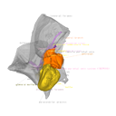
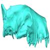
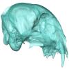
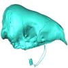
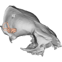
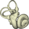
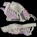
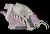
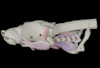
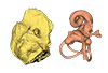
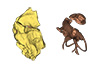
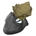
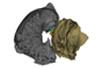
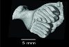
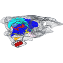
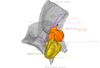
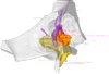
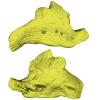
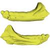
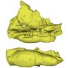
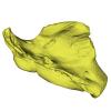
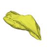
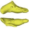
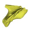
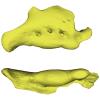
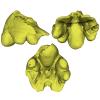
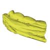
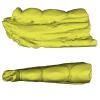
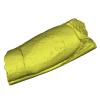
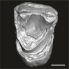
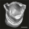
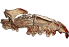
This project presents the osteological connexions of the petrosal bone of the extant Hippopotamidae Hippopotamus amphibius and Choeropsis liberiensis by a virtual osteological dissection of the ear region. The petrosal, the bulla, the sinuses and the major morphological features surrounding the petrosal bone are labelled, both in situ and in an exploded model presenting disassembly views. The directional underwater hearing mode of Hippopotamidae is discussed based on the new observations.
Choeropsis liberiensis UPPal-M09-5-005a View specimen
|
M3#1Labelled compact model of the right ear region of Choeropsis liberiensis (UPPal-M09-5-005a) Type: "3D_surfaces"doi: 10.18563/m3.sf1 state:published |
Download 3D surface file |

|
M3#2Labelled exploded model of the right ear region of Choeropsis liberiensis (UPPal-M09-5-005a) Type: "3D_surfaces"doi: 10.18563/m3.sf2 state:published |
Download 3D surface file |
Hippopotamus amphibius UM N179 View specimen
|
M3#3Labelled compact model of the right ear region of Hippopotamus amphibius (UM N 179) Type: "3D_surfaces"doi: 10.18563/m3.sf3 state:published |
Download 3D surface file |

|
M3#4Labelled exploded model of the right ear region of Hippopotamus amphibius (UM N 179) Type: "3D_surfaces"doi: 10.18563/m3.sf4 state:published |
Download 3D surface file |
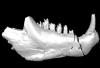
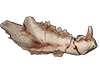
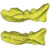
This contribution contains the 3D models described and figured in the following publication: Georgalis, G.L., K.T. Smith, L. Marivaux, A. Herrel, E.M. Essid, H.K. Ammar, W. Marzougui, R. Temani and R. Tabuce. 2024. The world’s largest worm lizard: a new giant trogonophid (Squamata: Amphisbaenia) with extreme dental adaptations from the Eocene of Chambi, Tunisia. Zoological Journal of the Linnean Society. https://doi.org/10.1093/zoolinnean/zlae133
Terastiodontosaurus marcelosanchezi ONM CBI-1-645 View specimen
|
M3#1561Holotype maxilla ONM CBI-1-645 of Terastiodontosaurus marcelosanchezi from the Eocene of Chambi Type: "3D_surfaces"doi: 10.18563/m3.sf.1561 state:published |
Download 3D surface file |
Terastiodontosaurus marcelosanchezi ONM CBI-1-646 View specimen
|
M3#1560Paratype dentary ONM CBI-1-646 of Terastiodontosaurus marcelosanchezi from the Eocene of Chambi Type: "3D_surfaces"doi: 10.18563/m3.sf.1560 state:published |
Download 3D surface file |
Terastiodontosaurus marcelosanchezi ONM CBI-1-648 View specimen
|
M3#1562Maxilla ONM CBI-1-648 of Terastiodontosaurus marcelosanchezi from the Eocene of Chambi Type: "3D_surfaces"doi: 10.18563/m3.sf.1562 state:published |
Download 3D surface file |
Terastiodontosaurus marcelosanchezi ONM CBI-1-649 View specimen
|
M3#1559Maxilla ONM CBI-1-649 of Terastiodontosaurus marcelosanchezi from the Eocene of Chambi Type: "3D_surfaces"doi: 10.18563/m3.sf.1559 state:published |
Download 3D surface file |
Terastiodontosaurus marcelosanchezi ONM CBI-1-650 View specimen
|
M3#1563Maxilla ONM CBI-1-650 of Terastiodontosaurus marcelosanchezi from the Eocene of Chambi Type: "3D_surfaces"doi: 10.18563/m3.sf.1563 state:published |
Download 3D surface file |
Terastiodontosaurus marcelosanchezi ONM CBI-1-651 View specimen
|
M3#1564Maxilla ONM CBI-1-651 of Terastiodontosaurus marcelosanchezi from the Eocene of Chambi Type: "3D_surfaces"doi: 10.18563/m3.sf.1564 state:published |
Download 3D surface file |
Terastiodontosaurus marcelosanchezi ONM CBI-1-653 View specimen
|
M3#1565Maxilla ONM CBI-1-653 of Terastiodontosaurus marcelosanchezi from the Eocene of Chambi Type: "3D_surfaces"doi: 10.18563/m3.sf.1565 state:published |
Download 3D surface file |
Terastiodontosaurus marcelosanchezi ONM CBI-1-654 View specimen
|
M3#1576Maxilla ONM CBI-1-654 of Terastiodontosaurus marcelosanchezi from the Eocene of Chambi Type: "3D_surfaces"doi: 10.18563/m3.sf.1576 state:published |
Download 3D surface file |
Terastiodontosaurus marcelosanchezi ONM CBI-1-657 View specimen

|
M3#1566Dentary ONM CBI-1-657 of Terastiodontosaurus marcelosanchezi from the Eocene of Chambi Type: "3D_surfaces"doi: 10.18563/m3.sf.1566 state:published |
Download 3D surface file |
Terastiodontosaurus marcelosanchezi ONM CBI-1-658 View specimen
|
M3#1567Premaxilla ONM CBI-1-658 of Terastiodontosaurus marcelosanchezi from the Eocene of Chambi Type: "3D_surfaces"doi: 10.18563/m3.sf.1567 state:published |
Download 3D surface file |
Terastiodontosaurus marcelosanchezi ONM CBI-1-659 View specimen
|
M3#1568Dentary ONM CBI-1-659 of Terastiodontosaurus marcelosanchezi from the Eocene of Chambi Type: "3D_surfaces"doi: 10.18563/m3.sf.1568 state:published |
Download 3D surface file |
Terastiodontosaurus marcelosanchezi ONM CBI-1-660 View specimen
|
M3#1569Dentary ONM CBI-1-660 of Terastiodontosaurus marcelosanchezi from the Eocene of Chambi Type: "3D_surfaces"doi: 10.18563/m3.sf.1569 state:published |
Download 3D surface file |
Terastiodontosaurus marcelosanchezi ONM CBI-1-661 View specimen
|
M3#1570Dentary ONM CBI-1-661 of Terastiodontosaurus marcelosanchezi from the Eocene of Chambi Type: "3D_surfaces"doi: 10.18563/m3.sf.1570 state:published |
Download 3D surface file |
Terastiodontosaurus marcelosanchezi ONM CBI-1-668 View specimen
|
M3#1571Dentary ONM CBI-1-668 of Terastiodontosaurus marcelosanchezi from the Eocene of Chambi Type: "3D_surfaces"doi: 10.18563/m3.sf.1571 state:published |
Download 3D surface file |
Terastiodontosaurus marcelosanchezi ONM CBI-1-670 View specimen
|
M3#1572Dentary ONM CBI-1-670 of Terastiodontosaurus marcelosanchezi from the Eocene of Chambi Type: "3D_surfaces"doi: 10.18563/m3.sf.1572 state:published |
Download 3D surface file |
Terastiodontosaurus marcelosanchezi ONM CBI-1-672 View specimen
|
M3#1573Premaxilla ONM CBI-1-672 of Terastiodontosaurus marcelosanchezi from the Eocene of Chambi Type: "3D_surfaces"doi: 10.18563/m3.sf.1573 state:published |
Download 3D surface file |
Terastiodontosaurus marcelosanchezi ONM CBI-1-711 View specimen
|
M3#1574Premaxilla ONM CBI-1-711 of Terastiodontosaurus marcelosanchezi from the Eocene of Chambi Type: "3D_surfaces"doi: 10.18563/m3.sf.1574 state:published |
Download 3D surface file |
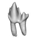
Todrasaurus gheerbranti UM THR 407 View specimen
|
M3#1575Holotype dentary UM THR 407 of Todrasaurus gheerbranti Type: "3D_surfaces"doi: 10.18563/m3.sf.1575 state:published |
Download 3D surface file |
This contribution contains the 3D model described and figured in the following publication: Martin, T., Averianov, A. O., Schultz, J. A., & Schwermann, A. H. (2023). A stem therian mammal from the Lower Cretaceous of Germany. Journal of Vertebrate Paleontology, e2224848.
Spelaeomolitor speratus WMNM P99101 View specimen

|
M3#12573D_model_Spelaeomolitor_lower_molar Type: "3D_surfaces"doi: 10.18563/m3.sf.1257 state:published |
Download 3D surface file |
|
M3#1258CT imagestack (jpgs) and info data sheet (pca file) in one zip folder Type: "3D_CT"doi: 10.18563/m3.sf.1258 state:published |
Download CT data |
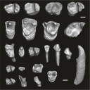
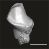
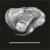
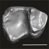
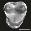
This contribution contains the three-dimensional digital models of the dental fossil material of anthropoid and strepsirrhine primates, discovered in Lower Oligocene detrital deposits outcropping in the Porto Rico and El Argoub areas, east of the Dakhla peninsula region (Atlantic Sahara; in the south of Morocco, near the northern border of Mauritania). These fossils were described, figured and discussed in the following publication: Marivaux et al. (2024), A new primate community from the earliest Oligocene of the Atlantic margin of Northwest Africa: Systematic, paleobiogeographic and paleoenvironmental implications. Journal of Human Evolution. https://doi.org/10.1016/j.jhevol.2024.103548
Catopithecus aff. browni DAK-Arg-087 View specimen

|
M3#1211Isolated right lower m3 (worn) Type: "3D_surfaces"doi: 10.18563/m3.sf.1211 state:published |
Download 3D surface file |
Catopithecus aff. browni DAK-Arg-088 View specimen
|
M3#1212Isolated right lower m2 (abraded/corroded) Type: "3D_surfaces"doi: 10.18563/m3.sf.1212 state:published |
Download 3D surface file |
Catopithecus aff. browni DAK-Arg-089 View specimen
|
M3#1213Isolated left lower m1 (worn) Type: "3D_surfaces"doi: 10.18563/m3.sf.1213 state:published |
Download 3D surface file |
Catopithecus aff. browni DAK-Pto-052 View specimen
|
M3#1214Isolated right lower m1 (pristine but lacking the mesiobuccal region) Type: "3D_surfaces"doi: 10.18563/m3.sf.1214 state:published |
Download 3D surface file |
Catopithecus aff. browni DAK-Arg-090 View specimen
|
M3#1215Isolated left upper P4 Type: "3D_surfaces"doi: 10.18563/m3.sf.1215 state:published |
Download 3D surface file |
Catopithecus aff. browni DAK-Arg-091 View specimen
|
M3#1216Isolated left upper M2 (worn and corroded) Type: "3D_surfaces"doi: 10.18563/m3.sf.1216 state:published |
Download 3D surface file |
Catopithecus aff. browni DAK-Pto-053 View specimen
|
M3#1217Isolated right upper M1 (lacking the buccal region) Type: "3D_surfaces"doi: 10.18563/m3.sf.1217 state:published |
Download 3D surface file |
Abuqatrania cf. basiodontos DAK-Arg-092 View specimen
|
M3#1218Isolated left lower c1 Type: "3D_surfaces"doi: 10.18563/m3.sf.1218 state:published |
Download 3D surface file |
?Propliopithecus sp. DAK-Pto-056 View specimen
|
M3#1219Isolated right lower m3 (fragment of talonid of a germ) Type: "3D_surfaces"doi: 10.18563/m3.sf.1219 state:published |
Download 3D surface file |
Abuqatrania cf. basiodontos DAK-Arg-093 View specimen
|
M3#1469Isolated right lower m1 Type: "3D_surfaces"doi: 10.18563/m3.sf.1469 state:published |
Download 3D surface file |
Abuqatrania cf. basiodontos DAK-Arg-094 View specimen
|
M3#1221Isolated left upper M1 or M2 (corroded, lacking the enamel cap [exposed dentine]) Type: "3D_surfaces"doi: 10.18563/m3.sf.1221 state:published |
Download 3D surface file |
Abuqatrania cf. basiodontos DAK-Arg-095 View specimen
|
M3#1222Isolated right lower i1 or i2 Type: "3D_surfaces"doi: 10.18563/m3.sf.1222 state:published |
Download 3D surface file |
Abuqatrania cf. basiodontos DAK-Arg-096 View specimen
|
M3#1223Isolated right lower p2 (worn apex) Type: "3D_surfaces"doi: 10.18563/m3.sf.1223 state:published |
Download 3D surface file |
Abuqatrania cf. basiodontos DAK-Arg-097 View specimen
|
M3#1224Isolated right lower p2 (worn apex and broken root) Type: "3D_surfaces"doi: 10.18563/m3.sf.1224 state:published |
Download 3D surface file |
Afrotarsius sp. DAK-Arg-098 View specimen
|
M3#1225Isolated left lower p3 Type: "3D_surfaces"doi: 10.18563/m3.sf.1225 state:published |
Download 3D surface file |
Afrotarsius sp. DAK-Pto-054 View specimen
|
M3#1226Isolated right lower m1 (abraded/corroded) Type: "3D_surfaces"doi: 10.18563/m3.sf.1226 state:published |
Download 3D surface file |
Orolemur mermozi DAK-Pto-055 View specimen
|
M3#1227Isolated right upper M1 or M2 (pristine, Holotype) Type: "3D_surfaces"doi: 10.18563/m3.sf.1227 state:published |
Download 3D surface file |
Wadilemur cf. elegans DAK-Arg-099 View specimen
|
M3#1228Isolated right lower m2 Type: "3D_surfaces"doi: 10.18563/m3.sf.1228 state:published |
Download 3D surface file |
cf. 'Anchomomys' milleri DAK-Arg-100 View specimen

|
M3#1229Isolated right lower c1 Type: "3D_surfaces"doi: 10.18563/m3.sf.1229 state:published |
Download 3D surface file |
Abuqatrania cf. basiodontos DAK-Arg-101 View specimen
|
M3#1396Isolated left upper M3 (abraded) Type: "3D_surfaces"doi: 10.18563/m3.sf.1396 state:published |
Download 3D surface file |
Orogalago saintexuperyi DAK-Arg-102 View specimen
|
M3#1397Isolated left lower m2 Type: "3D_surfaces"doi: 10.18563/m3.sf.1397 state:published |
Download 3D surface file |
Wadilemur cf. elegans DAK-Arg-103 View specimen

|
M3#1473Isolated right upper M1 or M2 (lacking the mesial and buccal regions) Type: "3D_surfaces"doi: 10.18563/m3.sf.1473 state:published |
Download 3D surface file |
This contribution contains the 3D model of an endocranial cast analyzed in “A 10 ka intentionally deformed human skull from Northeast Asia”. There are many studies on the morphological characteristics of intentional cranial deformation (ICD), but few related 3D models were published. Here, we present the surface model of an intentionally deformed 10 ka human cranium for further research on ICD practice. The 3D model of the endocranial cast of this ICD cranium was discovered near Harbin City, Province Heilongjiang, Northeast China. The fossil preserved only the frontal, parietal, and occipital bones. To complete the endocast model of the specimen, we printed a 3D model and used modeling clay to reconstruct the missing part based on the general form of the modern human endocast morphology.
Homo sapiens IVPP-PA1616 View specimen
|
M3#972The frontal region of the endocast is flattened, probably formed by the constant pressure on the frontal bone during growth. There is a well-developed frontal crest on the endocranial surface. The endocast widens posteriorly from the frontal lobe. The widest point of the endocast is at the lateral border of the parietal lobe. The lower parietal areas display a marked lateral expansion. The overall shape of the endocast is asymmetrical, with the left side of the parietal lobe being more laterally expanded than the right side. Like the frontal lobe, the occipital lobe is also anteroposteriorly flattened. Type: "3D_surfaces"doi: 10.18563/m3.sf.972 state:published |
Download 3D surface file |
|
M3#976The original endocranial cast model (with texture) of IVPP-PA1616. It shows the original structures of the specimen, and was not altered in any way. Type: "3D_surfaces"doi: 10.18563/m3.sf.976 state:published |
Download 3D surface file |
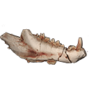
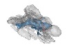
This contribution contains the 3D models described and figured in the following publication: Kassegne K. E., Mourlam M. J., Guinot G., Amoudji Y. Z., Martin J. E., Togbe K. A., Johnson A. K., Hautier L. 2021. First partial cranium of Togocetus from Kpogamé (Togo) and the protocetid diversity in the Togolese phosphate basin. Annales de Paléontologie, Issue 2, April–June 2021, 102488. https://doi.org/10.1016/j.annpal.2021.102488
Togocetus cf. traversei ULDG-KPO1 View specimen
|
M3#768The specimen consists of a partial cranium prepared out of a calcareous phosphate matrix. The partial cranium lacks the anterior part of the rostrum, the cranial roof, and most of the basicranium apart from the left zygomatic process of the squamosal. The maxilla, nasal, palatine, pterygoid, alisphenoid, and squamosal bones are preserved, as well as two incomplete dental rows described hereafter. Type: "3D_surfaces"doi: 10.18563/m3.sf.768 state:published |
Download 3D surface file |
|
M3#770µCT . Resolution: 0.3156mm. This scan can easily be opened with Fiji, MorphoDig, 3DSlicer, or any software that reads .MHD file format. Also, the .RAW file can be opened easily with other software such as Avizo/Amira when providing the correct dimensions (which are enclosed within the file name) Type: "3D_CT"doi: 10.18563/m3.sf.770 state:published |
Download CT data |