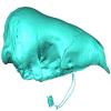
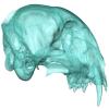
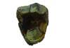
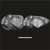
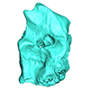
3D models of Ocnotherium skull
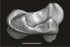
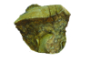
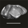
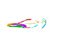
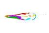
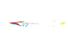
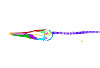
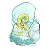
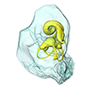
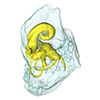
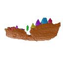
3D models of Kalakocetus, the earliest Cetacea
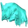
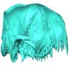
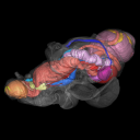
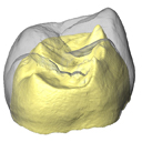
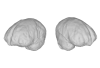
Explodable 3D Dog Skull for Veterinary Education
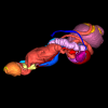
3D GM dataset of bird skeletal variation
Skeletal embryonic development in the catshark
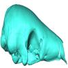
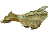
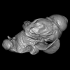
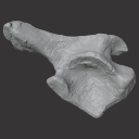
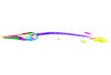
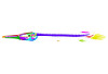
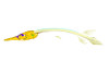
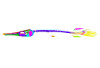
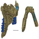
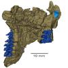
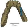
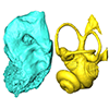
Bony connexions of the petrosal bone of extant hippos
bony labyrinth (14) , inner ear (11) , Eocene (11) , geometric morphometrics (10) , CT-scan (10) , Oligocene (9) , Micro-CT (9)
Maëva Judith Orliac (24) , Lionel Hautier (24) , Laurent Marivaux (18) , Renaud Lebrun (15) , Rodolphe Tabuce (14) , Pierre-Olivier Antoine (13) , Bastien Mennecart (13)
|
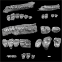
3D model related to the publication: An eosimiid primate of South Asian affinities in the Paleogene of Western Amazonia and the origin of New World monkeysLaurent Marivaux
Published online: 04/07/2023 |
|
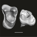
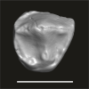
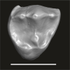
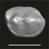
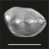
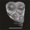
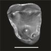
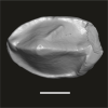
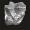
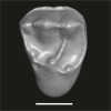
M3#1114Right first upper molar (rM1), pristine. Type: "3D_surfaces"doi: 10.18563/m3.sf.1114 state:published |
Download 3D surface file |
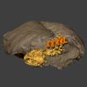
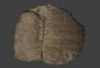
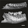
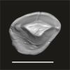
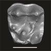
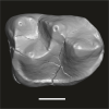
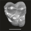
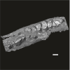
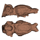
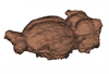
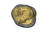
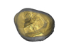
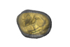
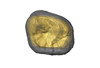
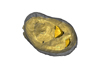
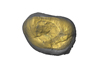
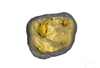
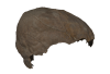
Turtles are one of the most impressive vertebrates. Much of the body is either hidden in a shell or can be drawn into it. Turtles impress with their individual longevity and their often peaceful disposition. Also, with their resilience, they have survived all extinction events since their emergence in the Late Triassic. Today's diversity of shapes is impressive and ranges from the large and high domed Galapagos turtles to the hamster-sized flat pancake turtles. The holotype of one of the oldest fossil turtles, Proganochelys quenstedtii, is housed in the paleontological collection in Tübingen/Germany. Since its discovery some years before 1873, P. quenstedtii has represented the 'prototype' of the turtle and has had an eventful scientific history. It was found in Neuenhaus (Häfner-Neuhausen in Schönbuch forest), Baden-Württemberg, Germany, and stems from Löwenstein-Formation (Weißer Keupersandstein), Late Triassic. The current catalogue number is GPIT-PV-30000. The specimen is listed in the historical inventory “Tübinger Petrefaktenverzeichnis 1841 bis 1896, [folio 326v.]“, as “[catalogue number: PV]16549, Schildkröte Weiser Keupersandstein Hafnerhausen” [turtle from White Keuper Sandstone]. Another, more recent synonym is “GPIT/RE/9396”. The same specimen was presented as uncatalogued by Gaffney (1990). Here we provide a surface scan of the steinkern for easier access of this famous specimen to the scientific community.
Proganochelys quenstedtii GPIT-PV-30000 View specimen
|
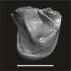
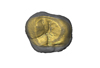
M3#967This the surface model of the steinkern of the shell of Proganochelys quenstedtii. Type: "3D_surfaces"doi: 10.18563/m3.sf.967 state:published |
Download 3D surface file |
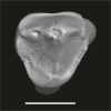
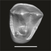
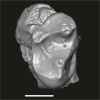
This contribution contains the 3D models described and figured in: The Neogene record of northern South American native ungulates. Smithsonian Contributions to Paleobiology. Doi: 10.5479/si.1943-6688.101
Hilarcotherium miyou IGMp 881327 View specimen
|
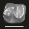
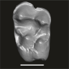
M3#318Right upper M2 Type: "3D_surfaces"doi: 10.18563/m3.sf.318 state:published |
Download 3D surface file |
Hilarcotherium miyou MUN-STRI 34216 View specimen
|
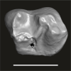
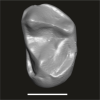
M3#319Right upper P4 Type: "3D_surfaces"doi: 10.18563/m3.sf.319 state:published |
Download 3D surface file |
|
M3#320Right upper M2 Type: "3D_surfaces"doi: 10.18563/m3.sf.320 state:published |
Download 3D surface file |
Falcontoxodon aguilerai AMU-CURS 585 View specimen
|
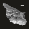
M3#321Maxilla with left M3-P2 and right I2 Type: "3D_surfaces"doi: 10.18563/m3.sf.321 state:published |
Download 3D surface file |
This contribution contains the 3D reconstruction of Canariomys bravoi, described and figured in the following publication: Michaux J., Hautier L., Hutterer R., Lebrun R., Guy F., García-Talavera F., 2012 : Body shape and life style of the extinct rodent Canariomys bravoi (Mammalia, Murinae) from Tenerife, Canary Islands (Spain). Comptes Rendus Palevol 11 (7), 485-494. DOI: 10.1016/j.crpv.2012.06.004
Canariomys bravoi TFMCV872-873 View specimen

|
M3#6This file contains the 3D reconstruction of Canariomys bravoi, described and figured in the following publication: Michaux J., Hautier L., Hutterer R., Lebrun R., Guy F., García-Talavera F., 2012 : Body shape and life style of the extinct rodent Canariomys bravoi (Mammalia, Murinae) from Tenerife, Canary Islands (Spain). Comptes Rendus Palevol 11 (7), 485-494. Type: "3D_surfaces"doi: 10.18563/m3.sf6 state:published |
Download 3D surface file |
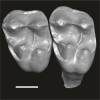
This contribution contains the three-dimensional digital models of the dental fossil material of strepsirrhine primates (Azibiidae and ?Djebelemuridae) from the late early to early middle Eocene of the Gour Lazib Complex in western Algeria and of Djebel Chambi in central-western Tunisia. These fossils were described, figured and discussed in the following publication: Marivaux et al. (2025), New insights into the diversity of strepsirrhine primates from the late early – early middle Eocene of North Africa (Algeria and Tunisia). Journal of Human Evolution, 103729. https://doi.org/10.1016/j.jhevol.2025.103729
Algeripithecus minimissimus ONM-CBI-1-38 View specimen
|
M3#1715Isolated right P3 Type: "3D_surfaces"doi: 10.18563/m3.sf.1715 state:published |
Download 3D surface file |
Algeripithecus minimissimus ONM-CBI-1-37 View specimen
|
M3#1716Isolated right P4 Type: "3D_surfaces"doi: 10.18563/m3.sf.1716 state:published |
Download 3D surface file |
Algeripithecus minimissimus ONM-CBI-1-1206 View specimen
|
M3#1717Isolated right p4 Type: "3D_surfaces"doi: 10.18563/m3.sf.1717 state:published |
Download 3D surface file |
Algeripithecus minimissimus ONM-CBI-1-1207 View specimen
|
M3#1718Isolated right p4 Type: "3D_surfaces"doi: 10.18563/m3.sf.1718 state:published |
Download 3D surface file |
Algeripithecus minimissimus ONM-CBI-1-1205 View specimen
|
M3#1719Fragment of right mandible bearing m1-3 (Holotype) Type: "3D_surfaces"doi: 10.18563/m3.sf.1719 state:published |
Download 3D surface file |
Algeripithecus minimissimus ONM-CBI-1-1209 View specimen
|
M3#1720Isolated left m2 Type: "3D_surfaces"doi: 10.18563/m3.sf.1720 state:published |
Download 3D surface file |
Algeripithecus minimissimus ONM-CBI-1-1208 View specimen
|
M3#1721Isolated right m2 Type: "3D_surfaces"doi: 10.18563/m3.sf.1721 state:published |
Download 3D surface file |
Algeripithecus minutus UM-HGL50-294 View specimen
|
M3#1722Left DP4 Type: "3D_surfaces"doi: 10.18563/m3.sf.1722 state:published |
Download 3D surface file |
Algeripithecus minutus UM-HGL50-297 View specimen
|
M3#1723Isolated right P2 Type: "3D_surfaces"doi: 10.18563/m3.sf.1723 state:published |
Download 3D surface file |
Algeripithecus minutus UM-HGL50-298 View specimen
|
M3#1724Isolated right P3 Type: "3D_surfaces"doi: 10.18563/m3.sf.1724 state:published |
Download 3D surface file |
Algeripithecus minutus UM-HGL50-299 View specimen
|
M3#1725Isolated right P4 Type: "3D_surfaces"doi: 10.18563/m3.sf.1725 state:published |
Download 3D surface file |
Algeripithecus minutus UM-HGL50-303 View specimen
|
M3#1726Isolated left P4 Type: "3D_surfaces"doi: 10.18563/m3.sf.1726 state:published |
Download 3D surface file |
Algeripithecus minutus UM-GZC-7 View specimen
|
M3#1727Isolated left M1 (lingually broken) Type: "3D_surfaces"doi: 10.18563/m3.sf.1727 state:published |
Download 3D surface file |
Algeripithecus minutus UM-GZC-1 View specimen
|
M3#1728Isolated left M2 (Holotype) Type: "3D_surfaces"doi: 10.18563/m3.sf.1728 state:published |
Download 3D surface file |
Algeripithecus minutus UM-HGL50-319 View specimen
|
M3#1729Isolated left M3 Type: "3D_surfaces"doi: 10.18563/m3.sf.1729 state:published |
Download 3D surface file |
Algeripithecus minutus UM-HGL50-397 View specimen
|
M3#1730Fragment of left mandible bearing p3-m3 Type: "3D_surfaces"doi: 10.18563/m3.sf.1730 state:published |
Download 3D surface file |
Azibius magnus UM-HGL50-258 View specimen
|
M3#1731Isolated right P3 or P4 Type: "3D_surfaces"doi: 10.18563/m3.sf.1731 state:published |
Download 3D surface file |
Azibius magnus UM-HGL50-260 View specimen
|
M3#1732Isolated right M2 Type: "3D_surfaces"doi: 10.18563/m3.sf.1732 state:published |
Download 3D surface file |
Azibius magnus UM-HGL50-261 View specimen
|
M3#1733Isolated left M3 Type: "3D_surfaces"doi: 10.18563/m3.sf.1733 state:published |
Download 3D surface file |
Azibius magnus UM-HGL50-263 View specimen
|
M3#1734Isolated left p3 Type: "3D_surfaces"doi: 10.18563/m3.sf.1734 state:published |
Download 3D surface file |
Azibius magnus UM-HGL50-264 View specimen
|
M3#1735Isolated right m1 (Holotype) Type: "3D_surfaces"doi: 10.18563/m3.sf.1735 state:published |
Download 3D surface file |
Azibius magnus UM-HGL50-265 View specimen
|
M3#1736Isolated right m1 (lingually broken) Type: "3D_surfaces"doi: 10.18563/m3.sf.1736 state:published |
Download 3D surface file |
Azibius magnus UM-HGL50-266 View specimen
|
M3#1738Isolated right m2 (corroded) Type: "3D_surfaces"doi: 10.18563/m3.sf.1738 state:published |
Download 3D surface file |
Azibius trerki UM-HGL50-166 View specimen
|
M3#1739Isolated right DP4 Type: "3D_surfaces"doi: 10.18563/m3.sf.1739 state:published |
Download 3D surface file |
Azibius trerki UM-HGL50-295 View specimen
|
M3#1740Isolated left DP4 Type: "3D_surfaces"doi: 10.18563/m3.sf.1740 state:published |
Download 3D surface file |
Azibius trerki UM-HGL51-46 View specimen
|
M3#1741Fragment of right maxillary bearing P3-4 Type: "3D_surfaces"doi: 10.18563/m3.sf.1741 state:published |
Download 3D surface file |
|
M3#1742Fragment of right maxillary bearing M3 Type: "3D_surfaces"doi: 10.18563/m3.sf.1742 state:published |
Download 3D surface file |
Azibius trerki UM-GZC-41 View specimen
|
M3#1743Isolated left P4 Type: "3D_surfaces"doi: 10.18563/m3.sf.1743 state:published |
Download 3D surface file |
Azibius trerki UM-HGL50-396 View specimen
|
M3#1744Boneless fragment of a left maxillary bearing M1-2 Type: "3D_surfaces"doi: 10.18563/m3.sf.1744 state:published |
Download 3D surface file |
Azibius trerki UM-HGL50-270 View specimen
|
M3#1745Fragment (talonid) of an isolated right dp4 Type: "3D_surfaces"doi: 10.18563/m3.sf.1745 state:published |
Download 3D surface file |
Azibius trerki UM-HGL50-248 View specimen
|
M3#1746Isolated left m1 Type: "3D_surfaces"doi: 10.18563/m3.sf.1746 state:published |
Download 3D surface file |
Azibius trerki UM-HGL50-256 View specimen
|
M3#1753Fragment of left mandible bearing p4-m3 Type: "3D_surfaces"doi: 10.18563/m3.sf.1753 state:published |
Download 3D surface file |
Lazibadapis anchomomyinopsis UM-HGL50-326 View specimen
|
M3#1747Isolated right M1 (buccally broken) Type: "3D_surfaces"doi: 10.18563/m3.sf.1747 state:published |
Download 3D surface file |
Lazibadapis anchomomyinopsis UM-HGL50-169 View specimen
|
M3#1748Isolated right M2 (corroded) Type: "3D_surfaces"doi: 10.18563/m3.sf.1748 state:published |
Download 3D surface file |
Lazibadapis anchomomyinopsis UM-HGL50-170 View specimen
|
M3#1749Isolated right M2 or M3 Type: "3D_surfaces"doi: 10.18563/m3.sf.1749 state:published |
Download 3D surface file |
Lazibadapis anchomomyinopsis UM-HGL50-325 View specimen

|
M3#1750Boneless fragment of left mandible preserving m2-3 (Holotype) -> m2 Type: "3D_surfaces"doi: 10.18563/m3.sf.1750 state:published |
Download 3D surface file |
|
M3#1751Boneless fragment of left mandible preserving m2-3 (Holotype) -> m3 Type: "3D_surfaces"doi: 10.18563/m3.sf.1751 state:published |
Download 3D surface file |
Lazibadapis anchomomyinopsis UM-HGL50-290 View specimen
|
M3#1752Isolated left m3 Type: "3D_surfaces"doi: 10.18563/m3.sf.1752 state:published |
Download 3D surface file |
The present 3D dataset contains the 3D models analyzed in the publication: Rosa, R. M., Salvador, R. B., & Cavallari, D. C. (2025). The disappearing act of the magician tree snail: anatomy, distribution, and phylogenetic relationships of Drymaeus magus (Gastropoda: Bulimulidae), a long-lost species hidden in plain sight. Zoological Journal of the Linnean Society.
Drymaeus magus CMRP 1049 View specimen
|
M3#1597Internal organs of Drymaeus magus Type: "3D_surfaces"doi: 10.18563/m3.sf.1597 state:published |
Download 3D surface file |
|
M3#1598External surface of Drymaeus magus Type: "3D_surfaces"doi: 10.18563/m3.sf.1598 state:published |
Download 3D surface file |
The present Dataset contains the micro-CT scan of the head of an anonymous 54 year old female donor, at a voxel resolution of 145µm. The skin of the face has been masked in order to avoid the donor to be recognized.
Homo sapiens UM_HS_2018_09_13 View specimen
|
M3#1152Micro-ct data set Type: "3D_CT"doi: 10.18563/m3.sf.1152 state:published |
Download CT data |
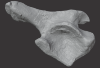
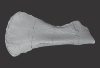
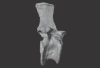
The present 3D Dataset contains the 3D models of an ilium, a vertebra, and a partial scapula of Prestosuchus sp. that were analyzed in “New Loricata remains from the Pinheiros-Chiniquá Sequence (Middle-Upper Triassic), southern Brazil”.
Prestosuchus sp. UFSM11603 View specimen
|
M3#1080Surface scan of a right ilium of Prestosuchus sp. with a 0.4 mm resolution. Type: "3D_surfaces"doi: 10.18563/m3.sf.1080 state:published |
Download 3D surface file |
Prestosuchus sp. UFSM11233 View specimen
|
M3#1081Surface scan of a partial right scapula of Prestosuchus sp. with a 0.4mm resolution. Type: "3D_surfaces"doi: 10.18563/m3.sf.1081 state:published |
Download 3D surface file |
Prestosuchus sp. UFSM11602a View specimen
|
M3#1082Surface scan of a anterior dorsal vertebra of Prestosuchus sp. with a 0.2 mm resolution. Type: "3D_surfaces"doi: 10.18563/m3.sf.1082 state:published |
Download 3D surface file |
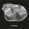
Our knowledge of the external brain morphology of the late Eocene artiodactyl ungulate Mixtotherium, relies on a plaster model realized on a specimen from the Victor Brun Museum in Montauban (France) and described by Dechaseaux (1973). Here, based on micro CT-scan data, we virtually reconstruct the 3D cast of the empty cavity of the partial cranium MA PHQ 716 from the Victor Brun Museum and compare it to the plaster model illustrated and described by Dechaseaux (1973). Indeed, the specimen from which the original plaster endocast originates was not identified by Dechaseaux by a specimen number. We confirm here that the studied specimen was indeed the one described and illustrated by Dechaseaux (1973). We also reconstruct a second, more detailed, model providing additional morphological and quantitative observations made available by micro CT scan investigation such as precisions on the neopallium folding and endocranial volumes.
Mixtotherium cuspidatum MA PHQ 716 View specimen
|
M3#857endocast of the brain cavity Type: "3D_surfaces"doi: 10.18563/m3.sf.857 state:published |
Download 3D surface file |
This contribution contains the 3D models described and figured in the following publications:
- Marini E., Lussu P., 2020. A virtual physical anthropology lab. Teaching in the time of coronavirus, in prep.;
- Lussu P., Bratzu D., Marini E., 2020. Cloud-based ultra close-range digital photogrammetry: validation of an approach for the effective virtual reconstruction of skeletal remains, in prep.
Homo sapiens MSAE 59 View specimen

|
M3#509MSAE 59 Type: "3D_surfaces"doi: 10.18563/m3.sf.509 state:published |
Download 3D surface file |
Homo sapiens MSAE 62 View specimen
|
M3#510MSAE 62 Type: "3D_surfaces"doi: 10.18563/m3.sf.510 state:published |
Download 3D surface file |
Homo sapiens MSAE 63 View specimen
|
M3#512MSAE 63 Type: "3D_surfaces"doi: 10.18563/m3.sf.512 state:published |
Download 3D surface file |
Homo sapiens MSAE 78 View specimen
|
M3#514MSAE 78 Type: "3D_surfaces"doi: 10.18563/m3.sf.514 state:published |
Download 3D surface file |
Homo sapiens MSAE 95 View specimen
|
M3#515MSAE 95 Type: "3D_surfaces"doi: 10.18563/m3.sf.515 state:published |
Download 3D surface file |
Homo sapiens MSAE 1852 View specimen
|
M3#516MSAE 1852 Type: "3D_surfaces"doi: 10.18563/m3.sf.516 state:published |
Download 3D surface file |
Homo sapiens MSAE 6426 View specimen
|
M3#517MSAE 6426 Type: "3D_surfaces"doi: 10.18563/m3.sf.517 state:published |
Download 3D surface file |
Homo sapiens MSAE 6428 View specimen
|
M3#518MSAE 6428 Type: "3D_surfaces"doi: 10.18563/m3.sf.518 state:published |
Download 3D surface file |
Homo sapiens MSAE 6992 View specimen
|
M3#519MSAE 6992 Type: "3D_surfaces"doi: 10.18563/m3.sf.519 state:published |
Download 3D surface file |
Homo sapiens MSAE 7688 View specimen
|
M3#520MSAE 7688 Type: "3D_surfaces"doi: 10.18563/m3.sf.520 state:published |
Download 3D surface file |
This contribution includes the 3D models of the reconstructed ossicular chain of the cainotheriid Caenomeryx filholi from the late Oligocene locality of Pech Desse (MP28, Quercy, France) described and figured in the publication of Assemat et al. (2020). It represents the oldest ossicular chain reconstruction for a Paleogene terrestrial artiodactyl species.
Caenomeryx filholi UM PDS 3353 View specimen
|
M3#508reconstruction of the middle ear with petrosal, bulla, stapes, incus, malleus Type: "3D_surfaces"doi: 10.18563/m3.sf.508 state:published |
Download 3D surface file |
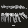
Using X-ray microtomography, we describe the ossification events during the larval development of a non-teleost actinopterygian species: the Cuban gar Atractosteus tristoechus from the order Lepisosteiformes. We provide a detailed developmental series for each anatomical structure, covering a large sequence of mineralization events going from an early stage (13 days post-hatching, 21mm total length) to an almost fully ossified larval stage (118dph or 87mm in standard length). With this work, we expect to bring new developmental data to be used in further comparative studies with other lineages of bony vertebrates. We also hope that the on-line publication of these twelve successive 3D reconstructions, fully labelled and flagged, will be an educational tool for all students in comparative anatomy.
Atractosteus tristoechus At1-13dph View specimen
|
M3#94At1-13dph : 13 dph larvae, 21 mm TL Type: "3D_surfaces"doi: 10.18563/m3.sf.94 state:published |
Download 3D surface file |
Atractosteus tristoechus At2-16dph View specimen
|
M3#95Atractosteus tristoechus larva, 16 dph, 26mm SL. Type: "3D_surfaces"doi: 10.18563/m3.sf.95 state:published |
Download 3D surface file |
Atractosteus tristoechus At3-19dph View specimen
|
M3#96Atractosteus tristoechus larva, 19 dph, 27mm SL. Type: "3D_surfaces"doi: 10.18563/m3.sf.96 state:published |
Download 3D surface file |
Atractosteus tristoechus At4-22dph View specimen
|
M3#97Atractosteus tristoechus larva, 22dph, 30mm SL. Type: "3D_surfaces"doi: 10.18563/m3.sf.97 state:published |
Download 3D surface file |
Atractosteus tristoechus At5-26dph View specimen
|
M3#98Atractosteus tristoechus larva, 26 dph, 32mm SL. Type: "3D_surfaces"doi: 10.18563/m3.sf.98 state:published |
Download 3D surface file |
Atractosteus tristoechus At6-31dph View specimen
|
M3#99Atractosteus tristoechus larva, 31 dph, 39mm SL. Type: "3D_surfaces"doi: 10.18563/m3.sf.99 state:published |
Download 3D surface file |
Atractosteus tristoechus At7-37dph View specimen
|
M3#100Atractosteus tristoechus larva, 37 dph, 43mm SL. Type: "3D_surfaces"doi: 10.18563/m3.sf.100 state:published |
Download 3D surface file |
Atractosteus tristoechus At8-52dph View specimen
|
M3#101Atractosteus tristoechus larva, 52 dph, 46mm SL. Type: "3D_surfaces"doi: 10.18563/m3.sf.101 state:published |
Download 3D surface file |
Atractosteus tristoechus At9-74dph View specimen
|
M3#102Atractosteus tristoechus larva, 74 dph, 61mm SL. Not all structures are colored, only newly ossified ones. Type: "3D_surfaces"doi: 10.18563/m3.sf.102 state:published |
Download 3D surface file |
Atractosteus tristoechus At10-89dph View specimen
|
M3#103Atractosteus tristoechus larva, 89 dph, 63mm SL. Not all structures are colored, only newly ossified ones. You may find the tag file in the At1-13dph reconstruction data. Type: "3D_surfaces"doi: 10.18563/m3.sf.103 state:published |
Download 3D surface file |
Atractosteus tristoechus At11-104dph View specimen
|
M3#104Atractosteus tristoechus larva, 104 dph, 70mm SL. Not all structures are colored, only newly ossified ones. Type: "3D_surfaces"doi: 10.18563/m3.sf.104 state:published |
Download 3D surface file |
Atractosteus tristoechus At12-118dph View specimen
|
M3#105Atractosteus tristoechus larva, 118 dph, 87mm SL. Type: "3D_surfaces"doi: 10.18563/m3.sf.105 state:published |
Download 3D surface file |
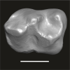
The present 3D Dataset contains the 3D models of external and internal aspects of human upper permanent second molars from the Neolithic necropolis analyzed in the following publication: Le Luyer M., Coquerelle M., Rottier S., Bayle P. (2016): Internal tooth structure and burial practices: insights into the Neolithic necropolis of Gurgy (France, 5100-4000 cal. BC). Plos One 11(7): e0159688. doi: 10.1371/journal.pone.0159688.
Homo sapiens GLN04-201-ULM2 View specimen
|
M3#74Outer enamel surface (OES) and enamel-dentine junction (EDJ) of Neolithic upper permanent left second molar Type: "3D_surfaces"doi: 10.18563/m3.sf.74 state:published |
Download 3D surface file |
Homo sapiens GLN04-206-ULM2 View specimen
|
M3#75Outer enamel surface (OES) and enamel-dentine junction (EDJ) of Neolithic upper permanent left second molar Type: "3D_surfaces"doi: 10.18563/m3.sf.75 state:published |
Download 3D surface file |
Homo sapiens GLN05-213-URM2 View specimen
|
M3#76Outer enamel surface (OES) and enamel-dentine junction (EDJ) of Neolithic upper permanent right second molar Type: "3D_surfaces"doi: 10.18563/m3.sf.76 state:published |
Download 3D surface file |
Homo sapiens GLN05-215A-URM2 View specimen
|
M3#77Outer enamel surface (OES) and enamel-dentine junction (EDJ) of Neolithic upper permanent right second molar Type: "3D_surfaces"doi: 10.18563/m3.sf.77 state:published |
Download 3D surface file |
Homo sapiens GLN06-215B-URM2 View specimen
|
M3#78Outer enamel surface (OES) and enamel-dentine junction (EDJ) of Neolithic upper permanent right second molar Type: "3D_surfaces"doi: 10.18563/m3.sf.78 state:published |
Download 3D surface file |
Homo sapiens GLN06-223-URM2 View specimen
|
M3#79Outer enamel surface (OES) and enamel-dentine junction (EDJ) of Neolithic upper permanent right second molar Type: "3D_surfaces"doi: 10.18563/m3.sf.79 state:published |
Download 3D surface file |
Homo sapiens GLN04-229-URM2 View specimen
|
M3#80Outer enamel surface (OES) and enamel-dentine junction (EDJ) of Neolithic upper permanent right second molar Type: "3D_surfaces"doi: 10.18563/m3.sf.80 state:published |
Download 3D surface file |
Homo sapiens GLN05-243B-ULM2 View specimen
|
M3#81Outer enamel surface (OES) and enamel-dentine junction (EDJ) with reconstructed dentine horn tip of Neolithic upper permanent left second molar Type: "3D_surfaces"doi: 10.18563/m3.sf.81 state:published |
Download 3D surface file |
Homo sapiens GLN04-248-ULM2 View specimen
|
M3#82Outer enamel surface (OES) and enamel-dentine junction (EDJ) with reconstructed dentine horn tip of Neolithic upper permanent left second molar Type: "3D_surfaces"doi: 10.18563/m3.sf.82 state:published |
Download 3D surface file |
Homo sapiens GLN04-252-ULM2 View specimen
|
M3#83Outer enamel surface (OES) and enamel-dentine junction (EDJ) of Neolithic upper permanent left second molar Type: "3D_surfaces"doi: 10.18563/m3.sf.83 state:published |
Download 3D surface file |
Homo sapiens GLN04-253-ULM2 View specimen
|
M3#84Outer enamel surface (OES) and enamel-dentine junction (EDJ) of Neolithic upper permanent left second molar Type: "3D_surfaces"doi: 10.18563/m3.sf.84 state:published |
Download 3D surface file |
Homo sapiens GLN05-257-URM2 View specimen
|
M3#85Outer enamel surface (OES) and enamel-dentine junction (EDJ) with reconstructed dentine horn tip of Neolithic upper permanent right second molar Type: "3D_surfaces"doi: 10.18563/m3.sf.85 state:published |
Download 3D surface file |
Homo sapiens GLN04-264-ULM2 View specimen
|
M3#86Outer enamel surface (OES) and enamel-dentine junction (EDJ) of Neolithic upper permanent left second molar Type: "3D_surfaces"doi: 10.18563/m3.sf.86 state:published |
Download 3D surface file |
Homo sapiens GLN04-277-URM2 View specimen
|
M3#87Outer enamel surface (OES) and enamel-dentine junction (EDJ) of Neolithic upper permanent right second molar Type: "3D_surfaces"doi: 10.18563/m3.sf.87 state:published |
Download 3D surface file |
Homo sapiens GLN04-289B-URM2 View specimen
|
M3#88Outer enamel surface (OES) and enamel-dentine junction (EDJ) of Neolithic upper permanent right second molar Type: "3D_surfaces"doi: 10.18563/m3.sf.88 state:published |
Download 3D surface file |
Homo sapiens GLN06-291-URM2 View specimen
|
M3#89Outer enamel surface (OES) and enamel-dentine junction (EDJ) with reconstructed dentine horn tip of Neolithic upper permanent right second molar Type: "3D_surfaces"doi: 10.18563/m3.sf.89 state:published |
Download 3D surface file |
Homo sapiens GLN05-292-URM2 View specimen
|
M3#90Outer enamel surface (OES) and enamel-dentine junction (EDJ) of Neolithic upper permanent right second molar Type: "3D_surfaces"doi: 10.18563/m3.sf.90 state:published |
Download 3D surface file |
Homo sapiens GLN05-294-ULM2 View specimen
|
M3#91Outer enamel surface (OES) and enamel-dentine junction (EDJ) with reconstructed dentine horn tip of Neolithic upper permanent left second molar Type: "3D_surfaces"doi: 10.18563/m3.sf.91 state:published |
Download 3D surface file |
Homo sapiens GLN05-308-URM2 View specimen
|
M3#93Outer enamel surface (OES) and enamel-dentine junction (EDJ) of Neolithic upper permanent right second molar Type: "3D_surfaces"doi: 10.18563/m3.sf.93 state:published |
Download 3D surface file |
Homo sapiens GLN05-301-ULM2 View specimen
|
M3#92Outer enamel surface (OES) and enamel-dentine junction (EDJ) of Neolithic upper permanent left second molar Type: "3D_surfaces"doi: 10.18563/m3.sf.92 state:published |
Download 3D surface file |
Current knowledge on the skeletogenesis of Chondrichthyes is scarce compared with their extant sister group, the bony fishes. Most of the previously described developmental tables in Chondrichthyes have focused on embryonic external morphology only. Due to its small body size and relative simplicity to raise eggs in laboratory conditions, the small-spotted catshark Scyliorhinus canicula has emerged as a reference species to describe developmental mechanisms in the Chondrichthyes lineage. Here we investigate the dynamic of mineralization in a set of six embryonic specimens using X-ray microtomography and describe the developing units of both the dermal skeleton (teeth and dermal scales) and endoskeleton (vertebral axis). This preliminary data on skeletogenesis in the catshark sets the first bases to a more complete investigation of the skeletal developmental in Chondrichthyes. It should provide comparison points with data known in osteichthyans and could thus be used in the broader context of gnathostome skeletal evolution.
Scyliorhinus canicula SC6_2_2015_03_20 View specimen

|
M3#50Mineralized skeleton of a 6,2 cm long embryo of Scyliorhinus canicula Type: "3D_surfaces"doi: 10.18563/m3.sf.50 state:published |
Download 3D surface file |
Scyliorhinus canicula SC6_7_2015_03_20 View specimen

|
M3#51Mineralized skeleton of a 6,7 cm long embryo of Scyliorhinus canicula Type: "3D_surfaces"doi: 10.18563/m3.sf.51 state:published |
Download 3D surface file |
Scyliorhinus canicula SC7_1_2015_04_03 View specimen

|
M3#52Mineralized skeleton of a 7,1 cm long embryo of Scyliorhinus canicula Type: "3D_surfaces"doi: 10.18563/m3.sf.52 state:published |
Download 3D surface file |
Scyliorhinus canicula SC7_5_2015_03_13 View specimen

|
M3#53Mineralized skeleton of a 7,5 cm long embryo of Scyliorhinus canicula Type: "3D_surfaces"doi: 10.18563/m3.sf.53 state:published |
Download 3D surface file |
Scyliorhinus canicula SC8_2015_03_20 View specimen

|
M3#54Mineralized skeleton of a 8 cm long embryo of Scyliorhinus canicula Type: "3D_surfaces"doi: 10.18563/m3.sf.54 state:published |
Download 3D surface file |
Scyliorhinus canicula SC10_2015_02_27 View specimen

|
M3#55Mineralized skeleton of a 10 cm long embryo of Scyliorhinus canicula Type: "3D_surfaces"doi: 10.18563/m3.sf.55 state:published |
Download 3D surface file |
This contribution contains the 3D models described and figured in the following publication: Gaetano, L. C., Abdala, F., Mancuso, C, and Vega N.2025. New traversodontid cynodont from the Late Triassic Chañares Formation. Publicación Electrónica de la Asociación Paleontológica Argentina.
Pontognathus ignotus PULR-V 287 View specimen
|
M3#1647partial snout preserving the lateralmost incisor, the base of the canine, and several postcanines Type: "3D_surfaces"doi: 10.18563/m3.sf.1647 state:published |
Download 3D surface file |
Massetognathus pascuali PULR-V 289 View specimen
|
M3#1646partial lower jaw Type: "3D_surfaces"doi: 10.18563/m3.sf.1646 state:published |
Download 3D surface file |
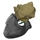
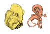
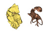
The present 3D Dataset contains the 3D models analyzed in Mennecart, B., Duranthon, F., & Costeur, L. 2024. Systematic contribution of the auditory region to the knowledge of the oldest European Bovidae (Mammalia, Ruminantia). Journal of Anatomy XXX. https://doi.org/10.1111/joa.14132
Pusillutragus montrealensis MHNT.PAL.2015.0.2261.4 View specimen
|
M3#1522Right petrosal, bony labyrinth, stapes Type: "3D_surfaces"doi: 10.18563/m3.sf.1522 state:published |
Download 3D surface file |
Pusillutragus montrealensis MHNT.PAL.2015.0.2261.9 View specimen
|
M3#1523Left petrosal and left bony labyrinth Type: "3D_surfaces"doi: 10.18563/m3.sf.1523 state:published |
Download 3D surface file |
Eotragus artenensis SMNS-P-41625 View specimen
|
M3#1524Petrosal (right), bony labyrinth (left) Type: "3D_surfaces"doi: 10.18563/m3.sf.1524 state:published |
Download 3D surface file |
Eotragus clavatus NMB San.15056 View specimen
|
M3#1528Right petrosal and right bony labyrinth Type: "3D_surfaces"doi: 10.18563/m3.sf.1528 state:published |
Download 3D surface file |
Eotragus clavatus NMB San.15055 View specimen
|
M3#1526Left Petrosal Type: "3D_surfaces"doi: 10.18563/m3.sf.1526 state:published |
Download 3D surface file |
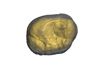
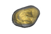
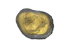
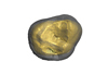
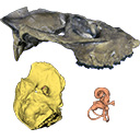
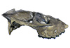
The holotype of Hamadasuchus rebouli Buffetaut 1994 from the Kem Kem beds of Morocco (Late Albian – Cenomanian) consists of a left dentary which is limited, fragmentary and reconstructed in some areas. To aid in assessing if the original diagnosis can be considered as valid, the specimen was CT scanned for the first time. This is especially important to resolve the taxonomic status of certain specimens that have been assigned to Hamadasuchus rebouli since then. The reconstructed structures in this contribution are in agreement with the original description, notably in terms of alveolar count; thus the original diagnosis of this taxon remains valid and some specimens are not referable to H. rebouli anymore.
Hamadasuchus rebouli MDE C001 View specimen

|
M3#1402Dentary and teeth Type: "3D_surfaces"doi: 10.18563/m3.sf.1402 state:published |
Download 3D surface file |

|
M3#1403Toothmarks Type: "3D_surfaces"doi: 10.18563/m3.sf.1403 state:published |
Download 3D surface file |
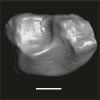
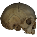
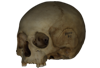
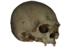
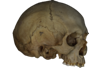
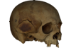
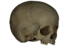
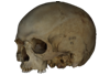
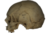
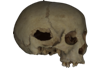
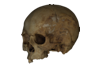
This contribution contains the 3D model of an endocranial cast analyzed in “A 10 ka intentionally deformed human skull from Northeast Asia”. There are many studies on the morphological characteristics of intentional cranial deformation (ICD), but few related 3D models were published. Here, we present the surface model of an intentionally deformed 10 ka human cranium for further research on ICD practice. The 3D model of the endocranial cast of this ICD cranium was discovered near Harbin City, Province Heilongjiang, Northeast China. The fossil preserved only the frontal, parietal, and occipital bones. To complete the endocast model of the specimen, we printed a 3D model and used modeling clay to reconstruct the missing part based on the general form of the modern human endocast morphology.
Homo sapiens IVPP-PA1616 View specimen
|
M3#972The frontal region of the endocast is flattened, probably formed by the constant pressure on the frontal bone during growth. There is a well-developed frontal crest on the endocranial surface. The endocast widens posteriorly from the frontal lobe. The widest point of the endocast is at the lateral border of the parietal lobe. The lower parietal areas display a marked lateral expansion. The overall shape of the endocast is asymmetrical, with the left side of the parietal lobe being more laterally expanded than the right side. Like the frontal lobe, the occipital lobe is also anteroposteriorly flattened. Type: "3D_surfaces"doi: 10.18563/m3.sf.972 state:published |
Download 3D surface file |
|
M3#976The original endocranial cast model (with texture) of IVPP-PA1616. It shows the original structures of the specimen, and was not altered in any way. Type: "3D_surfaces"doi: 10.18563/m3.sf.976 state:published |
Download 3D surface file |
The present 3D Dataset contains the 3D model of the skin of Allosaurus described in Hendrickx, C. et al. in press. Morphology and distribution of scales, dermal ossifications, and other non-feather integumentary structures in non-avialan theropod dinosaurs. Biological Reviews.
Allosaurus jimmadseni UMNH VP C481 View specimen
|
M3#902The material consists of a 3D reconstruction of the counterpart of a 30 cm2 patch of skin impression associated with the anterior dorsal ribs/pectoral region of the specimen of Allosaurus jimmadseni UMNH VP C481. The skin shows a semi-uniform basement of 1-2 mm diameter pebbles with a smaller number of slightly larger (up to 3 mm) ovoid scales. The irregular shape, distribution, and overall small size of these larger scales suggest that they are not classifiable as feature scales but rather as variations in the basement scales. Type: "3D_surfaces"doi: 10.18563/m3.sf.902 state:published |
Download 3D surface file |
The present 3D Dataset contains the 3D models analyzed in Mennecart B., Métais G., Costeur L., Ginsburg L, and Rössner G. 2021, Reassessment of the enigmatic ruminant Miocene genus Amphimoschus Bourgeois, 1873 (Mammalia, Artiodactyla, Pecora). PlosOne. https://doi.org/10.1371/journal.pone.0244661
Amphimoschus ponteleviensis MNHN.F.AR3266 View specimen
|
M3#701Surface scan of the cast of the skull of Amphimoschus ponteleviensis MNHN.F.AR3266 from Artenay (France) Type: "3D_surfaces"doi: 10.18563/m3.sf.701 state:published |
Download 3D surface file |
|
M3#702Right petrosal bone and bony labyrinth of the skull MNHN.F.AR3266 from Artenay (France) Type: "3D_surfaces"doi: 10.18563/m3.sf.702 state:published |
Download 3D surface file |
Amphimoschus ponteleviensis SMNS40693 View specimen
|
M3#704Left petrosal bone and bony labyrinth of the skull SMNS40693 from Langenau 1 (Germany) Type: "3D_surfaces"doi: 10.18563/m3.sf.704 state:published |
Download 3D surface file |