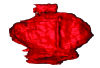
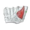
3D models of Cainotheriids Ossicular chain
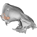
Explodable 3D Dog Skull for Veterinary Education
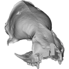
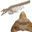
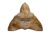
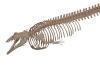
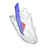
3D models of Kalakocetus, the earliest Cetacea
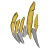
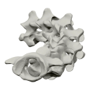
3D GM dataset of bird skeletal variation
Skeletal embryonic development in the catshark
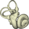
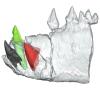
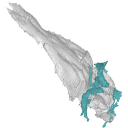
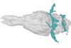
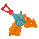
Bony connexions of the petrosal bone of extant hippos
bony labyrinth (14) , inner ear (11) , Eocene (11) , geometric morphometrics (10) , CT-scan (10) , Oligocene (9) , Micro-CT (9)
Lionel Hautier (25) , Maëva Judith Orliac (24) , Laurent Marivaux (18) , Renaud Lebrun (15) , Rodolphe Tabuce (15) , Pierre-Olivier Antoine (13) , Bastien Mennecart (13)
|
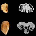
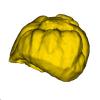
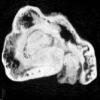
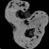
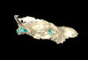
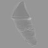
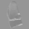
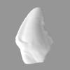
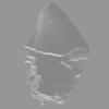
3D models related to the publication: Vitaceae seed and wood fossils from the Eocene–Oligocene phosphatic fissure fillings of Quercy, southwestern FranceYiyun Chen, Anaïs Boura
Published online: 10/06/2026 |
|
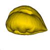
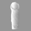
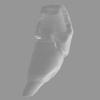
M3#1930Surface of the seed Type: "3D_surfaces"doi: 10.18563/m3.sf.1930 state:published |
Download 3D surface file |
|
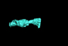
M3#1933Seed : 8.43 μm µCT scan Type: "3D_CT"doi: 10.18563/m3.sf.1933 state:published |
Download CT data |
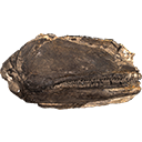
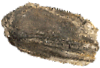
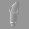
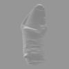
Vitis praerotundifolia UM-BAV-660 View specimen
|
M3#1931Seed : 8.43 μm µCT scan Type: "3D_CT"doi: 10.18563/m3.sf.1931 state:published |
Download CT data |
|
M3#1932Surface of the seed Type: "3D_surfaces"doi: 10.18563/m3.sf.1932 state:published |
Download 3D surface file |
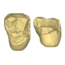
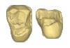
The present 3D Dataset contains the 3D model of a left dentary with m1-m3 analyzed in “A new fossil of Tayassuidae (Mammalia: Certartiodactyla) from the Pleistocene of northern Brazil”. The 3D model was generated using a laser scanning.
cf. Pecari tajacu UFSM 11606 View specimen
|
M3#498Left dentary with m1-m3 Type: "3D_surfaces"doi: 10.18563/m3.sf.498 state:published |
Download 3D surface file |
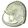
The present 3D Dataset contains the 3D model of the brain endocast of Neoepiblema acreensis analyzed in “Small within the largest: Brain size and anatomy of the extinct Neoepiblema acreensis, a giant rodent from the Neotropics”. The 3D model was generated using CT-Scanning and techniques of virtual reconstruction.
Neoepiblema acreensis UFAC 4515 View specimen
|
M3#502Brain endocast of Neoepiblema acreensis Type: "3D_surfaces"doi: 10.18563/m3.sf.502 state:published |
Download 3D surface file |
The present 3D Dataset contains 3D models of the cranium surface and of the bony labyrinth endocast of the stem bat Vielasia sigei. They are used by (Hand et al., 2023) to explore the phylogenetic position of this species, to infer its laryngeal echolocating capabilities, and to eventually discuss chiropteran evolution before the crown clade diversification.
Vielasia sigei UM VIE-250 View specimen
|
M3#1269External surface of the cranium Type: "3D_surfaces"doi: 10.18563/m3.sf.1269 state:published |
Download 3D surface file |
|
M3#1270Virtual endocast of the right bony labyrinth Type: "3D_surfaces"doi: 10.18563/m3.sf.1270 state:published |
Download 3D surface file |
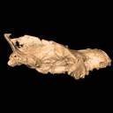
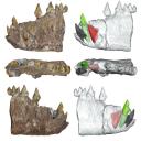
The presented dataset contains the 3D surface scan of the holotype of Birgeria americana, a partial skull described and depicted in: Romano, C., Jenks, J.F., Jattiot, R., Scheyer, T.M., Bylund, K.G. & Bucher, H. 2017. Marine Early Triassic Actinopterygii from Elko County (Nevada, USA): implications for the Smithian equatorial vertebrate eclipse. Journal of Paleontology. https://doi.org/10.1017/jpa.2017.36 .
Birgeria americana NMMNH P-66225 View specimen
|
M3#175NMMNH P-66225 is from upper lower Smithian to lower upper Smithian beds (Thaynes Group). The collecting site is located about 2.75 km south-southeast of the Winecup Ranch, east-central Elko County, Nevada, USA. P-66225 is a partial skull preserved within a large limestone nodule, with its right side exposed. It preserves the portion between the cleithrum posteriorly, and the level of the hind margin of the orbital opening anteriorly. The fossil has a length of 26 cm. Type: "3D_surfaces"doi: 10.18563/m3.sf.175 state:published |
Download 3D surface file |
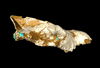
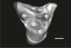
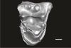
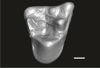
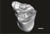
This contribution contains the 3D models of the isolated teeth of Canaanimico amazonensis, a new stem platyrrhine primate, described and figured in the following publication: Marivaux et al. (2016), Neotropics provide insights into the emergence of New World monkeys: new dental evidence from the late Oligocene of Peruvian Amazonia. Journal of Human Evolution. http://dx.doi.org/10.1016/j.jhevol.2016.05.011
Canaanimico amazonensis MUSM-2499 View specimen
|
M3#2893D model of left upper M2 Type: "3D_surfaces"doi: 10.18563/m3.sf.289 state:published |
Download 3D surface file |
Canaanimico amazonensis MUSM-2500 View specimen
|
M3#2903D model of left upper M1 (lingual part) Type: "3D_surfaces"doi: 10.18563/m3.sf.290 state:published |
Download 3D surface file |
This contribution contains the 3D model(s) described and figured in the following publication: Carolina A. Hoffmann, P. G. Rodrigues, M. B. Soares & M. B. Andrade. 2021. Brain endocast of two non-mammaliaform cynodonts from southern Brazil: an ontogenetic and evolutionary approach, Historical Biology, 33:8, 1196-1207, https://doi.org/10.1080/08912963.2019.1685512
Probelesodon kitchingi MCP 1600 PV View specimen
|
M3#9783D model of the brain endocast of Probelesodon kitchingi. Type: "3D_surfaces"doi: 10.18563/m3.sf.978 state:published |
Download 3D surface file |
Massetognathus ochagaviae MCP 3871 PV View specimen
|
M3#9793D model of the brain endocast of Massetognathus ochagaviae. Type: "3D_surfaces"doi: 10.18563/m3.sf.979 state:published |
Download 3D surface file |
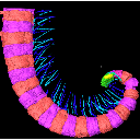
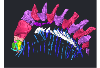
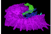
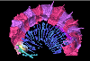
The present 3D Dataset contains the 3D models analyzed in the publication Fossils from the Montceau-les-Mines Lagerstätte (305 Ma) shed light on the anatomy, ecology and phylogeny of Carboniferous millipedes. Authors: Lheritier Mickael, Perroux Maëva, Vannier Jean, Escarguel Gilles, Wesener Thomas, Moritz Leif, Chabard Dominique, Adrien Jerome and Perrier Vincent. Journal of Systematics Palaeontology. https://doi.org/10.1080/14772019.2023.2169891
Amynilyspes fatimae MNHN.F.SOT.2134 View specimen
|
M3#1073Nearly complete specimen. Type: "3D_surfaces"doi: 10.18563/m3.sf.1073 state:published |
Download 3D surface file |
Amynilyspes fatimae MNHN.F.SOT.14983 View specimen
|
M3#1074Nearly complete specimen. Type: "3D_surfaces"doi: 10.18563/m3.sf.1074 state:published |
Download 3D surface file |
Amynilyspes fatimae MNHN.F.SOT.2129 View specimen
|
M3#1075Nearly complete specimen. Type: "3D_surfaces"doi: 10.18563/m3.sf.1075 state:published |
Download 3D surface file |
Blanzilius parriati MNHN.F.SOT.2114A View specimen
|
M3#1076Front part. Type: "3D_surfaces"doi: 10.18563/m3.sf.1076 state:published |
Download 3D surface file |
Blanzilius parriati MNHN.F.SOT.5148 View specimen
|
M3#1077Front part. Type: "3D_surfaces"doi: 10.18563/m3.sf.1077 state:published |
Download 3D surface file |
Blanzilius parriati MNHN.F.SOT.2113 View specimen
|
M3#1078Fragment with legs, sternites and possible tracheal openings. Type: "3D_surfaces"doi: 10.18563/m3.sf.1078 state:published |
Download 3D surface file |
Blanzilius parriati MNHN.F.SOT.81522 View specimen
|
M3#1079Nealry complete specimen. Type: "3D_surfaces"doi: 10.18563/m3.sf.1079 state:published |
Download 3D surface file |
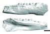
The present 3D Dataset contains the 3D models analyzed in Bianucci et al. 2023, A heavyweight early whale pushes the boundaries of vertebrate morphology, Nature. These include bones of the holotype of new species Perucetus colossus (MUSM 3248), as well as the articulated skeleton of Cynthiacetus peruvianus (holotype, MNHN.F.PRU10). The latter was used to estimate the total skeleton volume of P. colossus.
Perucetus colossus MUSM 3248 View specimen
|
M3#1131Thirteen vertebrae, rib, and innominate of Perucetus colossus (holotype, MUSM NNNN). Type: "3D_surfaces"doi: 10.18563/m3.sf.1131 state:published |
Download 3D surface file |
Cynthiacetus peruvianus MNHN.F.PRU10 View specimen
|
M3#1130Articulated skeleton of the holotype of Cynthiacetus peruvianus MNHN.F.PRU10 Type: "3D_surfaces"doi: 10.18563/m3.sf.1130 state:published |
Download 3D surface file |
This contribution contains 3D models of the holotype of a new species of long-nosed armadillos, the Guianan long-nosed armadillo (Dasypus guianensis) described in the following publication: Barthe M., Rancilhac L., Arteaga M. C., Feijó A., Tilak M.-K., Justy F., Loughry W. J., McDonough C. M., de Thoisy B., Catzeflis F., Billet G., Hautier L., Nabholz B., and Delsuc F. 2024. Exon capture museomics deciphers the nine-banded armadillo species complex and identifies a new species endemic to the Guiana Shield. Systematic Biology, syae027. https://doi.org/10.1093/sysbio/syae027
Dasypus guianensis MNHN-ZM-MO-2001-1317 View specimen
|
M3#1200Skeleton and carapace Type: "3D_surfaces"doi: 10.18563/m3.sf.1200 state:published |
Download 3D surface file |
|
M3#1201Frontal sinuses Type: "3D_surfaces"doi: 10.18563/m3.sf.1201 state:published |
Download 3D surface file |

The present 3D Dataset contains the 3D models of the skull of the holotype of Miocaperea pulchra.
Miocaperea pulchra SMNS-P-46978 View specimen

|
M3#1656Blender file containing two models (the skull being preserved in two parts) Type: "3D_surfaces"doi: 10.18563/m3.sf.1656 state:published |
Download 3D surface file |
We provide a 3D reconstruction of the skull of Latimeria chalumnae that can be easily accessed and visualized for a better understanding of its cranial anatomy. Different skeletal elements are saved as separate PLY files that can be combined to visualize the entire skull or isolated to virtually dissect the skull. We included some guidelines for a fast and easy visualization of the 3D skull.
Latimeria chalumnae MHNG 1080.070 View specimen

|
M3#1254the skeletal elements of the skull of Latimeria chalumnae included in 26 different PLY files Type: "3D_surfaces"doi: 10.18563/m3.sf.1254 state:published |
Download 3D surface file |
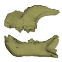
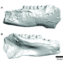
This contribution contains the 3D models described and figured in the following publication: Bonis et al. 2023. A new large pantherine and a sabre-toothed cat (Mammalia, Carnivora, Felidae) from the late Miocene hominoid-bearing Khorat sand pits, Nakhon Ratchasima Province, northeastern Thailand. The Science of Nature 110(5):42. https://doi.org/10.1007/s00114-023-01867-4
Pachypanthera piriyai CUF-KR-1 View specimen
|
M3#1209Holotype of Pachypanthera piriyai, a left hemi-mandible with alveoli for i1-i3 and canine, roots of p3, p4 and partially broken off m1 crown. Type: "3D_surfaces"doi: 10.18563/m3.sf.1209 state:published |
Download 3D surface file |
Pachypanthera piriyai CUF-KR-2 View specimen
|
M3#1210Paratype of Pachypanthera piriyai, a right hemi-maxilla with P3-P4, alveoli of C and M1, root of P2 Type: "3D_surfaces"doi: 10.18563/m3.sf.1210 state:published |
Download 3D surface file |
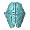
The present 3D Dataset contains the 3D model analyzed in the article : Dubied et al. (2021), Endocranium and ecology of Eurotherium theriodis, a European hyaenodont mammal from the Lutetian. Acta Palaeontologica Polonica 2021, https://doi.org/10.4202/app.00771.2020
Eurotherium theriodis NMB.Em12 View specimen
|
M3#381NMB.Em12 unprepared specimen Type: "3D_surfaces"doi: 10.18563/m3.sf.381 state:published |
Download 3D surface file |
|
M3#382NMB.Em12 cranium Type: "3D_surfaces"doi: 10.18563/m3.sf.382 state:published |
Download 3D surface file |
|
M3#383NMB.Em12 endocast Type: "3D_surfaces"doi: 10.18563/m3.sf.383 state:published |
Download 3D surface file |
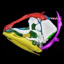
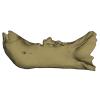
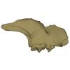
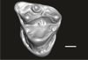
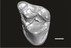
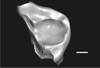
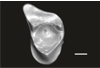
The present 3D dataset contains 3D models of new material from the middle Eocene of the Upper Subathu Formation in the Kalakot area (India), documenting the anterior dentition of the raoellid Indohyus indirae. Raoellidae are closely related to stem cetaceans and bring crucial information to understand the earliest phase of land to water transition in Cetacea.
Indohyus indirae GU/RJ/31 View specimen
|
M3#1505Right i1 Type: "3D_surfaces"doi: 10.18563/m3.sf.1505 state:published |
Download 3D surface file |
Indohyus indirae GU/RJ/32 View specimen
|
M3#1506Right i1 Type: "3D_surfaces"doi: 10.18563/m3.sf.1506 state:published |
Download 3D surface file |
Indohyus indirae GU/RJ/16 View specimen
|
M3#1507Left I3 Type: "3D_surfaces"doi: 10.18563/m3.sf.1507 state:published |
Download 3D surface file |
Indohyus indirae GU/RJ/23 View specimen
|
M3#1508Left I2 Type: "3D_surfaces"doi: 10.18563/m3.sf.1508 state:published |
Download 3D surface file |
Indohyus indirae GU/RJ/25 View specimen
|
M3#1509Left I1 Type: "3D_surfaces"doi: 10.18563/m3.sf.1509 state:published |
Download 3D surface file |
Indohyus indirae GU/RJ/26 View specimen
|
M3#1510Right I3 Type: "3D_surfaces"doi: 10.18563/m3.sf.1510 state:published |
Download 3D surface file |
Indohyus indirae GU/RJ/57 View specimen
|
M3#1511Left I1 Type: "3D_surfaces"doi: 10.18563/m3.sf.1511 state:published |
Download 3D surface file |
Indohyus indirae GU/RJ/61 View specimen
|
M3#1512Right upper canine Type: "3D_surfaces"doi: 10.18563/m3.sf.1512 state:published |
Download 3D surface file |
Indohyus indirae GU/RJ/63 View specimen

|
M3#1513Left upper canine Type: "3D_surfaces"doi: 10.18563/m3.sf.1513 state:published |
Download 3D surface file |
Indohyus indirae GU/RJ/74 View specimen
|
M3#1514Left upper canine Type: "3D_surfaces"doi: 10.18563/m3.sf.1514 state:published |
Download 3D surface file |
Indohyus indirae GU/RJ/439 View specimen
|
M3#1515Left upper canine Type: "3D_surfaces"doi: 10.18563/m3.sf.1515 state:published |
Download 3D surface file |
Indohyus indirae GU/RJ/457 View specimen
|
M3#1516Left upper canine Type: "3D_surfaces"doi: 10.18563/m3.sf.1516 state:published |
Download 3D surface file |
Indohyus indirae GU/RJ/846 View specimen
|
M3#1517Left upper canine Type: "3D_surfaces"doi: 10.18563/m3.sf.1517 state:published |
Download 3D surface file |
Indohyus indirae GU/RJ/822 View specimen
|
M3#1518Right fragmentary maxillary with decidual canine and I3 Type: "3D_surfaces"doi: 10.18563/m3.sf.1518 state:published |
Download 3D surface file |
Indohyus indirae GU/RJ/824 View specimen
|
M3#1519Right fragmentary mandible with lower canine and small part of the i3 Type: "3D_surfaces"doi: 10.18563/m3.sf.1519 state:published |
Download 3D surface file |
Indohyus indirae GU/RJ/838 View specimen
|
M3#1520Right fragmentary mandible with permanent i2, i3 and canine and small part of the root of the decidual i3 Type: "3D_surfaces"doi: 10.18563/m3.sf.1520 state:published |
Download 3D surface file |
Indohyus indirae GU/RJ/842 View specimen
|
M3#1521Left fragmentary mandible with decidual and permanent canine Type: "3D_surfaces"doi: 10.18563/m3.sf.1521 state:published |
Download 3D surface file |
Indohyus indirae GU/RJ/26,32,56,57,457,822,838,842 : Composite Anterior dentition View specimen
|
M3#15293D composite reconstruction of the anterior dentition of Indohyus indirae with GU/RJ/57 (I1), 56 (I2), 26 (I3), 457 (Upper canine), 822 (P1), 32 (i1), 838 (i2, i3 and lower canine) and 842 (p1) Type: "3D_surfaces"doi: 10.18563/m3.sf.1529 state:published |
Download 3D surface file |
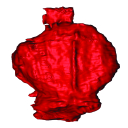
The present 3D Dataset contains the 3D models of the endocranial cast of two specimens of Indohyus indirae described in the article entitled “The endocranial cast of Indohyus (Artiodactyla, Raoellidae): the origin of the cetacean brain” (Orliac and Thewissen, 2021). They represent the cast of the main cavity of the braincase as well as associated intraosseous sinuses.
Indohyus indirae RR 207 View specimen
|
M3#710cast of the main endocranial cavity and associated intraosseous sinuses Type: "3D_surfaces"doi: 10.18563/m3.sf.710 state:published |
Download 3D surface file |
Indohyus indirae RR 601 View specimen
|
M3#711casts of the main endocranial cavity Type: "3D_surfaces"doi: 10.18563/m3.sf.711 state:published |
Download 3D surface file |

The present 3D Dataset contains the 3D model analyzed in the publication : On Roth’s “human fossil” from Baradero, Buenos Aires Province, Argentina: morphological and genetic analysis. The “human fossil” from Baradero, Buenos Aires Province, Argentina, is a collection of skeleton parts first recovered by Swiss paleontologist Santiago Roth and further studied by anthropologist Rudolf Martin. By the end of the 19th century and beginning of the 20th century it was considered as one of the oldest human skeletons from the southern cone. We studied the cranial anatomy and contextualized the ancient individual remains. We discuss the context of the finding, conducted an osteobiographical assessment and performed a 3D virtual reconstruction of the skull, using micro-CT-scans on selected skull fragments and the mandible. This was followed by the extraction of bone tissue and teeth samples for radiocarbon and genetic analyses, which brought only limited results due to poor preservation and possible contamination. We estimate that the individual from Baradero is a middle-aged adult male. We conclude that the revision of foundational collections with current methodological tools brings new insights and clarifies long held assumptions on the significance of samples that were recovered when archaeology was not yet professionalized.
Homo sapiens PIMUZ A/V 4217 View specimen
|
M3#11983D virtual reconstruction of the skull Type: "3D_surfaces"doi: 10.18563/m3.sf.1198 state:published |
Download 3D surface file |
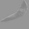
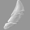
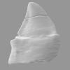
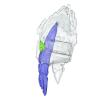
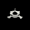
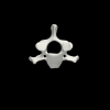
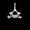
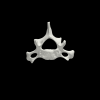
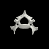
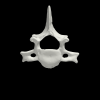
This contribution contains the 3D models of the ossicles of a protocetid archaeocete from the locality of Kpogamé, Togo, described and figured in the publication of Mourlam and Orliac (2019).
indet. indet. UM KPG-M 73 View specimen

|
M3#407stapes Type: "3D_surfaces"doi: 10.18563/m3.sf.407 state:published |
Download 3D surface file |

|
M3#408Incus Type: "3D_surfaces"doi: 10.18563/m3.sf.408 state:published |
Download 3D surface file |

|
M3#409Malleus Type: "3D_surfaces"doi: 10.18563/m3.sf.409 state:published |
Download 3D surface file |
The present 3D Dataset contains the 3D models analyzed in Merten, L.J.F, Manafzadeh, A.R., Herbst, E.C., Amson, E., Tambusso, P.S., Arnold, P., Nyakatura, J.A., 2023. The functional significance of aberrant cervical counts in sloths: insights from automated exhaustive analysis of cervical range of motion. Proceedings of the Royal Society B. doi: 10.1098/rspb.2023.1592
Ailurus fulgens PMJ_Mam_6639 View specimen
|
M3#1260cervical vertebral series (7 vertebrae) Type: "3D_surfaces"doi: 10.18563/m3.sf.1260 state:published |
Download 3D surface file |
Bradypus variegatus ZMB_Mam_91345 View specimen
|
M3#1261cervical vertebral series (8 vertebrae) + first thoracic vertebra Type: "3D_surfaces"doi: 10.18563/m3.sf.1261 state:published |
Download 3D surface file |
Bradypus variegatus ZMB_Mam_35824 View specimen

|
M3#1262cervical vertebral series (8 vertebrae) + first & second thoracic vertebra Type: "3D_surfaces"doi: 10.18563/m3.sf.1262 state:published |
Download 3D surface file |
Choloepus didactylus ZMB_Mam_38388 View specimen
|
M3#1263cervical vertebral series (7 vertebrae) Type: "3D_surfaces"doi: 10.18563/m3.sf.1263 state:published |
Download 3D surface file |
Choloepus didactylus ZMB_Mam_102634 View specimen
|
M3#1264cervical vertebral series (6 vertebrae) + first thoracic vertebra Type: "3D_surfaces"doi: 10.18563/m3.sf.1264 state:published |
Download 3D surface file |
Tamandua tetradactyla ZMB_Mam_91288 View specimen
|
M3#1266cervical vertebral series (7 vertebrae) + first thoracic vertebra Type: "3D_surfaces"doi: 10.18563/m3.sf.1266 state:published |
Download 3D surface file |
Glossotherium robustum MNHN_n/n View specimen
|
M3#1267cervical vertebral series (7 vertebrae) + first thoracic vertebra Type: "3D_surfaces"doi: 10.18563/m3.sf.1267 state:published |
Download 3D surface file |
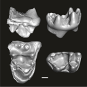
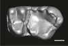
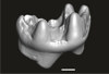
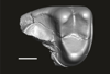
This contribution contains the three-dimensional digital models of eleven isolated fossil teeth of a merialine paroxyclaenid (Welcommoides gurki), discovered from lower Oligocene deposits of the Bugti Hills (Balochistan, Pakistan). These fossils were described, figured and discussed in the following publication: Solé et al. (2024), An unexpected late paroxyclaenid (Mammalia, Cimolesta) out of Europe: dental evidence from the Oligocene of the Bugti Hills, Pakistan. Papers in Palaeontology. https://doi.org/10.1002/spp2.1599
Welcommoides gurki UM-DBC 2225 View specimen
|
M3#1083Left m3 Type: "3D_surfaces"doi: 10.18563/m3.sf.1083 state:published |
Download 3D surface file |
Welcommoides gurki UM-DBC 2226 View specimen
|
M3#1084Right m3 Type: "3D_surfaces"doi: 10.18563/m3.sf.1084 state:published |
Download 3D surface file |
Welcommoides gurki UM-DBC 2227 View specimen
|
M3#1085Trigonid of a right lower molar Type: "3D_surfaces"doi: 10.18563/m3.sf.1085 state:published |
Download 3D surface file |
Welcommoides gurki UM-DBC 2230 View specimen
|
M3#1086Right DP4 Type: "3D_surfaces"doi: 10.18563/m3.sf.1086 state:published |
Download 3D surface file |
Welcommoides gurki UM-DBC 2228 View specimen
|
M3#1093Right M1 Type: "3D_surfaces"doi: 10.18563/m3.sf.1093 state:published |
Download 3D surface file |
Welcommoides gurki UM-DBC 2229 View specimen
|
M3#1087Right M2 Type: "3D_surfaces"doi: 10.18563/m3.sf.1087 state:published |
Download 3D surface file |
Welcommoides gurki UM-DBC 2236 View specimen
|
M3#1088Left M2 Type: "3D_surfaces"doi: 10.18563/m3.sf.1088 state:published |
Download 3D surface file |
Welcommoides gurki UM-DBC 2231 View specimen
|
M3#1089Left M3 Type: "3D_surfaces"doi: 10.18563/m3.sf.1089 state:published |
Download 3D surface file |
Welcommoides gurki UM-DBC 2232 View specimen
|
M3#1090Left M3 Type: "3D_surfaces"doi: 10.18563/m3.sf.1090 state:published |
Download 3D surface file |
Welcommoides gurki UM-DBC 2234 View specimen
|
M3#1091Left M3 Type: "3D_surfaces"doi: 10.18563/m3.sf.1091 state:published |
Download 3D surface file |
Welcommoides gurki UM-DBC 2233 View specimen
|
M3#1092Left M3 Type: "3D_surfaces"doi: 10.18563/m3.sf.1092 state:published |
Download 3D surface file |


